
Tuberculosis types with xray and findings
Reading File
Now let me search the library for TB types and X-ray findings in parallel.
Finding Sources
Finding Sources
Excellent - found great sources. Let me now read the radiology and respiratory medicine blocks on TB in parallel, plus grab the key X-ray images.
Reading File
Reading File
Finding Sources
Excellent content found. Now let me fetch the key X-ray images and read more from Murray & Nadel for miliary TB and the tuberculosis natural history diagram.
Loading Image
Loading Image
Loading Image
Reading File
Now let me fetch the miliary and HIV-TB X-ray images, and also fetch the primary TB X-ray from Grainger & Allison.
Loading Image
Loading Image
Loading Image
Reading File
I have all the key images and content I need. Now let me compile the comprehensive answer.
Tuberculosis: Types, X-ray Findings & Radiological Features
Overview - Natural History
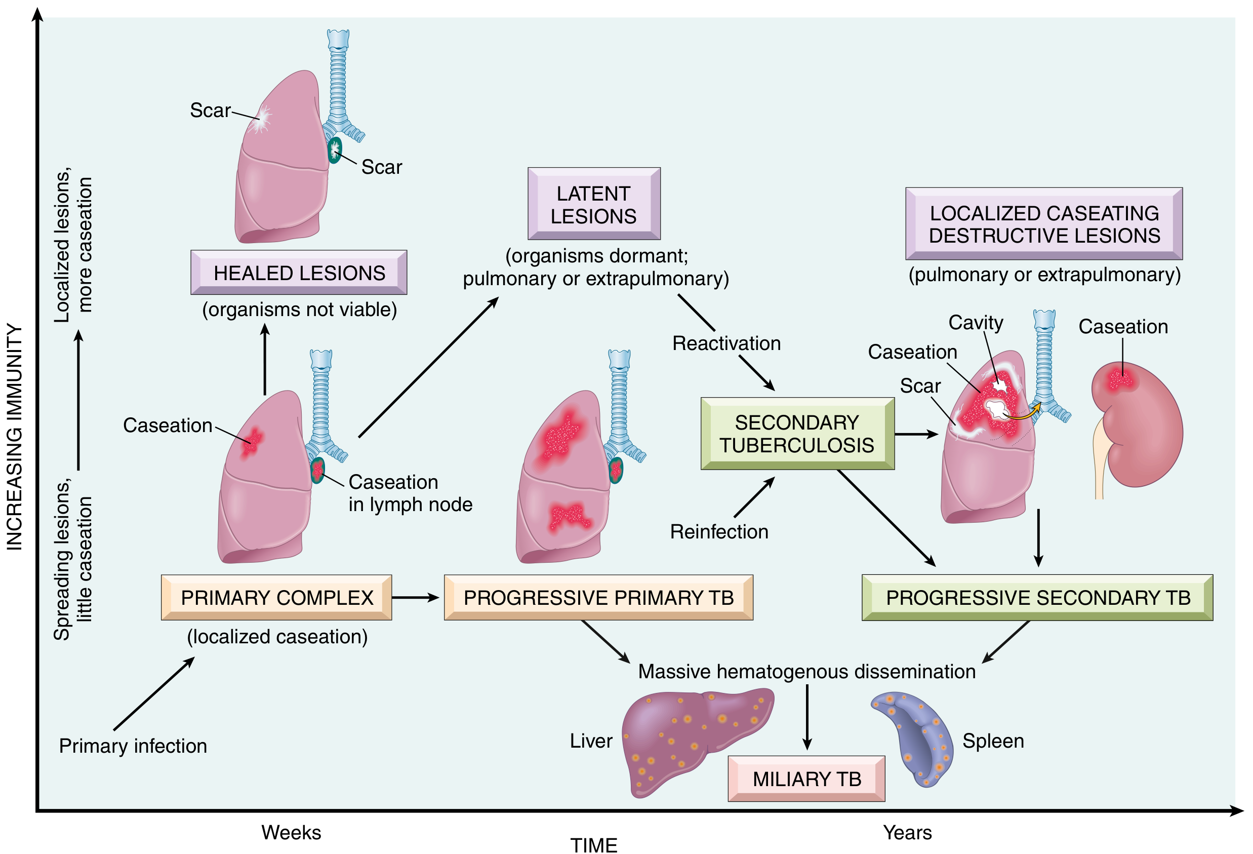
The natural history of TB - Primary Complex → Progressive Primary TB / Latent Lesions → Secondary (Reactivation) TB / Miliary TB (from Robbins & Kumar Basic Pathology)
1. Primary Tuberculosis
Definition: Results from the initial infection with Mycobacterium tuberculosis in a host with no prior immunity.
Pathology
- The inhaled bacilli settle in the mid/lower lung zones and form the Ghon focus (area of consolidation).
- Draining lymphatics carry bacilli to the ipsilateral hilar nodes → together these form the Ghon complex (primary complex).
- In most immunocompetent individuals, the infection is controlled; the lesions undergo caseous necrosis, calcification, and scarring.
- If immunity fails to contain it → Progressive Primary TB (see below).
Chest X-ray Findings
- Middle or lower lung zone opacity/consolidation (unlike reactivation TB, which is upper lobe)
- Ipsilateral hilar/paratracheal lymphadenopathy - this is the most characteristic feature
- Possible pleural effusion
- Possible atelectasis from lymph node compression of airways
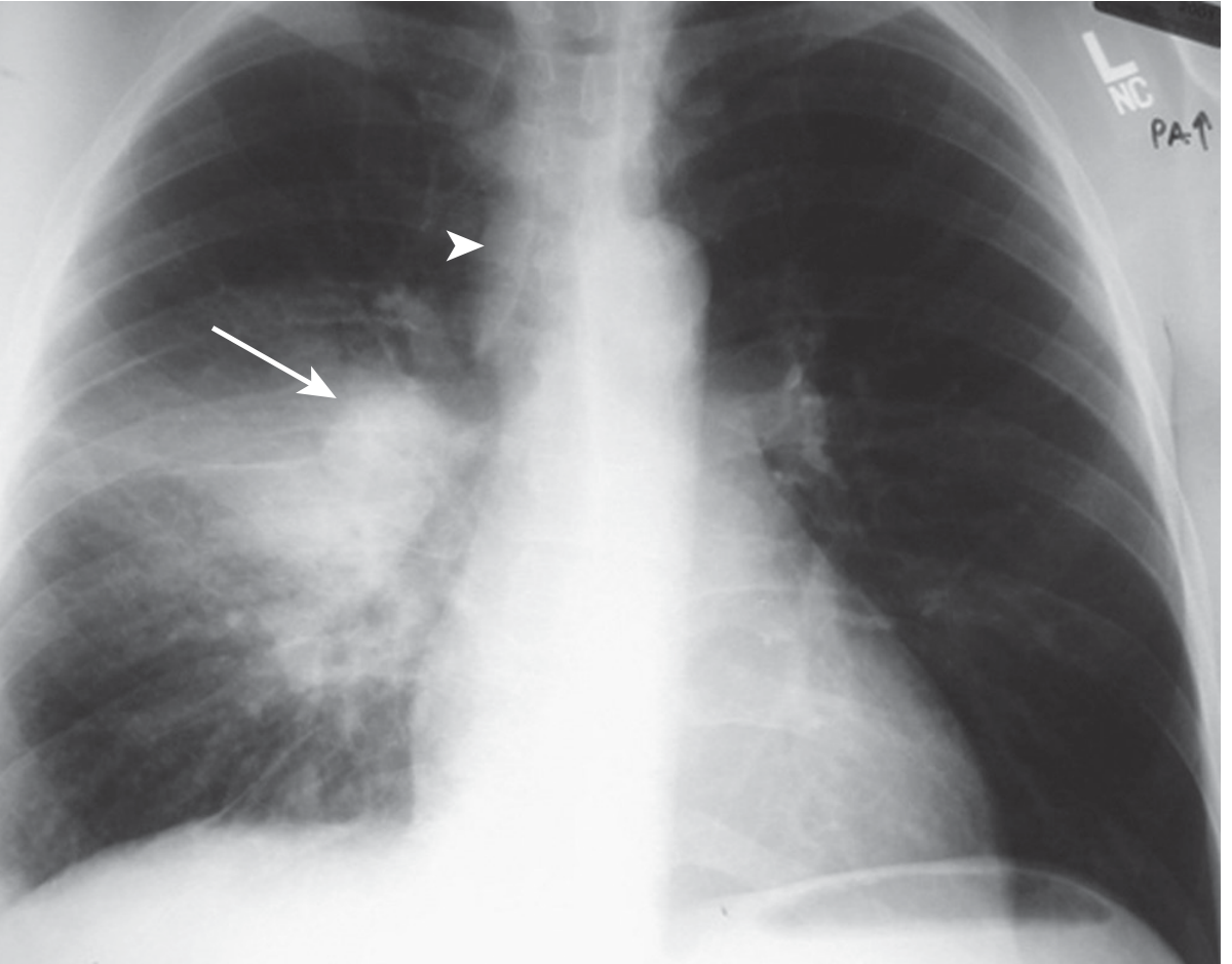
Figure 53.1 - Primary TB: Right lower lobe consolidation + right hilar lymphadenopathy (arrow) + right paratracheal lymph node enlargement (arrowhead). (Murray & Nadel's Respiratory Medicine)
From Grainger & Allison (shown below), primary TB in a child with hilar adenopathy:
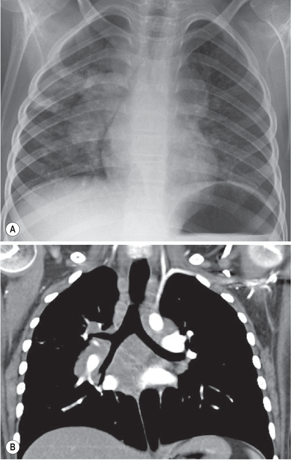
Fig. 70.28 Primary Tuberculosis - CXR (A) and coronal CT (B) demonstrating hilar adenopathy. (Grainger & Allison's Diagnostic Radiology)
2. Progressive Primary Tuberculosis
Definition: Primary infection that fails to be contained by developing cell-mediated immunity. More common in children, the elderly, and immunosuppressed.
X-ray Findings
- Rapidly enlarging pulmonary consolidation (lobar pattern)
- Cavitation can form in the primary focus
- Prominent hilar/mediastinal adenopathy
- May be indistinguishable from acute bacterial lobar pneumonia
3. Secondary (Reactivation) Tuberculosis
Definition: Develops when a previously infected individual undergoes reactivation of latent TB - either spontaneously (due to waning immunity) or through reinfection. This is the most common form in adults.
Pathology
- Reactivation driven by falling cell-mediated immunity (HIV, malnutrition, steroids, DM, old age, TNF inhibitors)
- Localizes to the lung apices because of higher oxygen tension there
- Hallmark: caseous necrosis with cavity formation
- Bronchogenic spread can carry infected material to other parts of the lung
Chest X-ray Findings
- Upper lobe involvement - predominantly apical and posterior segments of the right upper lobe (most common site), also apical-posterior segment of the left upper lobe
- Cavitation - thick-walled cavities, often in the upper lobes (highly characteristic)
- Fibronodular or patchy consolidation
- Volume loss and fibrosis with superior hilar retraction (old healed disease)
- Calcifications in healed lesions (Ranke complex)
- Bronchogenic spread pattern: new acinar/nodular opacities ("tree-in-bud") in ipsilateral lower lobe or contralateral lung
- Absence of hilar adenopathy (in contrast to primary TB)
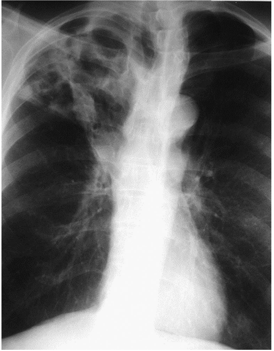
Figure 53.2 - Cavitary (Reactivation) TB: Extensive right upper lobe cavitation. (Murray & Nadel's Respiratory Medicine)
4. Miliary Tuberculosis
Definition: Results from massive hematogenous dissemination of M. tuberculosis - can occur as a complication of either primary or reactivation TB. The term "miliary" comes from the radiographic appearance resembling millet seeds (1-5 mm each).
At-risk groups
Children, elderly, immunocompromised (HIV, organ transplant, corticosteroids). Mortality rate is 20-30%.
Chest X-ray Findings
- Diffuse, bilateral, uniformly distributed tiny nodules (1-3 mm) throughout both lung fields
- Nodules are randomly distributed - no zone predilection
- "Snow-storm" or "millet seed" appearance
- May be subtle early on; CT is far more sensitive
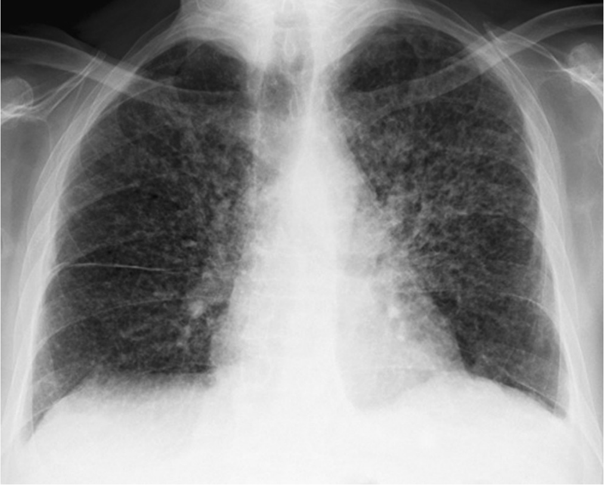
Figure 53.3 - Miliary TB: Numerous small, randomly distributed bilateral nodules ("miliary pattern"). (Murray & Nadel's Respiratory Medicine)
5. Tuberculosis in HIV/Immunocompromised Patients
The radiographic pattern depends on the degree of CD4 depletion:
| CD4 count | Pattern |
|---|---|
| Early HIV (CD4 relatively preserved) | Typical pattern - upper lobe infiltrates, cavitation |
| Advanced HIV (low CD4 / AIDS) | Atypical pattern - lower zone or diffuse opacities, prominent hilar/mediastinal adenopathy, NO cavitation |
| Some HIV patients | Normal chest radiograph despite active TB |
X-ray Findings (Advanced HIV)
- Bilateral hilar lymphadenopathy (prominent, unlike immunocompetent reactivation)
- Diffuse nodular or consolidative opacities
- Lower lung zone involvement
- Cavitation uncommon
- Pleural effusion more frequent
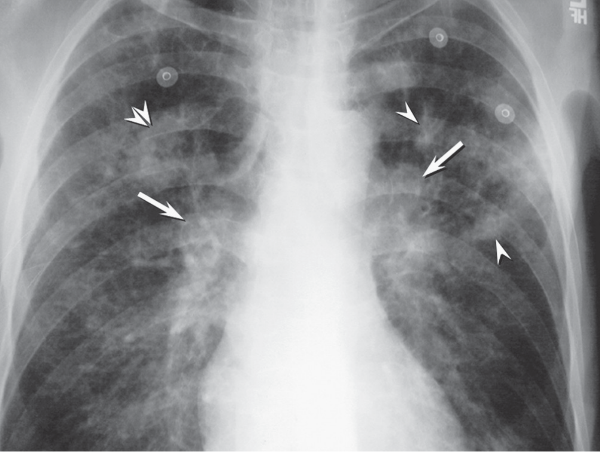
Figure 53.4 - TB + HIV: Bilateral hilar lymph node enlargement (arrows) + nodular opacities (arrowheads) + nodular consolidation (double arrowheads). (Murray & Nadel's Respiratory Medicine)
Summary Table
| Type | Location on CXR | Key Feature | Adenopathy | Cavitation |
|---|---|---|---|---|
| Primary TB | Mid/lower zones | Consolidation + hilar LN | Yes (ipsilateral) | Rare |
| Progressive Primary | Any zone, lobar | Rapid consolidation | Yes | Sometimes |
| Latent TB | Normal | No active lesion | No | No |
| Reactivation (Secondary) TB | Upper lobes (apical/posterior) | Cavitary fibronodular | Absent | Common |
| Miliary TB | Diffuse bilateral | 1-3 mm nodules ("millet seeds") | Variable | No |
| TB + HIV | Lower zones / diffuse | Atypical infiltrates | Yes (bilateral) | Uncommon |
Additional Radiological Notes
- Ranke complex: Calcified primary focus (Ghon focus) + calcified hilar lymph node = healed primary TB
- Simon foci: Small calcified apical nodules representing early hematogenous seeding - can reactivate years later
- CT advantages: Sensitivity 96% vs 48% for plain CXR in active TB. CT better detects lymphadenopathy, "tree-in-bud" pattern (bronchogenic spread), and cavity walls.
- "Tree-in-bud" sign on CT: Centrilobular nodules connected by branching linear densities - highly suggestive of endobronchial spread of TB.
- Activity cannot be determined from a single CXR alone - a cavity may be a sterile old lesion, and a fibrotic-appearing area may be active.
Sources: Murray & Nadel's Textbook of Respiratory Medicine, Grainger & Allison's Diagnostic Radiology, Robbins & Kumar Basic Pathology, Goldman-Cecil Medicine, Tintinalli's Emergency Medicine
This is a shared conversation. Sign in to Orris to start your own chat.