
Vitiligo
vitiligo
Vitiligo
Definition
- Fitzpatrick's Dermatology, Ch. 76
- Dermatology 2-Volume Set 5e, Ch. 66
Epidemiology
- Prevalence: ~0.5-1% worldwide; up to >8% in parts of India
- Peak onset: ages 10-30 years; ~50% of cases begin before age 20
- No definitive sex predilection, though females seek care more often
- Can occur in bone marrow transplant recipients from donors with vitiligo (supports immune transfer)
- Monozygotic twin concordance only ~23%, supporting an important environmental/non-genetic component
Pathogenesis
- Activated keratinocytes produce CXCL9 and CXCL10
- These recruit CD4+ helper and CD8+ cytotoxic T cells (which express CXCR3) to the skin
- CD8+ T cells produce IFN-γ and TNF, directly destroying melanocytes
- CD8+ T cells are both necessary and sufficient for melanocyte destruction
Clinical Features
Lesion Characteristics
- Well-defined, milk-white macules/patches with smooth, convex margins
- Surrounding skin may appear normal or hyperpigmented (normal or hyperpigmented border)
- Trichrome vitiligo: intermediate tan zones between normal skin and fully depigmented areas (especially in early disease)
- Leukotrichia: white hairs within depigmented patches
- Rarely: inflammatory erythematous border (can mimic tinea corporis)
Clinical Types / Distribution
| Type | Description |
|---|---|
| Generalized (most common) | Symmetric involvement; face, chest, hands, axillae, groin |
| Acrofacial | Distal fingers and facial orifices (lips, tips) |
| Segmental | Unilateral, Blaschkoid pattern; spreads for 1-2 years then stabilizes |
| Focal | Single or few macules in one area, often trigeminal (especially in children) |
| Mucosal | Oral/genital mucosa only |
| Universal | Nearly entire body surface depigmented |
Predilection Sites
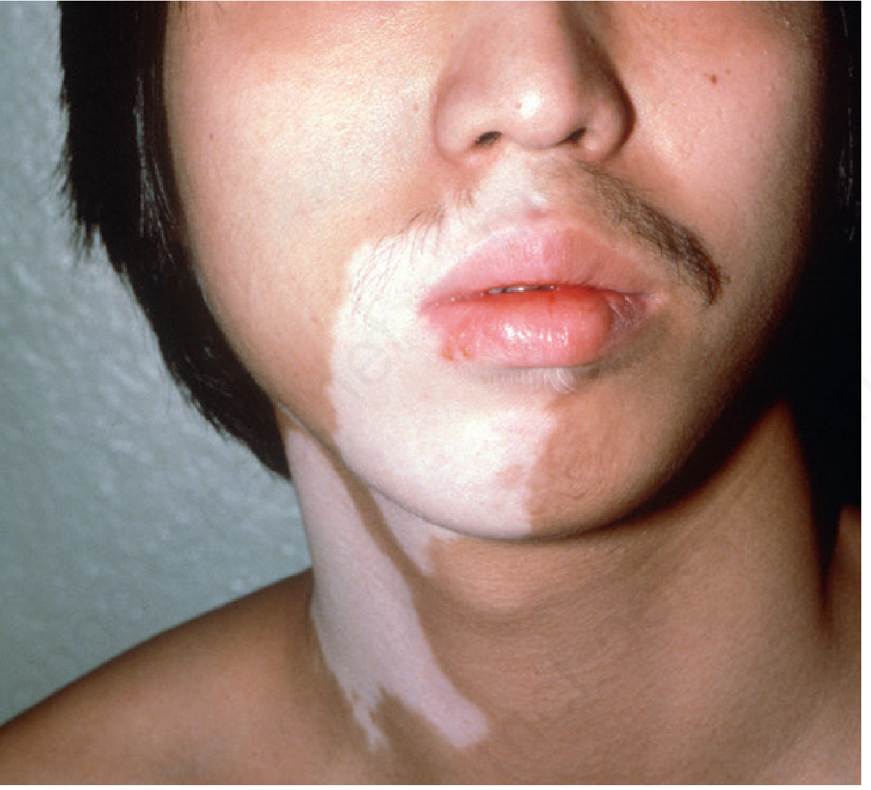
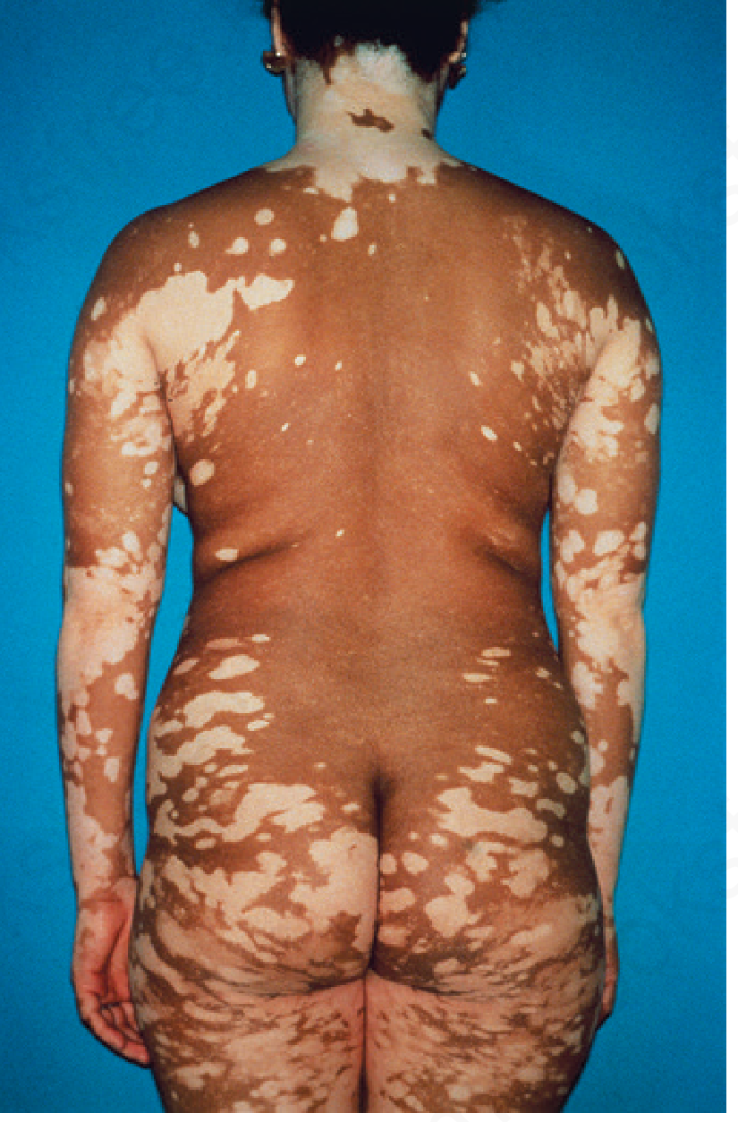
Signs of Active Disease
- Confetti-like lesions
- Hypomelanotic lesions with poorly defined borders
- Trichrome pattern
- Inflammatory (erythematous) border
- Koebnerization
Associated Conditions
- Autoimmune thyroid disease (~15% of adults, ~5-15% of children with vitiligo)
- Alopecia areata
- Pernicious anemia
- Type 1 diabetes mellitus
- Addison's disease
- Rheumatoid arthritis, lupus
- Up to 20% of patients have at least one comorbid autoimmune disorder
- Vogt-Koyanagi-Harada (VKH) syndrome: uveitis + aseptic meningitis + otic involvement + vitiligo/poliosis
- Alezzandrini syndrome: unilateral facial depigmentation + ipsilateral ocular changes
- Halo nevi: depigmented halo around melanocytic nevi - associated with vitiligo
Diagnosis
- Wood's lamp (UV-A light): Enhances contrast of depigmented lesions; particularly useful in fair-skinned patients - lesions fluoresce bright white
- Skin biopsy: Absence of melanocytes on DOPA stain; sparse lymphocytic infiltrate at active edges
- Lab workup for associated autoimmune diseases: TSH, anti-TPO antibodies, FBS, CBC, ANA
Disease Assessment Tools
- Vitiligo Area Scoring Index (VASI)
- Vitiligo European Task Force (VETF) score
- Vitiligo Extent Score (VES)
Treatment
1. Topical Therapies
| Agent | Notes |
|---|---|
| Topical corticosteroids | First-line for localized disease; moderate-to-potent class; risk of atrophy with prolonged use |
| Topical calcineurin inhibitors (tacrolimus 0.1%, pimecrolimus 1%) | Preferred for face and flexures; avoids atrophy; also used as maintenance (twice-weekly application) |
| Topical JAK inhibitors (ruxolitinib 1.5%) | Newer; FDA-approved for non-segmental vitiligo; targets JAK1/JAK2-IFN-γ pathway |
2. Phototherapy
- Narrowband UVB (NB-UVB): Most widely used phototherapy; 311-313 nm; effective for generalized disease; can combine with topical agents
- PUVA (psoralen + UVA): Oral or topical psoralen + UVA exposure; effective but more side effects (nausea, phototoxicity, cataract risk with oral PUVA); used less now with NB-UVB available
- Excimer laser (308 nm): Targeted NB-UVB; best for localized, resistant lesions
3. Systemic Therapies
- Oral corticosteroids (mini-pulse): Weekly low-dose dexamethasone or betamethasone; used to halt rapid progression
- Oral JAK inhibitors (ruxolitinib, baricitinib): Emerging; target CXCL9/10-CXCR3 and IFN-γ signaling; promising results in trials
- Oral PUVA (less commonly used now)
4. Surgical Options (for stable, localized vitiligo)
- Suction blister epidermal grafting - most widely used
- Split-thickness skin grafting
- Melanocyte-keratinocyte transplant procedure (MKTP)
- Punch grafting
5. Depigmentation
- Monobenzyl ether of hydroquinone (MBEH) 20% applied 1-2x/day for 9-12 months
- Requires lifelong strict photoprotection afterward
- Can cause contact dermatitis; may depigment distant sites
6. Cosmetic
- Camouflage cosmetics (cover-up makeup, self-tanning products)
- Micropigmentation (tattooing): Especially useful for lips, nipples, distal fingers - areas with poor repigmentation response
Prognosis & Course
- Course is unpredictable - may spread slowly, stabilize, or rarely progress rapidly
- Segmental vitiligo: reaches full extent within 1-2 years then usually stabilizes
- Generalized vitiligo: typically slow, chronic progression
- Spontaneous partial repigmentation can occur (especially with sun exposure); complete spontaneous repigmentation is rare
- After successful treatment, relapse rate ~40%
Mental Health Impact
Recent evidence note (PMID 40192197): A 2025 systematic review on off-label topical ruxolitinib in dermatology confirms its growing use in vitiligo, supporting the JAK inhibitor class as an important emerging treatment option.
Auscultation in front and back amd which condn also the sounds
Lung Auscultation: Front, Back & Clinical Correlations
Technique Basics
- Use the diaphragm of the stethoscope for higher-pitched sounds (breath sounds, wheezes, crackles)
- Use the bell for lower-pitched sounds (low-pitched rhonchi, cardiac sounds)
- Apply directly to skin, not through clothing - full skin contact is required
- Patient breathes through an open mouth with slow, deep breaths
- Room must be quiet
Auscultation Areas: Anterior (Front)
| Area | Lung Region Heard | Notes |
|---|---|---|
| Supraclavicular fossa (above clavicle) | Lung apex | Both sides |
| Infraclavicular area (below clavicle, 1st-2nd ICS) | Upper lobe | Both sides |
| 2nd-4th ICS, mid-clavicular line | Upper + mid lobe (R), Upper lobe (L) | |
| 4th-5th ICS, anterior axillary line | Right middle lobe (R) / Lingula (L) | ONLY heard anteriorly |
Key point: The right middle lobe and the lingula of the left lung can ONLY be auscultated anteriorly - they cannot be heard from the back.
Auscultation Areas: Posterior (Back)
| Area | Lung Region Heard | Notes |
|---|---|---|
| Suprascapular area (above spine of scapula) | Upper lobe apex | |
| Interscapular area (T3-T7, between scapulae) | Upper and middle lobes | Best heard with arms crossed to move scapulae apart |
| Infrascapular area (below T7) | Lower lobes | Bilateral - most important zone posteriorly |
| Costovertebral angle / lung bases | Lower lobe bases | Often the earliest site for basal crackles (e.g., heart failure) |
Compare left vs right at each level before moving down. Listen to at least 3 levels posteriorly (upper, mid, lower).
Systematic Sequence (Standard Approach)
- Upper zones (between neck and spine of scapula)
- Mid zones (interscapular)
- Lower zones (below scapulae, down to bases)
- Apices (supraclavicular)
- Upper zones (infraclavicular)
- Mid zones (right middle lobe / lingula)
- Mid-axillary line at 4th-6th ICS
- Important for detecting early pleural effusion and lower lobe pathology
Types of Breath Sounds
Normal Sounds
| Sound | Characteristics | Normal Location |
|---|---|---|
| Vesicular (normal breath sounds) | Soft, low-pitched; inspiration louder and longer than expiration (3:1 ratio); no gap between I and E | Peripheral lung fields (all zones except trachea) |
| Bronchial (tracheal) | Loud, high-pitched, hollow; expiration = inspiration or louder; gap between I and E | Over trachea/manubrium only (normal there) |
| Bronchovesicular | Intermediate intensity and pitch; I:E roughly equal | 1st and 2nd ICS anteriorly; between scapulae posteriorly |
Adventitious (Abnormal) Sounds - ATS Classification
| Sound | Characteristics | Mechanism | Clinical Condition |
|---|---|---|---|
| Fine crackles | Soft, high-pitched, short, discontinuous; mid-to-late inspiratory; like rubbing hair near the ear | Sudden opening of collapsed distal airways | Pulmonary fibrosis, early pulmonary edema, early pneumonia |
| Coarse crackles | Loud, low-pitched, longer, early inspiratory or expiratory; may clear with cough | Opening of secretion-laden distal airways | Bronchiectasis, COPD, pneumonia with secretions |
| Wheezes | Continuous (>250 ms), high-pitched (≥400 Hz), musical/hissing | Airway narrowing - oscillation of airway walls | Asthma, COPD (diffuse); endobronchial lesion (focal) |
| Rhonchi | Continuous (>250 ms), low-pitched (<200 Hz), snoring quality; may clear with cough | Secretions in large airways | COPD with secretions, chronic bronchitis |
| Stridor | Loud, high-pitched, mainly inspiratory; heard best over neck | Extrathoracic/upper airway obstruction | Croup, epiglottitis, anaphylaxis, foreign body - URGENT |
| Bronchial sounds in periphery | Abnormal when heard outside trachea/manubrium | Airless air spaces around a patent airway | Consolidation (pneumonia), atelectasis |
| Pleural friction rub | Creaking/grating, like leather rubbing; both inspiratory and expiratory; disappears when breath held | Inflamed pleural surfaces rubbing | Pleuritis, pulmonary infarction (PE), pleuropneumonia |
Vocal Resonance Tests
| Test | Method | Normal | Consolidation | Pleural Effusion |
|---|---|---|---|---|
| Bronchophony | Patient says "99" | Muffled | Loud and clear | Absent/muffled |
| Egophony | Patient says "E" | Sounds like "E" | Sounds like "A" (E-to-A change) | Absent (except at effusion upper border) |
| Whispered pectoriloquy | Patient whispers "1-2-3" | Faint/inaudible | Clearly heard | Absent |
Clinical Correlations: Auscultation Findings by Disease
| Disorder | Breath Sounds | Added Sounds | Other |
|---|---|---|---|
| Pneumonia (consolidation) | Bronchial breath sounds (periphery) | Crackles (fine/coarse) | Bronchophony, egophony, whispered pectoriloquy all increased; dullness on percussion |
| Pleural effusion (large) | Absent/decreased | None (or rub above effusion line) | Dullness; stony dullness; trachea shifted AWAY; decreased fremitus; egophony at upper border only |
| Pneumothorax | Absent on affected side | None | Hyperresonance; trachea shifted AWAY (tension PTX); absent fremitus |
| Bronchial asthma (acute) | Vesicular with prolonged expiration | Expiratory wheezes (may be pan-inspiratory/expiratory) | Hyperresonant; decreased expansion; decreased fremitus |
| COPD | Decreased/quiet breath sounds | Wheezes, rhonchi | Hyperinflation; pursed-lip breathing; barrel chest |
| Atelectasis | Absent/decreased | None | Dullness; trachea shifted TOWARD affected side; decreased fremitus |
| Pulmonary fibrosis (ILD) | Normal or reduced | Fine "Velcro" crackles (bilateral, basal, end-inspiratory) | Clubbing; restrictive spirometry |
| Pulmonary edema (CHF) | Decreased at bases | Bilateral basal fine crackles (initially), progressing upward | S3 gallop; JVD; orthopnea |
| Bronchiectasis | Coarse or bronchial | Coarse crackles, wheezes | Persistent; sputum production |
| Pulmonary embolism | Usually normal | Pleural rub (if infarction) | Tachycardia; may have minimal auscultatory findings |
| Foreign body (large airway) | Decreased unilaterally | Focal wheeze (unilateral, monophonic) | Stridor if upper airway |
| Lung collapse (lobar) | Absent over that lobe | None | Trachea shifted toward collapse |
Quick Memory Summary
- Bronchial sounds in periphery = consolidation or atelectasis (airless lung around open bronchus)
- Fine crackles at bases = pulmonary edema or fibrosis
- Coarse crackles clearing with cough = secretions (bronchiectasis, COPD)
- Diffuse expiratory wheeze = asthma or COPD
- Monophonic/unilateral wheeze = endobronchial obstruction
- Absent breath sounds + hyperresonance = pneumothorax
- Absent breath sounds + dull percussion = effusion or total atelectasis
- Pleural rub = pleuritis/infarct
- Stridor = upper airway emergency
Clot in brain
ischemic stroke CT brain infarct
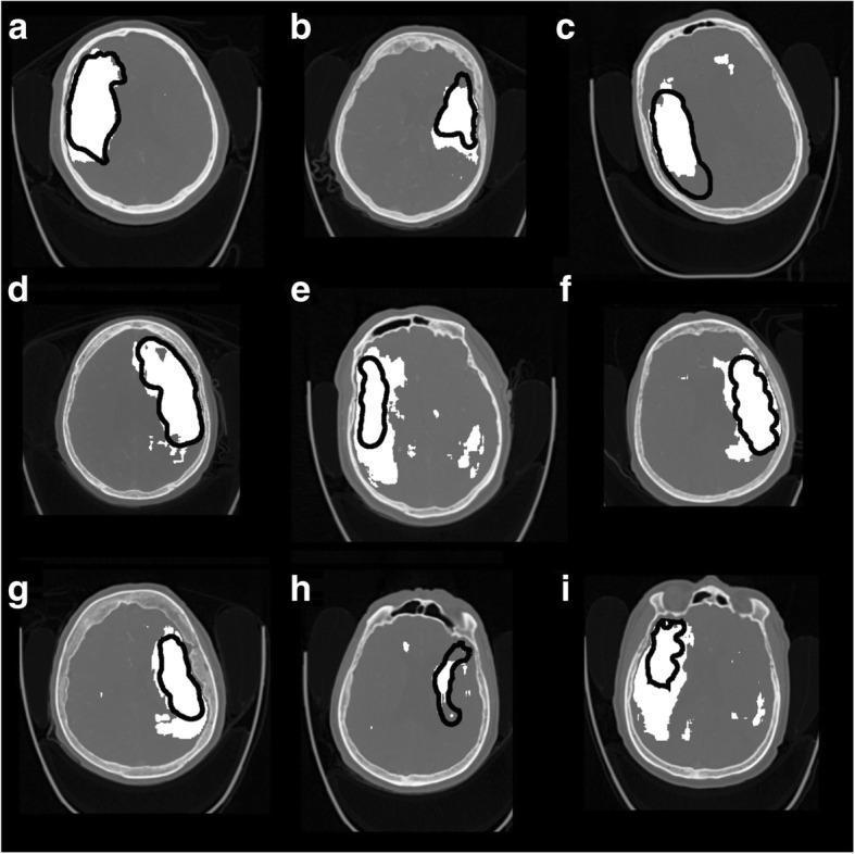
Diagnostic imaging series displaying nine axial Computed Tomography (CT) brain slices (labeled a-i) from patients with ischemic stroke. Each slice demonstrates the localization of an infarct through a comparison of human and automated segmentation. The brain tissue is presented in standard grayscale, showing normal anatomical structures such as the ventricles and cerebral hemispheres. Superimposed on these scans are two primary markers: solid white regions and black perimeter outlines. The solid white areas represent the output of a 3D Convolutional Neural Network (CNN) trained on CT angiography (CTA) and non-contrast CT (NCCT) features to identify ischemic lesions. The black perimeters delineate the 'ground truth' infarcted regions manually segmented by radiologists. Across the series, the images illustrate varying degrees of spatial overlap and discrepancy between the AI model and the human experts, highlighting clinical challenges in automated lesion quantification, particularly near the cortical surface and periventricular white matter. This comparison is used to evaluate the diagnostic accuracy, sensitivity, and specificity of deep learning algorithms in neuroradiology.
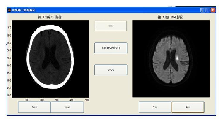
This Comparison Chart illustrates the side-by-side diagnostic evaluation of an ischemic stroke using a customized computer-aided assessment interface. The left panel shows an axial Non-Contrast Computed Tomography (NCCT) scan of the brain, demonstrating the characteristic hyperdense skull and low-contrast parenchyma where early subtle hypodensity might represent an acute infarct. The right panel displays a corresponding Magnetic Resonance Image (MRI), likely a Diffusion-Weighted Imaging (DWI) sequence, of the same patient. This MRI shows a distinct, localized hyperintense (bright) lesion in the left hemisphere adjacent to the lateral ventricle, which clearly delineates the area of acute cerebral infarction. The comparison highlights the higher sensitivity of MRI over CT in the hyperacute and acute phases of stroke. The interface includes navigational controls and analysis buttons ('RUN', 'Select Other DIR', 'SAVE'), indicating its use as a clinical decision support tool designed to assist radiologists in identifying and localizing stroke-affected regions.
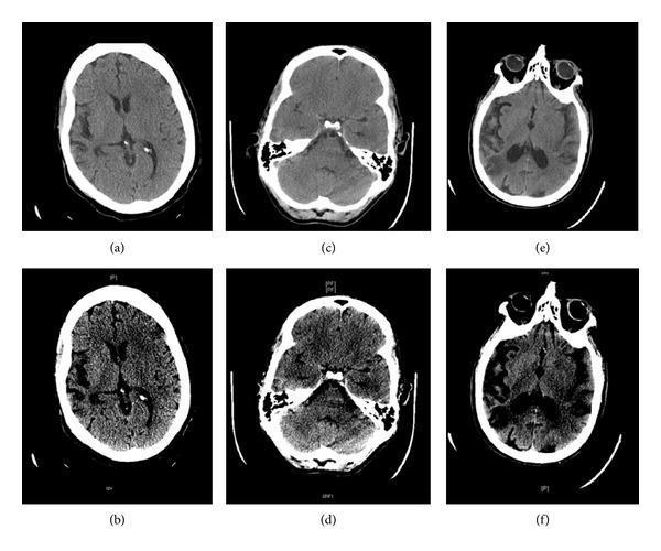
This diagnostic comparison contains six axial non-contrast head CT (NCCT) scans demonstrating the utility of 'Stroke Windows' in identifying acute ischemic stroke markers. The top row (a, c, e) shows head CTs in standard brain windows (level 35 HU, width 100 HU), while the bottom row (b, d, f) displays the corresponding levels in narrow stroke windows (level 35 HU, width 30 HU). The narrow windowing enhances tissue contrast to highlight early infarct signs. Key pathological findings visualized include: 1) Left sulcal effacement of the insular ribbon and hypodensity of the left basal ganglia (images a and b); 2) A right hyperdense middle cerebral artery (MCA) sign, indicative of acute thrombosis (images c and d); and 3) Hypodensity of the left basal ganglia (images e and f). These comparisons illustrate how adjusting window width and level can improve the visibility of subtle gray-white matter differentiation loss and vascular signs, facilitating earlier detection of ischemic territory in neuroradiology.
Clot in the Brain
| Type | Vessel Involved | Effect |
|---|---|---|
| Ischemic Stroke | Arteries (blocked) | Brain tissue dies from lack of blood supply |
| Cerebral Venous Thrombosis (CVT) | Veins/venous sinuses (blocked) | Blood backs up, causing venous infarction + raised ICP |
PART 1: ISCHEMIC STROKE (Arterial Clot)
Definition
Types / Mechanisms
- Clot forms directly at an atherosclerotic plaque, usually at vessel bifurcations (especially the internal carotid artery)
- Turbulent flow at bifurcations causes plaque ulceration → platelet adhesion → thrombus formation
- When stenosis exceeds 90% of vessel diameter, flow is critically reduced
- Involve small terminal vessels; infarcts 2-20 mm in size
- Affect: basal ganglia, thalamus, pons, internal capsule
- Mechanism: lipohyalinosis (from hypertension) or microemboli
- ~80-90% of lacunar stroke patients have hypertension
- Clot travels from heart to brain
- Atrial fibrillation is the most common cause (5x increased stroke risk)
- Also: mural thrombus from MI, valvular disease, patent foramen ovale
- Clot breaks off a proximal arterial plaque (e.g., carotid bifurcation) and travels distally
- Classic example: amaurosis fugax (carotid plaque embolizes to ophthalmic artery → transient monocular blindness)
- Arterial dissection (leading cause in young; carotid or vertebral; follows trauma, sneezing)
- Hypercoagulable states: antiphospholipid antibodies, protein C/S deficiency, sickle cell, polycythemia
- Oral contraceptives, pregnancy
- Vasospasm: cocaine, amphetamines, migraine
- Vasculitis, infections (varicella, fungal meningitis)
Risk Factors
| Modifiable | Non-modifiable |
|---|---|
| Hypertension | Age |
| Atrial fibrillation | Male sex |
| Diabetes | Race (Black, Hispanic > White) |
| Smoking | Family history |
| Dyslipidemia | Prior stroke/TIA |
| Obesity, sedentary lifestyle | |
| Carotid stenosis |
Clinical Features - FAST + MORE
- Face drooping (unilateral)
- Arm weakness (unilateral)
- Speech difficulty (dysarthria or aphasia)
- Time to call emergency services
| Territory | Artery | Symptoms |
|---|---|---|
| Middle cerebral artery (MCA) | MCA | Contralateral hemiplegia (face + arm > leg), hemisensory loss, homonymous hemianopia, aphasia (dominant), neglect (non-dominant) |
| Anterior cerebral artery (ACA) | ACA | Contralateral leg weakness > arm, personality change, urinary incontinence |
| Posterior circulation | Vertebrobasilar | Diplopia, dysphagia, vertigo, ataxia, crossed deficits (face ipsilateral, body contralateral), coma |
| Lacunar | Lenticulostriate | Pure motor hemiplegia, pure sensory stroke, ataxic hemiparesis, dysarthria-clumsy hand syndrome |
Imaging
- Normal in first 6 hours (early ischemia may be invisible)
- Early signs: loss of grey-white differentiation, hyperdense MCA sign (clot visible as white in vessel), sulcal effacement
- Later: hypodense (dark) area in infarcted territory
- Hyperdense MCA sign = acute thrombosis in the MCA
- Basal ganglia hypodensity = early infarct
- Bright (hyperintense) lesion visible within minutes of infarct
- Far more sensitive than CT in hyperacute phase
Management - Time is Brain!
| Step | Target |
|---|---|
| Door to doctor | 10 min |
| Door to CT completion | 25 min |
| Door to CT reading | 45 min |
| Door to treatment (tPA) | 60 min |
Acute Treatment
- Indicated within 4.5 hours of symptom onset (or last known well)
- Contraindications: hemorrhage on CT, recent surgery, anticoagulation, BP >185/110 (must treat first), blood glucose <50 or >400
- Dose: 0.9 mg/kg (max 90 mg); 10% as bolus, rest over 60 min
- For large vessel occlusion (ICA, MCA M1 segment)
- Window: up to 24 hours in selected patients (if salvageable penumbra on imaging)
- Superior to tPA alone for proximal occlusions
- Do NOT aggressively lower BP in acute ischemic stroke (collateral perfusion depends on it)
- Only lower if BP >220/120 (or >185/110 before tPA)
- Oxygen only if SpO2 <95% (avoid hyperoxia)
- Avoid dextrose solutions (hyperglycemia worsens infarct)
- Withhold oral intake until swallowing assessed (aspiration risk)
- Treat fever aggressively (even mild hyperthermia worsens outcome)
Secondary Prevention
| Mechanism | Drug |
|---|---|
| Cardioembolic (AF) | Anticoagulation (DOAC or warfarin) |
| Non-cardioembolic | Antiplatelet (aspirin ± clopidogrel, or ticagrelor) |
| Atherosclerosis | Statin (high-intensity), antihypertensive |
| Carotid stenosis >70% | Carotid endarterectomy or stenting |
PART 2: CEREBRAL VENOUS THROMBOSIS (CVT)
What It Is
Sites Involved (Most to Least Common)
- Superior sagittal sinus (most common)
- Transverse sinus
- Sigmoid sinus
- Cavernous sinus (usually septic - from facial/orbital infection)
- Deep cerebral veins (internal cerebral veins, vein of Galen)
- Cortical veins
Causes / Risk Factors
- Facial/orbital infection → cavernous sinus thrombosis
- Otitis media/mastoiditis → lateral sinus thrombosis
- Oral contraceptive pill (most common in young women)
- Pregnancy and puerperium
- Thrombophilia (factor V Leiden, protein C/S deficiency, antiphospholipid syndrome)
- Dehydration
- Malignancy
- Behcet's disease, vasculitis
- Head trauma
- ~15-20% idiopathic
Clinical Features
- Headache (most common; gradual or thunderclap onset) - can be the only symptom early
- Seizures (focal or generalized)
- Focal neurological deficits (hemiparesis, aphasia) - often fluctuating and bilateral
- Papilledema (raised ICP)
- Altered consciousness / coma (severe cases)
- Cavernous sinus thrombosis specifically: proptosis, chemosis, painful ophthalmoplegia, fever
Key distinction from arterial stroke: CVT symptoms are often subacute (days to weeks), bilateral, and may have seizures + headache + raised ICP together.
Imaging
- Hyperdense (white) sinus/vein = acute thrombus
- "Cord sign" = hyperdense cortical vein
- Low-density parenchymal swelling
- "Empty delta sign" = hypodense clot within an enhancing superior sagittal sinus (triangular shape)
- Loss of normal flow void in sinus
- T1 and T2 hyperintensity in thrombosed vessel
- SWI: "blooming" hypointensity of thrombosed vein; prominent serpiginous draining veins (venous congestion)
- MR Venography: loss of flow signal in affected sinus
- Often hemorrhagic (venous infarcts bleed)
- Bilateral, parasagittal distribution (SSS thrombosis)
- Crosses arterial territories (unlike arterial strokes)
Treatment
- Anticoagulation - first line:
- IV heparin or subcutaneous LMWH → followed by warfarin
- Indicated even in the presence of hemorrhagic infarction (unique - unlike arterial stroke)
- DOACs: emerging evidence, not yet definitively superior to warfarin
- Reduce ICP: acetazolamide, repeated LP (for isolated intracranial hypertension), head elevation
- Antiepileptics: if seizures occur
- Antibiotics: if septic etiology
- Thrombectomy / local thrombolysis: for patients deteriorating despite anticoagulation
- Decompressive surgery: in severe cases with herniation risk
- Thrombophilia screening in all patients
Key Comparison: Arterial vs Venous Stroke
| Feature | Ischemic Stroke (Arterial) | CVT (Venous) |
|---|---|---|
| Onset | Sudden (seconds-minutes) | Subacute (days-weeks) |
| Headache | Less common | Very common, often prominent |
| Seizures | Less common | Common |
| Distribution | Arterial territory | Crosses arterial territories; bilateral |
| Hemorrhage | Less common | Frequently hemorrhagic |
| CT finding | Hypodense infarct; hyperdense artery | Hyperdense sinus; empty delta sign |
| Treatment | tPA, thrombectomy, antiplatelets | Anticoagulation (even with bleed!) |
| Common cause | AF, atherosclerosis | OCP, thrombophilia, pregnancy |