
Total abdominal hysterectomy
Reading File
Total abdominal hysterectomy (TAH) is a major gynecological surgery involving removal of the uterus (and cervix) through an abdominal incision. Let me search the textbook library for detailed coverage.
Searching sources...
Finding Sources
Finding Sources
Reading File
Reading File
Reading File
Reading File
Reading File
Finding Sources
Reading File
Reading File
Now let me fetch the key surgical anatomy diagram and get the indications/postoperative management sections.
Loading Image
Finding Sources
Finding Sources
Reading File
I now have comprehensive information from the textbooks. Let me also fetch the surgical steps diagram.
Loading Image
I now have all the information needed for a thorough response. Here is the complete overview of Total Abdominal Hysterectomy (TAH):
Total Abdominal Hysterectomy (TAH)
A total abdominal hysterectomy is the surgical removal of both the uterine corpus and cervix through an open abdominal incision (as opposed to vaginal or laparoscopic routes). It is one of the most common major gynecologic procedures.
- Berek & Novak's Gynecology, p. 1420
- Sabiston Textbook of Surgery, p. 2799
Definition and Terminology
| Term | Meaning |
|---|---|
| Total hysterectomy | Removal of uterus + cervix |
| Subtotal (supracervical) hysterectomy | Removal of uterus only, cervix left in place |
| TAH-BSO | Total abdominal hysterectomy + bilateral salpingo-oophorectomy |
| Radical hysterectomy | Extended removal including parametria and upper vaginal cuff (for cancer) |
Indications
The most common indications for hysterectomy include:
- Uterine leiomyomas (fibroids) - the most common indication in the United States; abnormal uterine bleeding from fibroids is the leading reason for surgery
- Abnormal uterine bleeding (refractory to medical management)
- Pelvic organ prolapse
- Endometriosis / adenomyosis
- Chronic pelvic pain
- Gynecologic malignancies - endometrial, cervical, ovarian cancer
- Obstetric emergencies - uterine rupture, intractable postpartum hemorrhage
Abdominal hysterectomy is preferred (over vaginal/laparoscopic) when:
- Large uterus or large pelvic masses are present
- Extensive adhesions or endometriosis are expected
- Simultaneous abdominal exploration is needed
- Adnexal pathology requires assessment
Route Comparison
| Comparison | Key Finding |
|---|---|
| Vaginal vs. abdominal | Vaginal: ~9.5 days faster return to normal activities |
| Laparoscopic vs. abdominal | Laparoscopic: ~13.6 days faster; but higher urinary tract injury risk (OR 2.4) |
| Vaginal vs. laparoscopic | No significant difference in outcomes |
Vaginal hysterectomy is the preferred route when feasible. Abdominal hysterectomy carries the highest rate of perioperative complications among the three approaches.
Berek & Novak's Gynecology, p. 1417-1418
Preoperative Preparation
- Antibiotics: Cefazolin 1-2 g IV (3 g in obese patients) given 30-60 minutes before skin incision; redose after 3 hours of surgery or with excessive bleeding
- Skin prep: Chlorhexidine-alcohol preparation preferred over povidone-iodine
- Hair removal: Clipping only (no shaving) at time of surgery
- Bowel prep: No longer routinely recommended for most cases
- Positioning: Dorsal supine; Foley catheter placed after anesthesia; pelvic exam under anesthesia to confirm findings
- Infection risk factors to optimize: Glycemic control (diabetes), smoking cessation, nutritional status
Surgical Anatomy
Key structures to identify and protect during TAH:
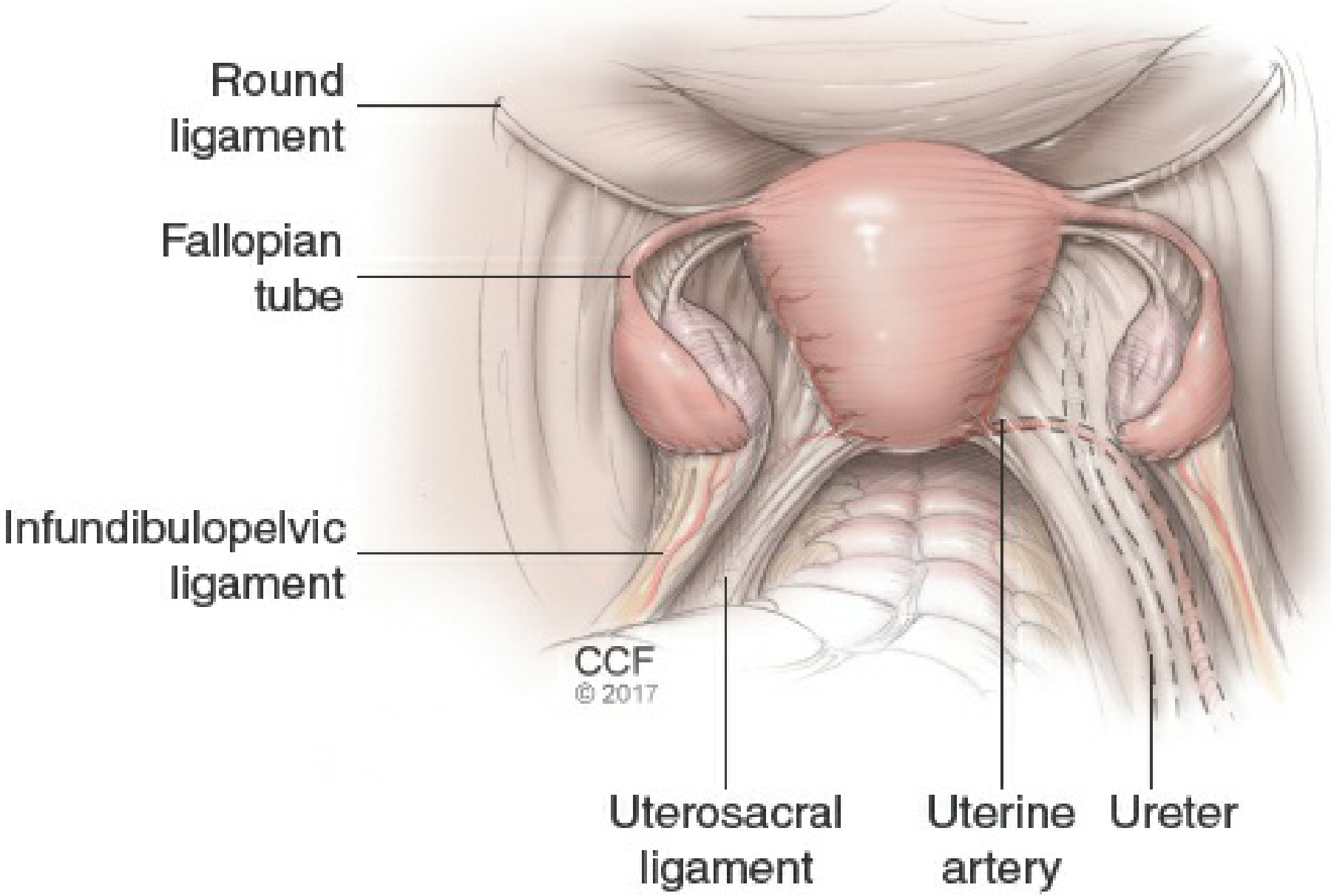
Figure 27-1: Pertinent anatomy including round ligament, fallopian tube, infundibulopelvic ligament, uterosacral ligament, uterine artery, and ureter.
Surgical Steps (Sequential)
1. Incision
- Low transverse (Pfannenstiel): Preferred for benign disease; better cosmesis
- Vertical midline: Used for large uteri, malignancy, need for exploration, or expected adhesions
- Chosen based on indication, body habitus, surgical history, size of pathology, and surgeon preference
- Fascia opened with traction on lateral edges to avoid enterotomy
2. Abdominal Exploration
- Peritoneal cytology if needed (oncologic cases)
- Systematic examination: liver, gallbladder, stomach, kidneys, para-aortic nodes, bowel
3. Retraction
- Balfour or O'Connor-O'Sullivan retractors most commonly used
- Bookwalter retractor useful in obese patients
4. Elevation of the Uterus
- Broad ligament clamps placed at each cornu, crossing the round ligament
- Uterus deviated laterally to place tension on contralateral structures
5. Round Ligament Division
- Uterus deviated to patient's left; right round ligament placed under tension
- Ligated with delayed absorbable suture or transected with electrocautery
- Anterior broad ligament incised medially toward the internal cervical os
- Bladder flap developed to separate bladder from lower uterine segment
6. Ureter Identification
- Retroperitoneum entered by extending incision cephalad on posterior broad ligament
- External iliac artery identified along medial psoas; traced cephalad to iliac bifurcation
- Ureter crosses common iliac artery at its bifurcation
- Ureter left attached to medial broad ligament leaf to protect its blood supply
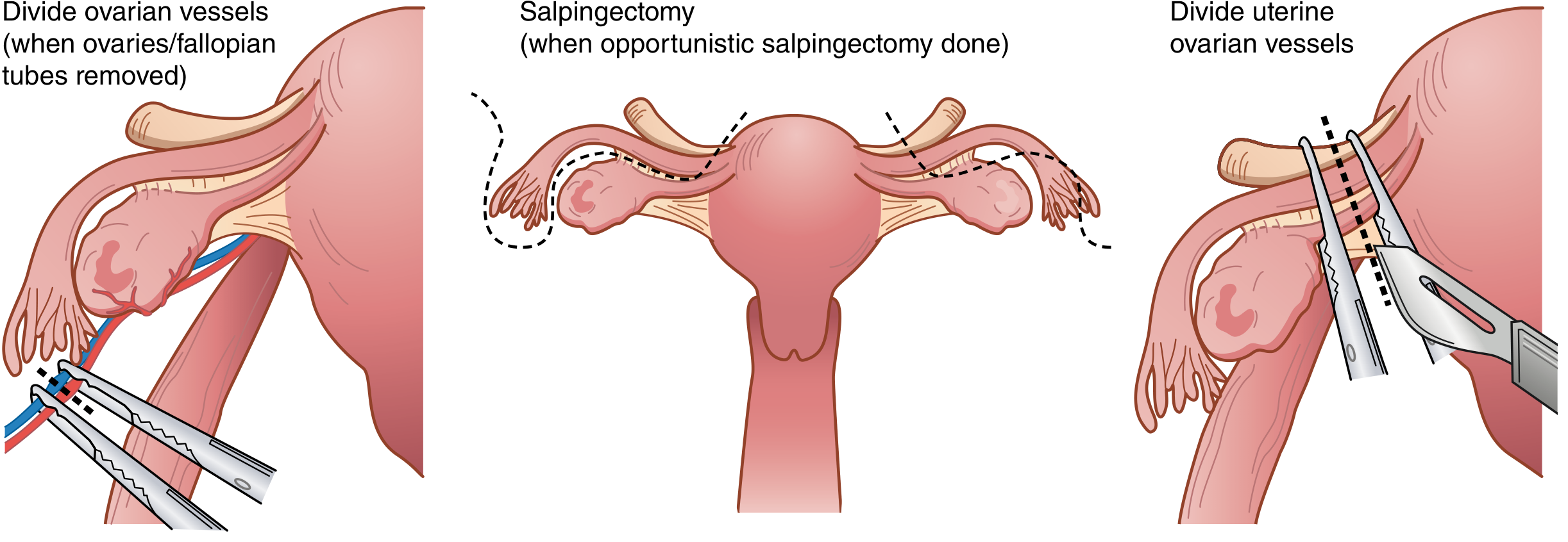
7. Vascular Ligation
If ovaries are removed (BSO):
- Posterior broad ligament leaves incised; infundibulopelvic (IP) ligaments doubly clamped with curved Heaney or Zeppelin clamps and divided
If ovaries preserved:
- Two Kelly or Heaney clamps placed across the utero-ovarian vessels, which are divided and doubly ligated
8. Bladder Dissection
- Bladder dissected off cervix sharply (Metzenbaum scissors) or with electrocautery
- Bladder retracted inferiorly completely below the level of the cervix
9. Uterine Vessel Ligation
- Curved Heaney or Zeppelin clamp placed across uterine vessels at the level of the internal cervical os
- Pedicle cut and suture ligated
10. Cardinal and Uterosacral Ligament Division
- Straight Heaney or Zeppelin clamps placed on cardinal and uterosacral ligaments in serial steps bilaterally
- Pedicles cut and suture ligated until the level of the external cervical os
11. Colpotomy and Specimen Removal
- Curved clamps placed across vagina just below cervix from both sides
- Curved scissors cut above clamps to separate the uterus and cervix from the vagina
- Alternatively: direct sharp circumferential incision around the cervix, extended with curved scissors
12. Vaginal Cuff Closure
- Heaney transfixion stitches placed beneath vaginal corner clamps
- Interrupted figure-of-eight stitches or running suture used to close vagina in midline
- Uterosacral and cardinal ligaments incorporated into vaginal cuff angles for support
13. Hemostasis and Irrigation
- Pelvis irrigated with saline
- Meticulous hemostasis of all vascular pedicles
- Ureters checked for integrity and absence of dilation
14. Closure
- Pelvic peritoneum is not reapproximated as a separate layer
- Fascia closed with interrupted or continuous 0 or 1 monofilament absorbable suture; bites ~1 cm from edge, ~1 cm apart
- Subcutaneous fat closed if ≥2 cm thick (reduces wound disruption)
- Skin closed with staples or subcuticular sutures
Intraoperative Complications
Ureteral Injury
- Three most common sites:
- Pelvic brim where ureter crosses over common iliac vessels
- Midpelvis where uterine artery crosses over it ("water under the bridge")
- Near the cervix at the internal os
- Best prevention: direct visualization via retroperitoneal dissection (NOT reliance on ureteral catheters alone)
- Ureteral catheters may cause hematuria and urinary retention, and may give false sense of security in fibrotic/scarred pelvis
- If obstruction suspected: IV indigo carmine or methylene blue injection + cystoscopy
Hemorrhage
- Identify and ligate all vascular pedicles; back-bleeding from specimen pedicles controlled by double ligation
Bladder Injury
- Most common visceral injury; recognized by direct visualization intraoperatively
- Repaired in two layers; Foley catheter left for prolonged drainage
Bowel Injury
- Risk increased with adhesions, endometriosis, prior surgery
Postoperative Management
Bladder
- Foley catheter removed within 24 hours unless concurrent urogynecologic procedure performed
- Postvoid residuals should be <100 mL before catheter removal
Pain Management
- Multimodal preemptive analgesia: gabapentin, acetaminophen, COX-2 inhibitors started perioperatively
- Reduces postoperative opioid use and improves pain scores
- Preemptive fentanyl lowers postoperative pain scores
Diet
- Resume diet as soon as alert; solids as tolerated
- Early postoperative feeding is safe and speeds return of bowel function
Activity
- Ambulation begins on postoperative day 1 if possible
- No lifting >20 pounds for 6 weeks
- No sexual intercourse for at least 6 weeks (until vaginal cuff fully healed)
- No driving until full mobility returns
Wound Care
- Dressing left on for 24 hours; then daily cleaning with mild soap and water
Postoperative Complications
| Complication | Notes |
|---|---|
| Wound infection | Risk 3-10%; higher with abdominal vs. vaginal approach; obesity, diabetes, long OR time increase risk |
| Vaginal cuff cellulitis/hematoma/abscess | Common early complication |
| Urinary tract infection | Very common; linked to catheter use |
| Bladder/ureteral injury | Most common within the first 2 postoperative weeks |
| Thromboembolism (DVT/PE) | Early ambulation is key prophylaxis |
| Ileus / bowel obstruction | Especially with adhesions |
| Hemorrhage | Pedicle back-bleeding; vaginal cuff hematoma |
| Pneumonia / atelectasis | Prevented by early ambulation |
| Ovarian remnant syndrome | If ovarian tissue inadvertently left |
Overall complication rate: 7.9% for benign vs. 19.4% for malignant indications.
Key Anatomic Relationships to Remember
- The ureter is ~1-2 cm lateral to the cervix as it passes under the uterine artery - classic site of injury ("ureter is the water under the uterine artery bridge")
- The uterine artery arises from the internal iliac (hypogastric) artery
- The cardinal (Mackenrodt) ligament is the primary support of the cervix and upper vagina - its division is the critical step in gaining access to the vagina
- The uterosacral ligaments provide posterior support and are reattached to the vaginal cuff to prevent vault prolapse
Sources: Berek & Novak's Gynecology, pp. 1417-1429 | Sabiston Textbook of Surgery, pp. 2799-2800 | Tintinalli's Emergency Medicine, p. 706 | Schwartz's Principles of Surgery
This is a shared conversation. Sign in to Orris to start your own chat.