
Sglt2 inhibitor drug causing fourniers
The SGLT2 inhibitor most associated with Fournier's gangrene is canagliflozin, though the FDA warning covers the entire class. Let me pull the full details from the medical library.
Searching sources...
Reading File
Finding Sources
Searching PubMed
SGLT2 inhibitor Fournier gangrene
Reading File
Reading File
Reading File
Here is a complete summary of SGLT2 inhibitors and Fournier's gangrene, drawn from multiple authoritative textbooks:
SGLT2 Inhibitors and Fournier's Gangrene
The Drug Class
All SGLT2 inhibitors carry this risk as a class effect. The approved agents in the US are:
- Canagliflozin
- Dapagliflozin
- Empagliflozin
- Ertugliflozin
No single agent is uniquely implicated - the FDA warning applies to the entire class.
What is Fournier's Gangrene?
Fournier's gangrene (also called necrotizing fasciitis of the perineum) is:
-
A severe, rapidly progressing necrotizing infection of the external genitalia, perineum, or perianal area
-
Polymicrobial in nature (mixed aerobic and anaerobic organisms)
-
Life-threatening without urgent surgical intervention
-
Dermatology 2-Volume Set, 5e
FDA Warning (2018)
In August 2018, the FDA released a safety communication based on 12 post-marketing cases of Fournier's gangrene associated with SGLT2 inhibitor use - a rare but serious adverse effect. This prompted labeling updates for all drugs in the class.
- Fuster and Hurst's The Heart, 15th Edition
Mechanism / Why SGLT2 Inhibitors Cause This
SGLT2 inhibitors work by causing glycosuria (spilling glucose into the urine). This creates a glucose-rich perineal environment that:
- Promotes bacterial and fungal overgrowth in the genital/perineal area
- Increases susceptibility to urogenital infections (a well-known class effect)
- In predisposed patients (especially diabetics, immunocompromised), this can progress to deep-tissue necrotizing infection
The underlying diabetes itself is an independent risk factor for Fournier's gangrene, as is immunosuppression.
- Katzung's Basic and Clinical Pharmacology, 16th Edition
- Lippincott Illustrated Reviews: Pharmacology
Other Risk Factors That Compound the Risk
| Risk Factor | Notes |
|---|---|
| Diabetes mellitus | Independent predisposing condition |
| Immunosuppression | Reduces ability to fight infection |
| Poor perineal hygiene | Direct risk factor |
| Combination with sulfonylurea/insulin | Increases hypoglycemia risk, worsens metabolic milieu |
Management of Fournier's Gangrene
- Emergency surgical debridement - wide and extensive
- Prolonged systemic antibiotics (broad-spectrum, covering polymicrobial flora)
- Discontinue the SGLT2 inhibitor immediately
- Supportive care / ICU level management
Other Notable Adverse Effects of SGLT2 Inhibitors (for context)
- Genital mycotic infections (most common; same glycosuria mechanism)
- UTIs
- Volume depletion / hypotension
- DKA at near-normal glucose levels ("euglycemic DKA")
- Bone fractures (canagliflozin)
- Modest LDL increase (canagliflozin, empagliflozin: 4-8%)
- Renal insufficiency (especially in CKD)
Key exam point: Fournier's gangrene with SGLT2 inhibitors is a class effect, not specific to one drug. The mnemonic to remember: glycosuria → perineal glucose → necrotizing infection. Patients should be counseled on perineal hygiene and to seek urgent care for any genital/perineal pain, swelling, or fever.
Stain of hsv
The classic stain for HSV is the Tzanck smear, which shows multinucleated giant cells. Let me pull the full details from the medical library.
Searching sources...
Finding Sources
Reading File
Reading File
Loading Image
Staining for HSV - Complete Summary
1. Tzanck Smear (Classic / Most Tested)
How it is done:
- Scrape the base of a freshly ruptured vesicle
- Stain with Giemsa or Wright's stain (Papanicolaou stain can also be used)
- Examine microscopically for multinucleated giant cells (Tzanck cells)
What you see:
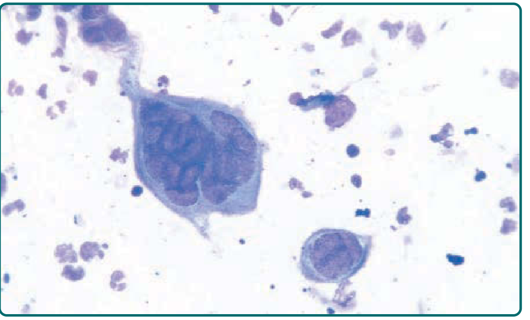
Giemsa-stained smear from a vesicle base showing a giant multinucleated keratinocyte (Tzanck cell). Compare its size with surrounding neutrophils.
- Fitzpatrick's Dermatology, 5e
Key Points to Remember
| Feature | Detail |
|---|---|
| Test name | Tzanck smear / Tzanck test |
| Stains used | Giemsa (most common), Wright's, Papanicolaou |
| Finding | Multinucleated giant cells (Tzanck cells) + intranuclear eosinophilic inclusions |
| Sensitivity | Low - positive in <40% of culture-proven cases; even lower (<30%) for mucosal swabs |
| Specificity problem | Cannot distinguish HSV from VZV - both cause identical Tzanck cell changes |
| Specimen | Scraping from base of freshly ruptured vesicle |
2. Direct Fluorescent Antibody (DFA) Staining
- Stains lesion scrapings with fluorescent-labeled antibodies
- More sensitive than Tzanck smear, but less sensitive than viral culture
- Can be used as an alternative rapid test
3. Skin Biopsy (Histopathology)
On H&E or special stains, biopsy shows:
- Enlarged, swollen epithelial cells that are often separated (ballooning degeneration)
- Multinucleated cells
- Intranuclear eosinophilic inclusion bodies (Cowdry type A inclusions)
4. Gold Standard: PCR + Viral Culture
| Method | Notes |
|---|---|
| PCR | Most sensitive; preferred for CNS/neonatal HSV; can type HSV-1 vs HSV-2 |
| Viral culture | Cytopathic effect visible in 48-96 hours; spin-amplified culture can give result in <24 hours; needed for antiviral sensitivity testing |
Summary Hierarchy (Sensitivity)
PCR > Viral culture > DFA staining > Tzanck smear
Exam pearl: The Tzanck smear is positive in HSV and VZV - it does NOT differentiate between the two. For HSV-1 vs HSV-2 typing, PCR or culture is required. - Harrison's Principles of Internal Medicine, 22e
This is a shared conversation. Sign in to Orris to start your own chat.