
steps of erytropoiesis
Reading File
Now let me search the medical library for erythropoiesis content.
Finding Sources
Finding Sources
Good results. Let me read the most relevant sections - from Histology textbooks and Biochemistry which will have the best step-by-step coverage.
Reading File
Reading File
Reading File
Now let me read the detailed erythropoiesis steps from the Histology textbook and fetch the diagram image.
Reading File
Loading Image
Now let me fetch the maturation stages diagram and read the detailed cell descriptions.
Loading Image
Reading File
I now have excellent content and images. Let me compile the full answer.
Here is a complete breakdown of erythropoiesis, the process by which red blood cells (RBCs) are produced, primarily in the red bone marrow of adults.
Erythropoiesis - Steps of Red Blood Cell Formation
Stimulus and Regulation
When tissue oxygen delivery falls (hypoxia), the kidney senses it and releases erythropoietin (EPO), a 34-kDa glycoprotein hormone. EPO acts on specific receptors on erythroid progenitor cells, stimulating their proliferation and maturation. The entire process - from stem cell to circulating RBC - takes about 7 days.
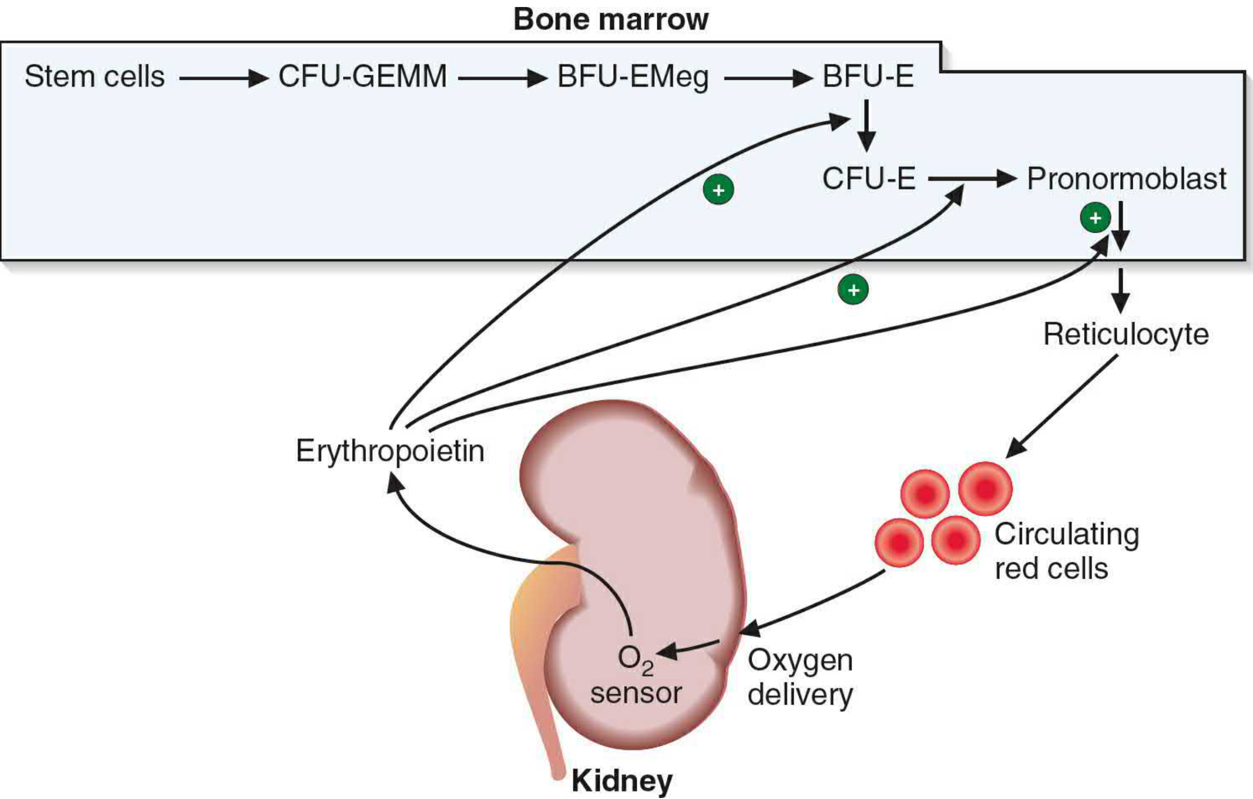
Stages of Erythropoiesis
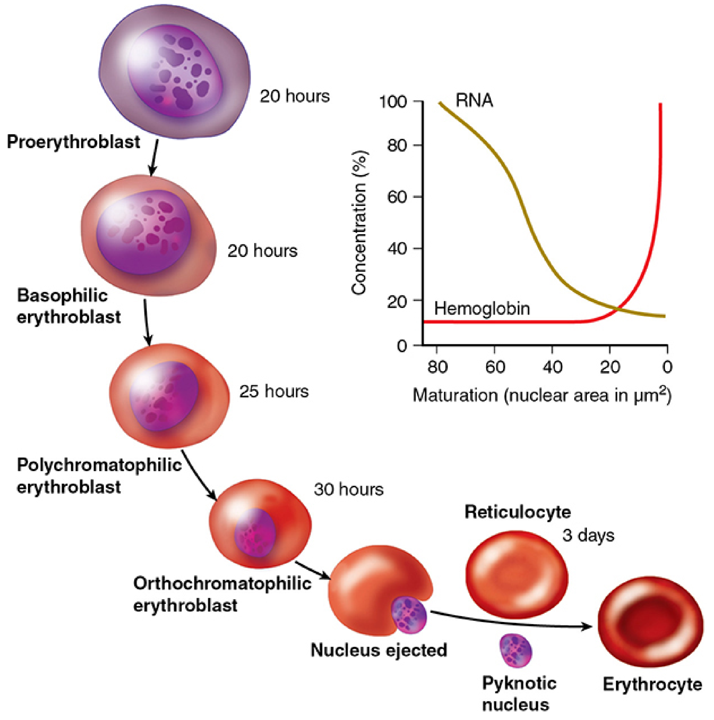
Phase 1 - Progenitor Cells (not morphologically identifiable)
These are identified only by their behavior in cell culture, not by microscopy:
| Cell | Key Features |
|---|---|
| Pluripotent hematopoietic stem cell (HSC) | Self-renewing; gives rise to all blood lineages |
| Common myeloid progenitor (CFU-GEMM) | Colony-forming unit for Granulocyte, Erythroid, Monocyte, Megakaryocyte |
| BFU-EMeg | Bipotent erythroid-megakaryocyte progenitor |
| BFU-E (Burst-Forming Unit - Erythroid) | Large burst of colonies; EPO-sensitive |
| CFU-E (Colony-Forming Unit - Erythroid) | Highly EPO-sensitive; last progenitor before recognizable precursors |
Phase 2 - Morphologically Recognizable Precursors (in bone marrow)
All stages below undergo mitosis and show a progressive pattern: cell size decreases, nucleus condenses then is extruded, RNA decreases, hemoglobin increases.
Step 1: Proerythroblast (Pronormoblast)
- Largest cell in the series (~14-19 µm)
- Large nucleus with open, lacy chromatin and prominent nucleoli
- Cytoplasm is deeply basophilic (rich in RNA/polyribosomes, little Hb)
- First recognizable erythroid precursor
- Duration: ~20 hours
Step 2: Basophilic Erythroblast (Early Normoblast)
- Smaller than proerythroblast
- Nucleus begins to condense; nucleoli less visible
- Cytoplasm remains intensely basophilic - large number of free polysomes actively synthesizing hemoglobin
- Actively divides (mitosis occurs here)
- Duration: ~20 hours
Step 3: Polychromatophilic Erythroblast (Intermediate Normoblast)
- Further reduced cell size
- Nucleus more condensed with coarser chromatin
- Cytoplasm shows both blue (basophilic) and pink (eosinophilic) areas - hence "polychromatophilic" - because Hb is accumulating while polyribosomes are declining
- Last stage that actively undergoes mitosis
- Duration: ~25 hours
Step 4: Orthochromatophilic Erythroblast (Late Normoblast)
- Small cell; nucleus is pyknotic (intensely condensed, dark, small)
- Cytoplasm is now uniformly eosinophilic (pink) - Hb has largely replaced the polyribosomes
- No further mitosis
- The pyknotic nucleus is ejected (extruded) and phagocytosed by nearby macrophages
- Duration: ~30 hours
Phase 3 - Post-Enucleation Stages
Step 5: Reticulocyte
- Nucleus has been extruded - the cell is anucleate
- Still retains a few polyribosomes and mRNA, capable of some hemoglobin synthesis
- When stained with brilliant cresyl blue, residual RNA appears as a faint bluish network (reticulum) - hence the name
- Released from bone marrow into circulation
- Circulates for 1-2 days, matures in the spleen where ribosomes and mRNA are removed
- Normally constitutes ~1% of circulating RBCs
- Duration in circulation: ~3 days
Step 6: Mature Erythrocyte (Red Blood Cell)
- Biconcave disc, ~7-8 µm
- No nucleus, no organelles, no ribosomes
- Packed with hemoglobin (~33 g/dL)
- Life span: ~120 days
- Removed by macrophages in spleen, bone marrow, and liver (reticuloendothelial system) when senescent
Key Changes During Maturation - Summary
| Feature | Early (Proerythroblast) | Late (Orthochromatophilic) |
|---|---|---|
| Cell size | Large | Small |
| Nucleus | Large, open chromatin, nucleoli present | Pyknotic, then extruded |
| Cytoplasm color | Deep blue (basophilic) | Pink (eosinophilic) |
| RNA / Ribosomes | High | Very low / absent |
| Hemoglobin | Absent | High |
| Mitosis | Yes | No |
Fate of Extruded Nucleus and Senescent RBCs
When the orthochromatophilic erythroblast ejects its nucleus, it is phagocytosed by macrophages in the bone marrow. When RBCs reach ~120 days, they too are phagocytosed; heme and globin dissociate - iron is recycled via ferritin/hemosiderin in the spleen and bone marrow for new Hb synthesis, and the porphyrin ring is degraded to bilirubin, bound to albumin, transported to the liver, conjugated, and excreted in bile.
Sources: Junqueira's Basic Histology, Text and Atlas, 17e; Histology: A Text and Atlas with Correlated Cell and Molecular Biology; Basic Medical Biochemistry - A Clinical Approach, 6e
This is a shared conversation. Sign in to Orris to start your own chat.