
Teach me about TIA i am an MBBS intern so use reference books like HARRISSON MEDICINE
"transient ischemic attack" management
Transient Ischemic Attack (TIA) - Complete MBBS Intern Guide
1. Definition (The Modern Tissue-Based Definition)
"A TIA is defined as transient neurologic dysfunction caused by focal ischemia of brain tissue, spinal cord, or retina without acute infarction."
- Frameworks for Internal Medicine
"Patients with symptoms less than 24 hours can have representative ischemic lesions on diffusion-weighted or perfusion-weighted MRI, so TIA has moved from time-based to tissue-based definition."
- Rosen's Emergency Medicine
2. Pathophysiology & Mechanisms
| Mechanism | Description |
|---|---|
| Atherothrombosis | Thrombus on a diseased vessel (carotid, MCA, vertebrobasilar) transiently occludes flow |
| Embolism | Most TIAs are truly embolic - a clot lodges then lyses spontaneously |
| Lacunar | Small penetrating vessel disease; "capsular warning syndrome" - escalating limb weakness episodes culminating in lacunar stroke |
| Cardioembolism | Clot from heart (atrial fibrillation, recent MI, valvular disease) |
| Hematologic | Polycythemia vera, sickle cell, thrombocytosis, leukemia, hyperviscosity states |
"It has been realized that many TIAs previously attributed to atherothrombosis are truly embolic strokes that leave a trace of infarction but have resolved clinically."
- Adams & Victor's Principles of Neurology
3. Clinical Features
Anterior Circulation (Carotid territory) TIA:
- Hemiparesis / hemiplegia (contralateral)
- Hemisensory loss (contralateral face, arm, leg)
- Aphasia (if dominant hemisphere)
- Amaurosis fugax - transient monocular blindness ("curtain coming down"), from ophthalmic artery (branch of ICA)
Posterior Circulation (Vertebrobasilar) TIA:
- Diplopia, dysarthria, dysphagia
- Vertigo + ataxia (cerebellar)
- Crossed deficits (ipsilateral cranial nerve + contralateral limb weakness)
- Drop attacks (sudden loss of postural tone without LOC)
- Bilateral visual field defects
"TIAs may present as transient spells of hemiparesis, aphasia, numbness or tingling on one side, dysarthria, diplopia, ataxia, obscuration of a visual field, or combinations thereof that replicate the stroke syndromes. Even limb shaking can represent a TIA."
- Adams & Victor's Principles of Neurology
4. Risk of Subsequent Stroke (Why TIA is a Neurological Emergency)
-
Up to 10% stroke risk within 2 days
-
Up to 15% stroke risk at 90 days (Frameworks for Internal Medicine)
-
~6% of strokes after TIA occur within one month of the first attack
-
~6% more in the following year (Adams & Victor)
5. Risk Stratification - The ABCD² Score
| Clinical Factor | Score |
|---|---|
| A - Age ≥60 years | 1 |
| B - BP >140 mmHg systolic OR >90 mmHg diastolic | 1 |
| C - Clinical symptoms: Unilateral weakness | 2 |
| C - Clinical symptoms: Speech disturbance without weakness | 1 |
| D - Duration >60 min | 2 |
| D - Duration 10-59 min | 1 |
| D - Diabetes (oral meds or insulin) | 1 |
| Total | 0-7 |
- Score 0: 0% | Score 4: 8% | Score 5: 12% | Score 6: 17% | Score 7: 22%
- 0-3: Low risk (1% stroke in 48h)
- 4-5: Moderate risk (4.1% in 48h)
- ≥6: High risk (8% in 48h)
Note: More recent studies have found the ABCD² score alone is insufficient - it should be combined with DWI-MRI/MRA (ABCD²-I or ABCD³-I scoring) for better prediction. - Rosen's
6. Differential Diagnosis
| Condition | Distinguishing Feature |
|---|---|
| Migraine aura | Symptoms "march" along a limb (vs. abrupt TIA onset); headache follows as deficit resolves; positive symptoms (scintillations, sensory spread) |
| Focal seizure | Positive motor symptoms (jerking), shorter duration, may have postictal weakness (Todd's paralysis); EEG may be needed |
| Hypoglycemia | Always check blood glucose; can mimic focal deficits |
| Subdural hematoma | History of head trauma; progressive symptoms |
| Brain tumour | Insidious onset, ± seizures, ± headache |
| Multiple sclerosis | Subacute onset, young patient, prior episodes, CSF/MRI findings |
| Conversion disorder | Non-anatomical pattern, inconsistent exam, psychological context |
7. Investigations
Immediate (Emergency) Workup:
- Non-contrast CT brain - Rule out haemorrhage (hemorrhagic stroke contraindication to antiplatelets)
- MRI brain with DWI - Detects early infarction (DWI positive = stroke, not TIA); gold standard
- CT angiography / MR angiography - Carotid and intracranial vessels
- ECG - Screen for atrial fibrillation
- Blood glucose - Rule out hypoglycemia
- CBC, coagulation, lipids, renal function
- Echocardiogram - Screen for cardiac source (if cardioembolic mechanism suspected)
- Carotid Doppler - Ipsilateral carotid stenosis assessment
8. Management
A. Antiplatelet Therapy (Cornerstone of Treatment)
"The combination of aspirin and clopidogrel was found to prevent stroke following TIA better than aspirin alone in a large Chinese randomized trial [CHANCE] and the NIH-sponsored POINT trial."
- Harrison's 22E
- DAPT started within 24 hours and continued for 21 days is the current standard
- After 21 days, switch to single antiplatelet (aspirin or clopidogrel alone)
- The benefit of DAPT is confined to the first 21 days (pooled POINT + CHANCE analysis)
"Failure to respond to clopidogrel is linked to carriage of a common CYP2C19 polymorphism...this mutation is common, particularly in Asians."
- Harrison's 22E
B. Anticoagulation
- Indicated when the mechanism is cardioembolism (e.g., atrial fibrillation)
- Direct oral anticoagulants (DOACs) are preferred over warfarin for AF
- Thrombolysis (tPA) is contraindicated if symptoms are improving (a defining feature of TIA)
C. Statin Therapy
- Atorvastatin 80 mg/day (SPARCL trial): significant reduction in secondary stroke and TIA
- Target LDL <70 mg/dL for secondary prevention
- All patients with prior ischemic TIA should receive a statin
D. Blood Pressure Control
- Target <130/80 mmHg (all hypertension should be treated)
- SPRINT trial: SBP <120 mmHg reduced stroke + MI by 43%
- Preferred agents: thiazide diuretics and ACE inhibitors (Harrison's 22E)
9. Carotid Endarterectomy (CEA) - Surgical Prevention
| Stenosis | Recommendation |
|---|---|
| ≥70% symptomatic stenosis | Strong indication for CEA - reduces 2-year stroke risk from 26% (medical) to 9% (surgical) = 65% relative risk reduction |
| 50-70% symptomatic stenosis | Significant but less robust benefit |
| <30% stenosis | Harm shown with surgery |
- Symptomatic hemispheric ischemia
- High-grade ipsilateral ICA stenosis
- Institutional perioperative morbidity/mortality ≤6%
- Most beneficial when performed within 2 weeks of symptom onset
- Greater benefit in patients >75 years and in men
10. Secondary Prevention Summary
| Risk Factor | Target/Intervention |
|---|---|
| Hypertension | <130/80 mmHg; thiazides, ACE inhibitors preferred |
| Dyslipidemia | LDL <70 mg/dL; atorvastatin 80 mg |
| Atrial fibrillation | Anticoagulation (DOAC preferred) |
| Carotid stenosis ≥70% | CEA within 2 weeks |
| Diabetes | Tight glucose control; prevent new DM |
| Smoking | Cessation strongly recommended |
| Antiplatelet | Aspirin + clopidogrel x 21 days, then monotherapy |
11. Memory Aids for Internship
- Age ≥60 (1 pt)
- Blood pressure ≥140/90 (1 pt)
- Clinical features: weakness (2 pts), speech only (1 pt)
- Duration: >60 min (2 pts), 10-59 min (1 pt)
- Diabetes (1 pt)
Key References
- Harrison's Principles of Internal Medicine 22E (2025), Chapters 438-439
- Adams & Victor's Principles of Neurology 12E, Chapter 33
- Rosen's Emergency Medicine 10E, Chapter 87
- Frameworks for Internal Medicine, Chapter 43
Does yellow phosphorus ingestion have a good or bad prognosis
"yellow phosphorus" poisoning prognosis
PMID: 39132751
Yellow Phosphorus Ingestion: Prognosis
Short Answer: BAD - Very Poor Prognosis
Why It's So Dangerous - Mechanism of Toxicity
"Phosphorus is a protoplasmic poison. It affects cellular oxidation. Its effect on cellular metabolism is comparable to ischaemia... This is known as necrobiosis, which is classically manifested in the liver."
- Parikh's Textbook of Medical Jurisprudence, Forensic Medicine and Toxicology
- Local irritant action on the GI tract
- Absorbed phosphorus stays in blood in elemental form for 1-2 days
- Gets oxidised to hypophosphorous and phosphorous acids - acting as a hepatotoxic, protoplasmic poison
- Causes widespread fatty infiltration and degeneration, especially in liver and cerebral cortex
- Disturbs carbohydrate, fat, and protein metabolism - fat deposits in liver at the cost of glycogen (necrobiosis)
- Leads to acute yellow atrophy of the liver - the pathognomonic end-stage finding
Fatal Dose and Fatal Period
| Parameter | Value |
|---|---|
| Fatal dose (adults) | 60-120 mg |
| Fatal dose (children) | 10-25 mg |
| Fatal period | Death from collapse within 24 hours (early); in the usual course, 6-7 days or longer |
The Triphasic Clinical Course (Key Feature for Exams)
Stage 1 - GI Irritation (within 2-6 hours, lasts 1-2 days)
- Garlic taste in mouth, garlic odour in breath
- Luminous vomitus and phosphorescent faeces (glows in the dark) - pathognomonic
- Burning pain throat to stomach
- Profuse vomiting (bile-stained, blood-tinged)
- Diarrhoea, intense thirst
- Cardiac and respiratory depression, cold clammy skin
- Patient may die at this stage from cardiovascular collapse
Stage 2 - Apparent Recovery / "Honeymoon Phase" (2-4 days)
- Symptoms reduce in intensity, vitals seem to improve
- Patient and doctor may feel reassured
- This is deceptive - phosphorus is being absorbed and oxidised, silently damaging the liver
Stage 3 - Hepatorenal Failure (starts around Day 4-7)
- Vomiting and diarrhoea return more severely
- Jaundice sets in and deepens rapidly
- Liver enlarged, soft, tender → later shrinks (acute yellow atrophy = necrosis)
- Haemorrhagic manifestations: epistaxis, haematemesis, haematuria, melaena, petechiae - from coagulopathy (hypoprothrobinaemia)
- Renal failure: oliguria → anuria; urine contains blood, albumin, bile, amino acids (leucine, tyrosine, cysteine)
- CNS: headache, tinnitus, vertigo, insomnia, delirium, priapism (frequent), cramps, paralysis
- Hypoglycaemia, weak irregular pulse, falling BP, pulmonary oedema, cyanosis
- Death from hepatic and renal insufficiency
"The clinical picture is suggestive of acute yellow atrophy of the liver."
- Parikh's Textbook
Prognostic Markers (Recent Evidence)
- Overall mortality: 36.5%
- Mean survival among those who died: 5.26 days after ingestion
- Symptoms (GI + neurological) typically appear ~48 hours post ingestion
| Marker | Finding in Non-survivors |
|---|---|
| Elevated aminotransferases | 74.3% of non-survivors |
| Prolonged prothrombin time | 65.7% |
| Hyperbilirubinaemia | 65.7% |
| Hyperlactataemia (lactate >2 mmol/L) | 97.1% (most sensitive) |
"As progression to death occurs within a week of yellow phosphorus ingestion in most cases, determining prognosis as early as possible enables swift referral to a liver transplant centre."
- Ramkumar et al., Clin Toxicol 2024 [PMID: 39132751]
Treatment (and Why Prognosis Remains Poor Despite Treatment)
| Measure | Details |
|---|---|
| Gastric lavage | 0.5% KMnO₄ - oxidises phosphorus to harmless phosphoric acid + phosphates; repeat till no garlic smell |
| Copper sulphate | 250 mg in water every 5 min until emesis; coats phosphorus particles with inert copper phosphide |
| Activated charcoal | Adsorbs poison |
| Liquid paraffin | Retards absorption, hastens elimination |
| CONTRAINDICATED | Oils, fats, milk - dissolve phosphorus and promote absorption |
| Liver support | ICU-level hepatic failure management |
| Liver transplantation | Only definitive option in fulminant failure |
Summary Table
| Aspect | Fact |
|---|---|
| Prognosis | Very poor; high mortality |
| Mortality in studies | ~36% (modern ICU care) |
| Fatal dose | 60-120 mg (adults) |
| Cause of death | Fulminant hepatic failure + multi-organ dysfunction |
| Key pathology | Acute yellow atrophy of liver (necrobiosis) |
| Clinical hallmark | Triphasic course with deceptive middle "recovery" phase |
| Diagnostic clue | Luminous vomitus/stool, garlic odour |
| Best early prognostic marker | 24-hour serum lactate ≥2.67 mmol/L |
| Contraindicated in treatment | Milk, oils, fats |
Teach me about vit b12 deficiency
"vitamin B12 deficiency" cobalamin
Vitamin B12 (Cobalamin) Deficiency - Complete Guide for MBBS Intern
1. Biochemistry Basics
| Reaction | Enzyme | Deficiency Effect |
|---|---|---|
| Homocysteine → Methionine | Methionine synthase (requires methylcobalamin) | ↑ Homocysteine; folate "trap"; impaired DNA synthesis |
| Methylmalonyl-CoA → Succinyl-CoA | Methylmalonyl-CoA mutase (requires adenosylcobalamin) | ↑ Methylmalonic acid (MMA); toxic to myelin |
- The folate trap: B12 deficiency "traps" folate as 5-methylTHF, making it unavailable for DNA synthesis → megaloblastic changes
- MMA accumulation damages myelin directly → explains the neurological syndrome independent of anemia
2. Absorption of Vitamin B12 (Must Know for Exams)
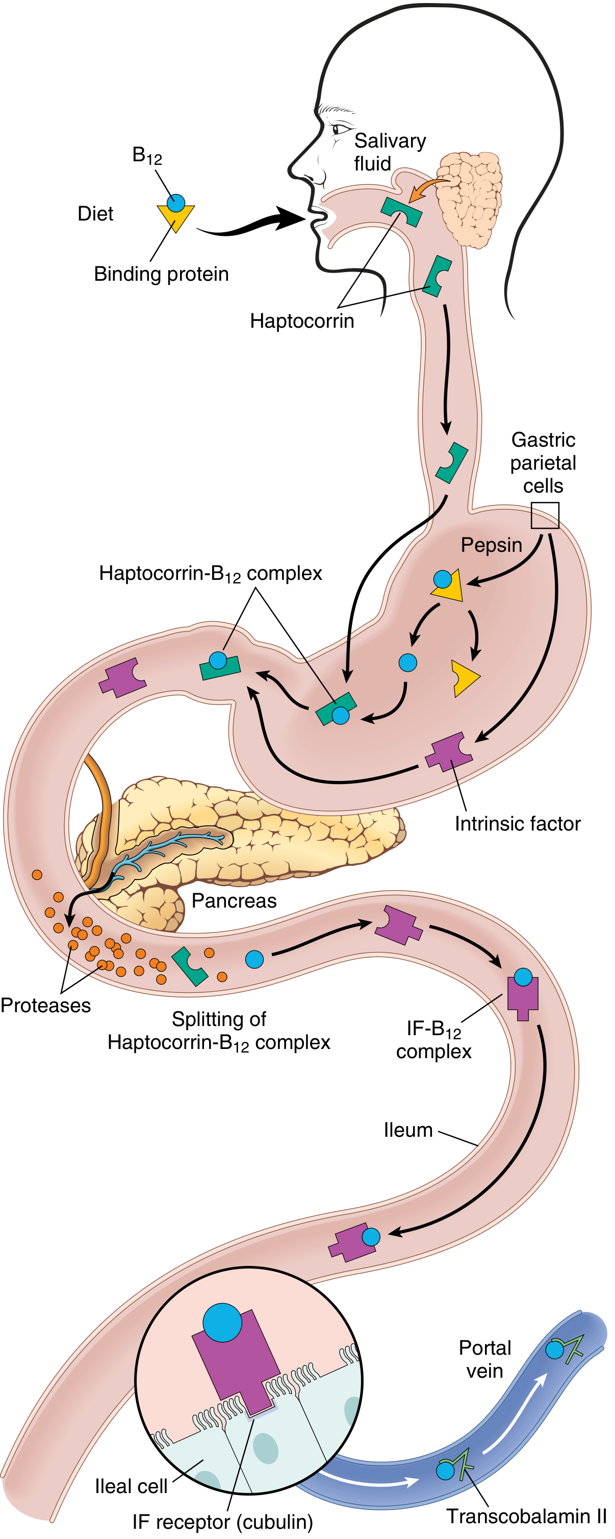
- Mouth/Stomach: Dietary B12 (bound to food protein) is freed by pepsin; binds to salivary haptocorrin (R-protein)
- Stomach: Gastric parietal cells secrete Intrinsic Factor (IF) simultaneously
- Duodenum: Pancreatic proteases cleave the haptocorrin-B12 complex; free B12 binds to IF → IF-B12 complex
- Terminal ileum: IF-B12 complex binds to cubilin receptor on ileal enterocytes and is endocytosed
- Systemic: B12 binds to Transcobalamin II and is transported to liver and bone marrow
3. Causes / Etiology
A. Decreased Intrinsic Factor (Most Common in Adults)
| Cause | Mechanism |
|---|---|
| Pernicious anemia | Autoimmune destruction of gastric parietal cells; anti-IF antibodies |
| Gastrectomy / gastric bypass | Loss of parietal cell mass |
| Type A atrophic gastritis | Autoimmune; fundal gland destruction |
| Congenital IF deficiency | Rare; infant presentation |
B. Food-Cobalamin Malabsorption
- Type B atrophic gastritis (H. pylori, age-related achlorhydria)
- PPI or H2-blocker use (reduce acid needed to release B12 from food)
- Metformin - reduces absorption via unknown mechanism (very common, clinically important!)
C. Ileal Disorders (Loss of Absorption Site)
- Terminal ileal resection or Crohn's disease
- Imerslund-Gräsbeck syndrome (congenital cubilin receptor defect)
- Ileal conduit surgery
D. Usurpation / Competition in Gut
- Bacterial overgrowth (blind loop syndrome)
- Fish tapeworm - Diphyllobothrium latum (competes for B12)
- Tropical sprue
E. Nutritional Deficiency
- Strict vegans (no animal products)
- Breast-fed infant of a B12-deficient mother
- Elderly with poor intake
F. Drug-Induced
- Nitrous oxide (N₂O) - irreversibly oxidises cobalamin; causes acute SCD especially in marginally deficient patients ("anesthesia paresthetica" in operating room staff and whippet abusers)
- Metformin, PPIs, H2 blockers
"Vitamin B12 deficiency is virtually never caused by inadequate intake except in vegetarians who scrupulously avoid milk and eggs."
- Robbins Basic Pathology
4. Clinical Features
A. Haematological (Megaloblastic Anaemia)
- Insidious onset: pallor, easy fatigability, dyspnoea on exertion
- Severe anaemia → congestive cardiac failure
- Mild jaundice (from ineffective erythropoiesis - intramedullary haemolysis)
- Glossitis - beefy red, smooth, sore tongue (Hunter's glossitis)
- Anorexia, weight loss
B. Neurological - Subacute Combined Degeneration (SCD) of Spinal Cord
"The spinal cord, brain, optic nerves, and peripheral nerves are all affected by vitamin B12 deficiency, giving rise to a classic neurologic syndrome in which the spinal cord is usually affected first and often exclusively."
- Adams & Victor's Principles of Neurology
- Posterior columns (dorsal columns) - first and most severely affected
- Lateral corticospinal tracts - hence "combined"
- Paresthesias - symmetrical tingling, "pins and needles" in hands and feet (often hands first)
- Sensory ataxia - unsteady gait, worsens in the dark; loss of vibration and position sense
- Motor signs - spasticity, hyperreflexia, Babinski positive (but early on reflexes may be absent due to concurrent neuropathy)
- Cognitive/psychiatric - irritability, apathy, dementia, depression, psychosis ("megaloblastic madness")
- Optic neuropathy - centrocecal scotomas, optic atrophy (less common)
- Autonomic dysfunction - urinary incontinence, impotence
"Importantly, folate administration does not prevent and may worsen neurologic symptoms that are specific to vitamin B12 deficiency."
- Robbins Basic Pathology
C. Other Features
- Increased risk of gastric carcinoma in pernicious anemia patients
- Thrombocytopenia, leukopenia (pancytopenia in severe cases)
- Hyperhomocysteinemia → increased thrombotic and cardiovascular risk
5. Investigations
Blood Count and Film
| Finding | Details |
|---|---|
| MCV | >100 fL (macrocytosis) |
| Anaemia | Normochromic macrocytic |
| Peripheral smear | Oval macrocytes + hypersegmented neutrophils (>5% with ≥5 lobes, or any with 6 lobes) |
| WBC | Leukopenia possible |
| Platelets | Thrombocytopenia in severe cases |
| Reticulocyte count | Decreased (ineffective erythropoiesis) |
Serum Levels
| Test | B12 Deficiency | Comments |
|---|---|---|
| Serum B12 | Low (<200 pg/mL) | Can be falsely normal in some |
| Serum folate | Normal or elevated | Folate "trapped" in methylTHF form |
| Methylmalonic acid (MMA) | Elevated | Most sensitive/specific for functional B12 deficiency |
| Homocysteine | Elevated | Elevated in BOTH B12 and folate deficiency |
| LDH | Elevated (intramedullary haemolysis) | |
| Indirect bilirubin | Mildly elevated |
Key distinction: MMA is elevated in B12 deficiency but NOT in folate deficiency. Homocysteine is elevated in both.
For Pernicious Anemia Specifically
- Anti-intrinsic factor antibodies (highly specific, ~50% sensitive)
- Anti-parietal cell antibodies (sensitive ~90%, but less specific)
- Schilling test (now rarely done): shows malabsorption corrected by oral IF administration
Imaging (Neurology)
- MRI spine: T2 hyperintensity in posterior columns ± lateral columns of cervical and upper thoracic cord - pathognomonic when present
6. B12 vs Folate Deficiency - Distinguishing Features
| Feature | B12 Deficiency | Folate Deficiency |
|---|---|---|
| Macrocytic anaemia | Yes | Yes |
| Hypersegmented neutrophils | Yes | Yes |
| Neurological involvement (SCD) | Yes | No |
| Serum MMA | Elevated | Normal |
| Serum homocysteine | Elevated | Elevated |
| Serum folate | Normal/elevated | Low |
| Common causes | Pernicious anemia, malabsorption | Poor diet, alcohol, pregnancy, antifolates |
| Giving folate alone | Corrects anaemia but WORSENS neuro | Corrects all features |
7. Treatment
Parenteral B12 (First-line for Pernicious Anemia and SCD)
"The diagnosis of pernicious anemia demands the administration of vitamin B12 and the continuation of treatment for the rest of the patient's life."
- Adams & Victor's Principles of Neurology
- Cyanocobalamin or hydroxocobalamin 1000 μg IM daily for 1 week
- Then 1000 μg IM weekly for 1 month
- Then 1000 μg IM monthly for life (in pernicious anemia)
Oral High-Dose B12 (Alternative for dietary/mild cases)
- 500-1000 μg oral daily can work even in malabsorption (via passive diffusion ~1% absorption without IF)
- Effective for vegans, elderly with dietary deficiency, maintenance therapy
- Caution: Reserve parenteral therapy for manifest neurological disease (SCD)
Monitoring Response
- Reticulocyte count rises within 2-3 days of starting B12 - first sign of response
- Hb begins rising in ~1 week; normalises over 6-8 weeks
- Neurological recovery: greatest in patients with symptoms <3 months; if treated early (within weeks), recovery is usually complete
"All neurologic symptoms and signs may improve, mostly during the first 3 to 6 months of therapy... In practically all instances there is some degree of improvement, although in cases of longest duration, the best that can be accomplished is an arrest of progression."
- Adams & Victor
Important Cautions in Treatment
- Never give folate alone in unconfirmed megaloblastic anaemia - it will correct the anaemia but allow neurological disease to progress untreated
- Give both B12 and folate if unsure, until results confirm which is deficient
- In severe anaemia, if blood transfusion is needed, give slowly to avoid precipitating heart failure
8. Pernicious Anemia - Special Note
- Type A autoimmune (fundal) atrophic gastritis
- Autoantibodies: anti-parietal cell antibodies AND anti-intrinsic factor antibodies
- Associated with other autoimmune diseases: Hashimoto's thyroiditis, type 1 DM, Addison's disease, vitiligo
- Increased risk of gastric adenocarcinoma and gastric carcinoid tumours - requires endoscopic surveillance
- Predominantly affects females, middle age to elderly
- Can be congenital (rare, presents in infancy)
9. Memorable Summary
B12 Deficiency = 3 A's (from MBBS exam perspective):
1. Anaemia (megaloblastic)
2. Axonal/cord disease (SCD = posterior + lateral columns)
3. Autoimmune (pernicious anemia as the main cause)
Lab clue: ↑MMA + ↑homocysteine = B12 deficiency
Normal MMA + ↑homocysteine = folate deficiency
Treatment trap: NEVER correct anaemia with folate alone
- masks B12 deficiency while neurology worsens
Explain GVHD to someone whos an intern in a medical college
"graft-versus-host disease" pathophysiology management
Graft-versus-Host Disease (GVHD) - Complete Guide for Medical Intern
The Core Concept - Think of It This Way
1. Definition
"GVHD occurs when immunologically competent cells or their precursors are transplanted into immunologically depleted recipients, and the transferred cells recognize alloantigens in the host and attack host tissues."
- Robbins Basic Pathology
2. When Does GVHD Occur?
- The transplanted cells must be immunologically competent (contain functioning T cells)
- The recipient must express tissue antigens not present in the donor (HLA or minor histocompatibility antigen mismatch)
- The recipient must be unable to reject the transplanted cells (because they've been deliberately immunosuppressed and irradiated)
- Solid organ transplants containing lymphoid tissue (small bowel, liver, lung)
- Blood transfusion of non-irradiated blood to an immunocompromised patient
- Intrauterine or neonatal transfusion
- Even autologous HSCT can cause a mild, self-limited GVHD (loss of self-tolerance from conditioning)
3. Pathophysiology - Step by Step
Step 1: The Conditioning Regimen Primes the Battlefield
- Destroy their diseased bone marrow
- Suppress immunity enough to accept the donor graft
Step 2: Donor T Cells Recognise Host as Foreign
- See host minor histocompatibility antigens presented by host APCs
- Become activated (via TCR + MHC interaction)
- Clonally expand
"In most cases, the reaction is directed against minor histocompatibility antigens of the host because bone marrow transplantation is not usually performed when there are differences in MHC alleles."
- Cellular & Molecular Immunology
Step 3: Effector Attack on Host Tissues
- CD8+ CTLs directly kill host epithelial cells
- NK cells attack dying host epithelium (seen on histology)
- Cytokines (TNF-α, IFN-γ) amplify tissue destruction
- Result: epithelial cell apoptosis and necrosis in the three main target organs: skin, liver, gut
4. Classification: Acute vs Chronic GVHD
Acute GVHD
| Organ | What Happens | Clinical Presentation |
|---|---|---|
| Skin | Lymphocytic infiltration + epidermal cell apoptosis | Erythematous morbilliform rash; starts at neck, ears, palms, soles → generalises |
| Liver | Destruction of small bile ducts (biliary epithelium) | Jaundice, elevated bilirubin, cholestatic hepatitis |
| GI tract | Mucosal ulceration | High-volume watery or bloody diarrhoea, nausea, vomiting, abdominal pain |
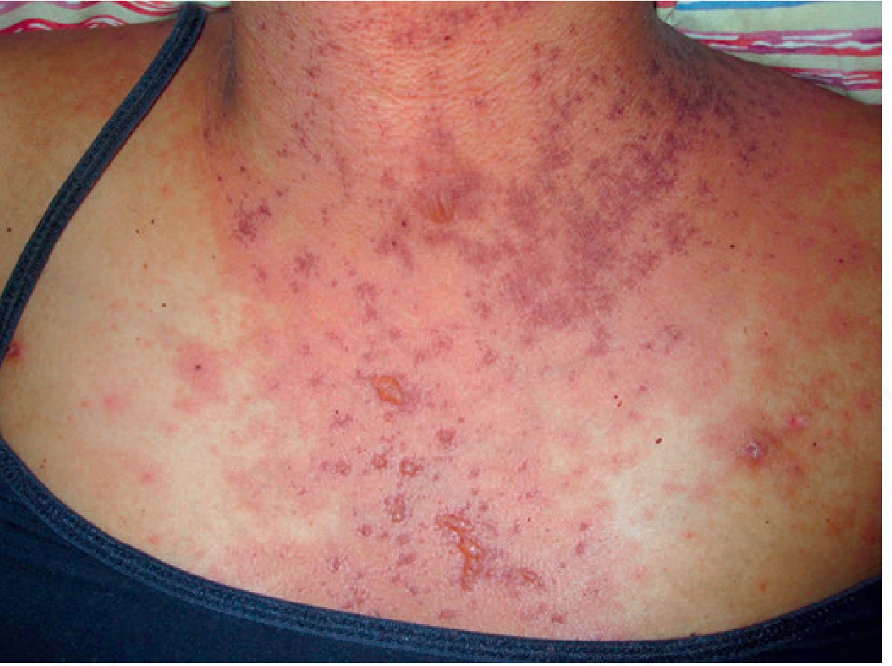
| Stage | BSA Involved |
|---|---|
| Stage 1 | <25% BSA |
| Stage 2 | 25-50% BSA |
| Stage 3 | >50% BSA (generalised erythroderma) |
| Stage 4 | Erythroderma + bullae (resembles TEN - Toxic Epidermal Necrolysis) |
"Grade IV GVHD is characterised by full-thickness slough and may resemble toxic epidermal necrolysis, and it may be impossible to distinguish the two clinically or histologically."
- Andrews' Diseases of the Skin
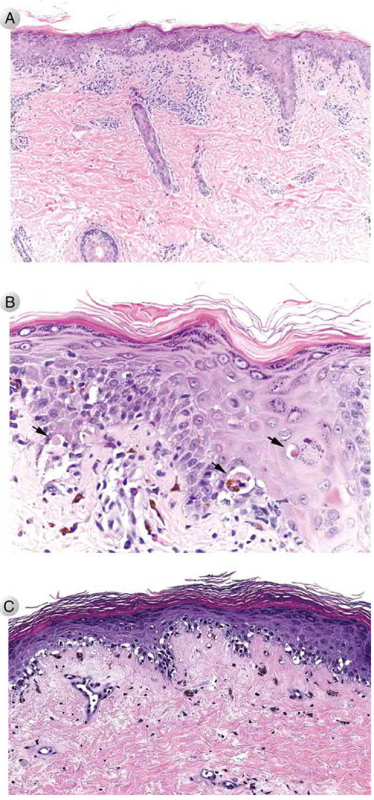
Chronic GVHD
- Skin: Dermal fibrosis resembling systemic sclerosis (scleroderma); lichenoid changes, dry skin, hair loss, nail dystrophy
- Liver: Biliary fibrosis, cholestasis
- Lungs: Bronchiolitis obliterans (obliteration of small airways) - a feared, often irreversible complication
- Eyes: Dry eyes (sicca syndrome)
- Mouth: Dry mouth, lichen planus-like lesions
- Joints: Contractures from fibrosis
- Mimics: Can look like Sjögren's, scleroderma, lichen planus, polymyositis, or other autoimmune diseases
"Patients develop skin lesions with dermal fibrosis resembling those of systemic sclerosis and manifestations mimicking other autoimmune disorders."
- Robbins Basic Pathology
5. The GVHD-GVT Paradox (Extremely Important Concept)
| Effect | What it does | Desirable? |
|---|---|---|
| GVHD | Donor T cells attack host normal tissues | Bad |
| Graft-versus-Tumour (GVT) | Donor T cells attack remaining host cancer cells | Good - prevents relapse |
- If you deplete T cells from the graft → GVHD is eliminated but GVT effect is lost → cancer relapses
- Also, T-cell depleted grafts engraft poorly and increase risk of EBV-related B-cell lymphoma
"Depletion of donor T cells before transplantation virtually eliminates the disease. This approach, however, is a mixed blessing: GVHD is reduced, but the recurrence of tumor in patients with leukemia, as well as the incidence of graft failures and EBV-related B-cell lymphoma, increase."
- Robbins Basic Pathology
6. Prevention
| Drug | Mechanism |
|---|---|
| Cyclosporine / Tacrolimus | Calcineurin inhibitors - block T cell IL-2 production |
| Methotrexate | Antimetabolite - inhibits T cell proliferation |
| Sirolimus (Rapamycin) | mTOR inhibitor - blocks T cell activation/proliferation |
| MMF (Mycophenolate) | Antimetabolite - blocks purine synthesis in lymphocytes |
7. Treatment
- High-dose corticosteroids (prednisolone 1-2 mg/kg/day) - mainstay of treatment
- Steroid-refractory acute GVHD carries a very poor prognosis; options include ruxolitinib (JAK1/2 inhibitor), anti-thymocyte globulin, basiliximab
- Corticosteroids + calcineurin inhibitors (cyclosporine/tacrolimus)
- Ibrutinib (BTK inhibitor) - FDA approved for steroid-refractory chronic GVHD
- Ruxolitinib - approved for both steroid-refractory acute and chronic GVHD
- Phototherapy: Narrow-band UVB, PUVA, UVA1 - used as second-line agents, especially for sclerodermoid skin disease
8. Summary Table - Acute vs Chronic GVHD
| Feature | Acute GVHD | Chronic GVHD |
|---|---|---|
| Timing | Days to weeks (classically <100 days) | After 100 days (but can overlap) |
| Pathology | Epithelial cell death/apoptosis | Fibrosis and atrophy |
| Skin | Morbilliform rash, erythroderma, bullae | Scleroderma-like, lichenoid changes |
| Liver | Cholestatic jaundice | Biliary fibrosis |
| GI tract | Bloody diarrhoea, ulceration | Strictures, malabsorption |
| Lungs | Rare | Bronchiolitis obliterans |
| Eyes/Mouth | Rare | Sicca syndrome (dry eyes/mouth) |
| Effectors | CD4+, CD8+ T cells, NK cells, cytokines | Fibrosis pathway, B cells also involved |
| Treatment | IV steroids | Steroids + CNI ± ruxolitinib/ibrutinib |
9. Intern-Level Memory Hook
GVHD = "The Gift That Fights Back"
Who gets it? → Allogeneic HSCT recipients
Who causes it? → Donor T cells
Who they attack? → Skin, Liver, Gut (the SLiG organs)
Acute = KILL (apoptosis, necrosis, bloody diarrhoea)
Chronic = SCAR (fibrosis, looks like autoimmune disease)
Key paradox: Kill T cells → no GVHD but cancer comes back (lose GVT effect)
Prevention: HLA match + cyclosporine/tacrolimus + methotrexate
Treatment: Steroids first; ruxolitinib/ibrutinib if refractory
Explain the anatomy of liver and gallbladder to an intern
Anatomy of the Liver and Gallbladder - Complete Guide for Medical Intern
PART 1: THE LIVER
1. Gross Features - The Basics
- Largest solid organ and largest gland in the body
- Weighs 1400-1600 g in the healthy adult (Robbins)
- Dimensions roughly 15 cm x 20 cm x 10 cm
- Right hypochondrium and epigastric region
- Protected under the right costal margin
- The upper border reaches up to the 4th intercostal space (right nipple level)
- Lower border follows the right costal margin, crosses the epigastrium to the left
- Covered by Glisson's capsule (fibrous capsule) which sends septa into the parenchyma
- Mostly covered by peritoneum except the bare area - a triangular area on the posterior surface that is directly in contact with the diaphragm (no peritoneum)
2. Surfaces and Impressions
- Convex, faces upward and anteriorly
- In contact with the diaphragm
- Divided into right and left lobes by the falciform ligament
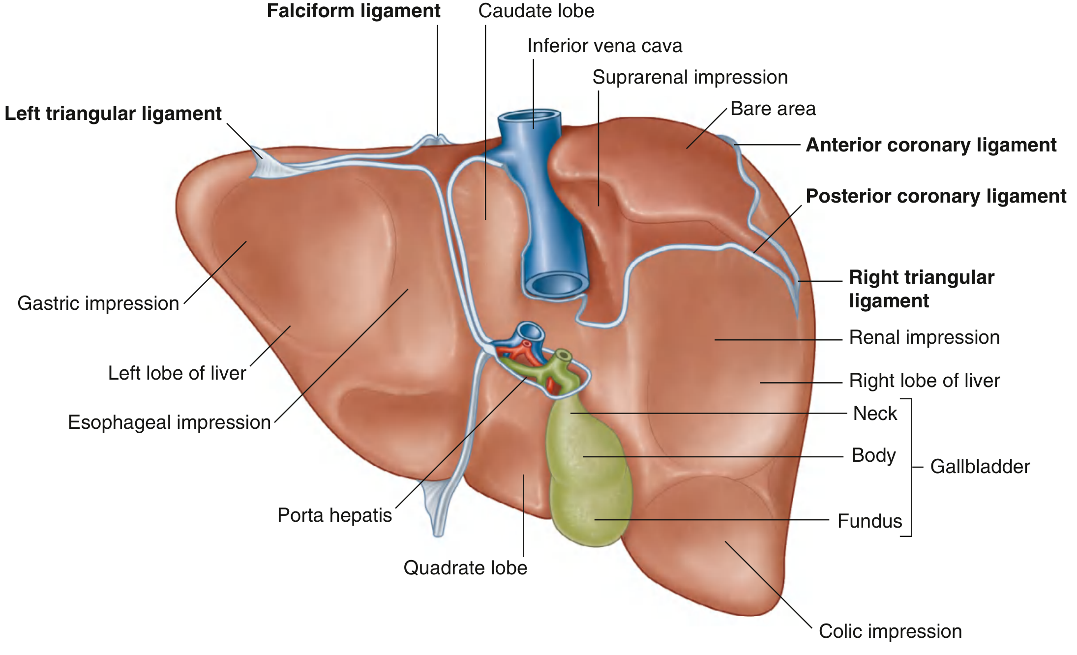
- Left limb: Fissure for ligamentum teres (round ligament) anteriorly + fissure for ligamentum venosum posteriorly
- Right limb: Gallbladder fossa anteriorly + groove for IVC posteriorly
- Crossbar: Porta hepatis - the gateway of the liver
| Region | Structure |
|---|---|
| Right lobe | Renal impression (right kidney) |
| Right lobe | Colic impression (right colic flexure) |
| Right lobe | Suprarenal impression (right adrenal) |
| Left lobe | Gastric impression (stomach) |
| Left lobe | Oesophageal impression |
| Near porta hepatis | Duodenal impression |
3. Lobes of the Liver - Morphological (Gross)
| Lobe | Position | Boundaries |
|---|---|---|
| Right lobe | Large, right side | Right of falciform (anterior), right of fissure for GB + IVC groove (posterior) |
| Left lobe | Smaller, left side | Left of falciform (anterior) |
| Caudate lobe | Posterior, between IVC groove and fissure for lig. venosum | Visible on posterior surface |
| Quadrate lobe | Inferior, square-shaped | Between GB fossa (right) and fissure for lig. teres (left), anterior to porta hepatis |
Exam tip: Caudate and quadrate lobes are anatomically part of the right lobe but functionally belong to the left lobe (they receive left portal blood and drain via left hepatic duct).
4. Functional Segmental Anatomy - Couinaud's Segments (SURGICAL ANATOMY)
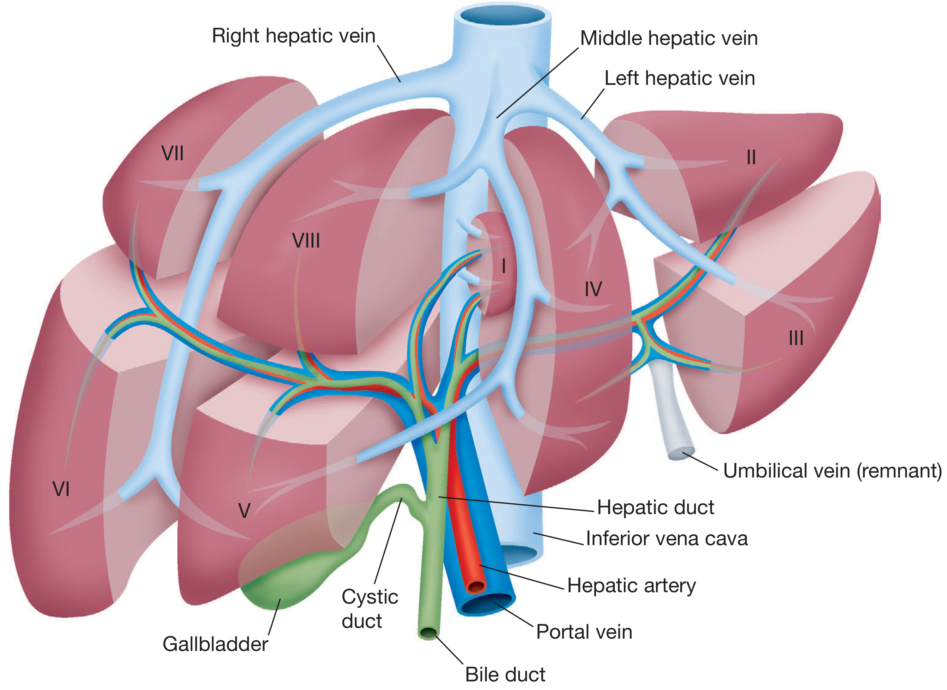
- Portal vein branch
- Hepatic artery branch
- Bile duct
- Hepatic vein drainage
"The liver is composed of 8 segments, each supplied by terminal branches of the portal vein (80% of blood flow) and hepatic artery (20%), and drained by bile ducts and hepatic veins."
- Bailey & Love's Surgery
- Left hepatic vein - between segments II/III and IV
- Middle hepatic vein - the main lobar fissure (Cantlie's line) dividing true right from left hemiliver
- Right hepatic vein - between anterior (V, VIII) and posterior (VI, VII) right sectors
| Segment | Location | Lobe |
|---|---|---|
| I | Caudate lobe (posterior, between IVC and lig. venosum) | Independent - both sides |
| II | Left posterior superior | Left lobe |
| III | Left anterior inferior | Left lobe |
| IV (IVa/IVb) | Quadrate area (medial left lobe) | Left lobe |
| V | Right anterior inferior | Right lobe |
| VI | Right posterior inferior | Right lobe |
| VII | Right posterior superior | Right lobe |
| VIII | Right anterior superior | Right lobe |
Clinical pearl: Segment I (caudate) has direct venous drainage into the IVC - it is spared in Budd-Chiari syndrome because its venous drainage is independent of the hepatic veins!
5. Blood Supply - The Dual Supply (Most Important Concept)
| Vessel | Contribution | Source |
|---|---|---|
| Portal vein | 60-70% of blood flow | Nutrient-rich venous blood from gut and spleen |
| Hepatic artery | 30-40% of blood flow | Oxygenated arterial blood |
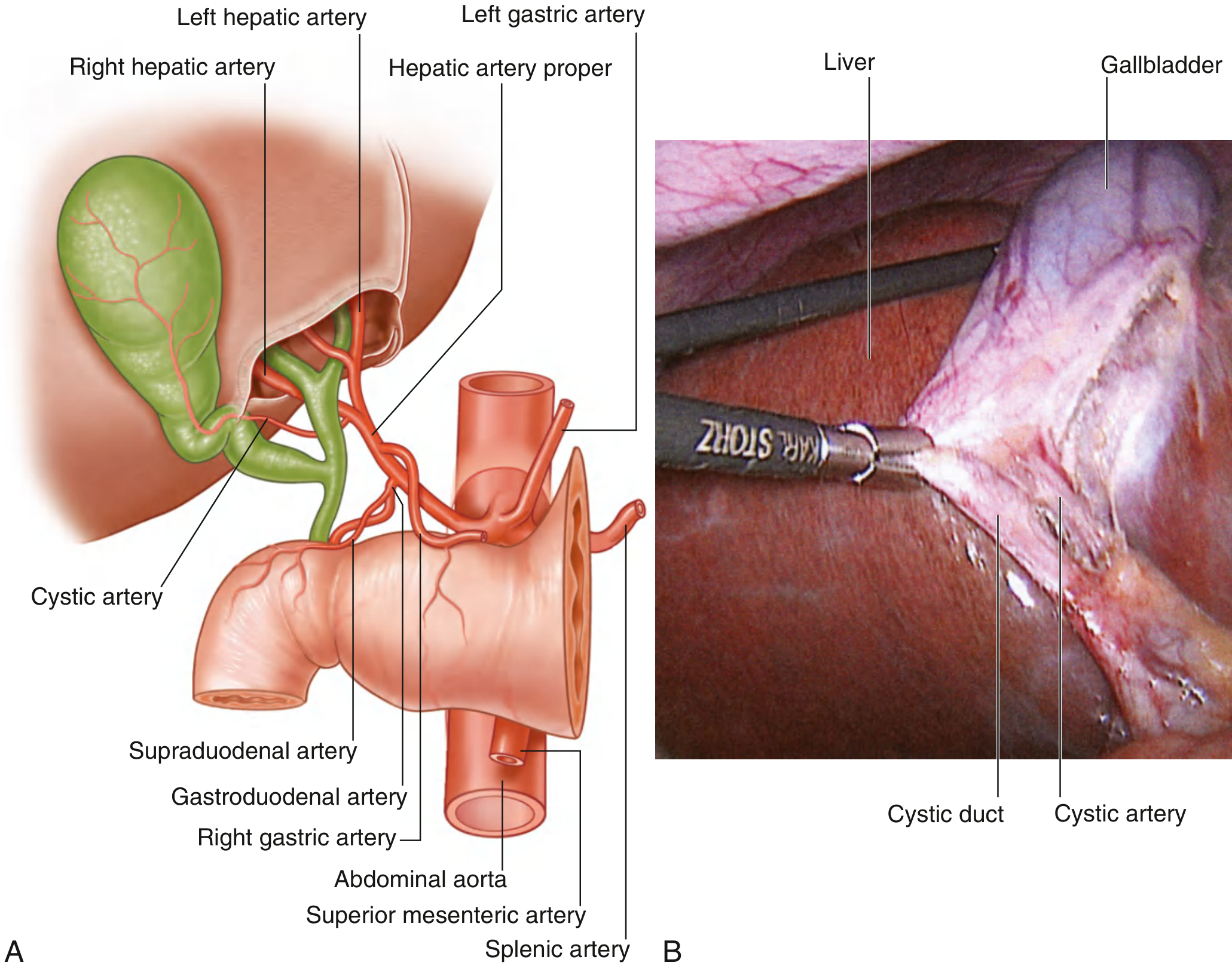
Abdominal aorta
↓
Coeliac trunk
↓
Common hepatic artery
├── Gastroduodenal artery (descends behind duodenum)
└── Hepatic artery proper
├── Right hepatic artery → cystic artery → gallbladder
└── Left hepatic artery → left lobe
- Right hepatic artery may arise from the superior mesenteric artery (17% of cases) - "replaced right hepatic artery"
- Left hepatic artery may arise from the left gastric artery (running in lesser omentum)
- These variations are critical to know before cholecystectomy or hepatic surgery!
- Formed behind the neck of the pancreas by union of superior mesenteric vein + splenic vein
- Drains: gut (stomach to rectum), spleen, pancreas
- Carries nutrients, toxins, and bacteria from the gut → liver for first-pass metabolism
- Normal portal pressure: 5-10 mmHg (portal hypertension >12 mmHg)
Liver sinusoids
↓
Central veins (terminal hepatic veins)
↓
Three hepatic veins (Right, Middle, Left)
↓
Inferior Vena Cava (IVC)
↓
Right atrium
6. Porta Hepatis - The Gateway
Portal triad at porta hepatis:
- Bile duct → RIGHT lateral
- Hepatic artery proper → LEFT lateral (medial)
- Portal vein → POSTERIOR (largest, deepest)
7. Ligaments of the Liver
| Ligament | Formed from | Connects | Contains |
|---|---|---|---|
| Falciform ligament | Ventral mesentery | Liver to anterior abdominal wall and diaphragm | Ligamentum teres (remnant of left umbilical vein) |
| Coronary ligament (anterior + posterior layers) | Peritoneal reflections | Liver to diaphragm | Encloses the bare area |
| Right triangular ligament | Fusion of coronary layers | Liver to diaphragm (right) | - |
| Left triangular ligament | Fusion of coronary layers | Liver to diaphragm (left) | - |
| Lesser omentum | Ventral mesentery | Liver (porta hepatis/lig. venosum) to stomach/duodenum | Portal triad in free edge (hepatoduodenal ligament) |
| Ligamentum teres | Obliterated umbilical vein | Runs in falciform ligament | Used as landmark for segment IV |
| Ligamentum venosum | Obliterated ductus venosus | In fissure on visceral surface | - |
8. Microanatomy (Histology) - The Liver Lobule
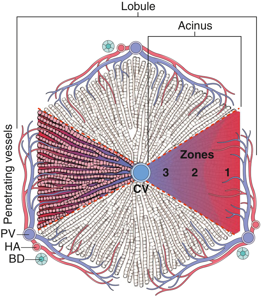
- Hexagonal unit, 1-2 mm diameter
- Central vein (terminal hepatic vein) at the center
- Portal tracts at the periphery (corners of hexagon) - each contains portal vein branch + hepatic artery branch + bile duct
- Hepatocytes arranged in plates (cords) radiating from centre
- Sinusoids - specialised capillaries between hepatocyte plates
- Blood flows: portal tract → sinusoids → central vein → hepatic vein
- Based on blood supply rather than vein drainage
- Three zones based on oxygen and nutrient gradient:
| Zone | Location | Blood supply | Injury pattern |
|---|---|---|---|
| Zone 1 (periportal) | Around portal tract | First to receive blood; most oxygenated | Phosphorus poisoning, eclampsia |
| Zone 2 | Mid-zone | Intermediate | Yellow fever |
| Zone 3 (centrilobular) | Around central vein | Last; least oxygenated | Paracetamol toxicity, ischaemia, alcohol |
| Cell | Location | Function |
|---|---|---|
| Kupffer cells | Lining sinusoids (luminal face) | Tissue macrophages; phagocytosis of bacteria, old RBCs |
| Hepatic stellate cells (Ito cells) | Space of Disse | Vitamin A storage; become activated in fibrosis → produce collagen |
| Space of Disse | Between sinusoidal endothelium and hepatocytes | Allows exchange; hepatocyte microvilli project into it |
PART 2: THE GALLBLADDER
1. Gross Anatomy
- Shape: Pear-shaped sac
- Size: 7 cm long, 3 cm wide
- Capacity: 30-50 mL (Sleisenger)
- Position: In the gallbladder fossa on the visceral (inferior) surface of the liver, between the right and quadrate lobes
- Function: Stores, concentrates (5-10x), and releases bile
| Part | Features |
|---|---|
| Fundus | Rounded free end; projects below liver edge; surface marking = tip of 9th right costal cartilage at lateral border of rectus abdominis |
| Body | Main part; lies against transverse colon and duodenum |
| Neck | Narrow part with spiral fold (Spiral valve of Heister); connects to cystic duct; Hartmann's pouch is the bulging at the infundibulum |
Clinical pearl: Gallstones can impact in Hartmann's pouch, obstructing the cystic duct and causing cholecystitis. If inflammation spreads to compress the adjacent common hepatic duct, this causes Mirizzi syndrome (obstructive jaundice).
2. Biliary System - The Complete Ductal Tree
Right and Left hepatic ducts (emerge from liver at porta hepatis)
↓
Common Hepatic Duct (CHD)
↓
+ Cystic Duct (from gallbladder neck, ~4 cm long)
↓
Common Bile Duct (CBD) (~8 cm long, 6-8 mm diameter)
↓
Joined by Main Pancreatic Duct (of Wirsung)
↓
Ampulla of Vater → opens into 2nd part of duodenum
(Sphincter of Oddi controls flow)
- Runs in the free edge of lesser omentum (hepatoduodenal ligament) - right side
- Passes behind the 1st part of duodenum
- Runs in a groove on the posterior surface of the pancreatic head
- Carcinoma of head of pancreas compresses the CBD here → painless obstructive jaundice (Courvoisier's law)
3. Blood, Lymph, and Nerve Supply of the Gallbladder
- Cystic artery - usually a branch of the right hepatic artery
- Divides near the gallbladder neck into superficial (serosal) and deep (intramural) branches
- Cystic artery is an end artery → ischaemic necrosis occurs easily in severe cholecystitis!
- Arterial variations are common and critically important in laparoscopic cholecystectomy
- Cystic vein → portal vein (or directly into hepatic sinusoids)
- Some small veins drain directly into the liver from the gallbladder bed
- Lymph nodes at the neck of the gallbladder (Lund's node / Calot's node)
- → Coeliac nodes → cisterna chyli
- Sympathetic: Coeliac axis (T7-T9) - travels with hepatic artery and portal vein
- Parasympathetic: Vagus nerve - regulates gallbladder motility (contraction in response to CCK)
- Visceral pain from gallbladder → referred to right subcostal, epigastric, and right scapular (shoulder tip) regions via sympathetic fibres
4. Triangle of Calot (Hepatocystic Triangle) - SURGICAL LANDMARK
Triangle of Calot boundaries:
- Superior border: Inferior surface of the liver (right lobe)
- Medial border: Common hepatic duct
- Lateral border: Cystic duct
Contents:
- Cystic artery (must be identified and ligated)
- Cystic lymph node (node of Calot/Lund)
Exam tip: Before clipping and dividing, surgeons must achieve the "critical view of safety" - identifying only 2 structures entering the gallbladder (cystic duct and cystic artery) in the cleared triangle. Failure to do so is the most common cause of bile duct injury during cholecystectomy.
5. Wall Structure of the Gallbladder
- Mucosa - columnar epithelium with folds (rugae) that allow expansion; no muscularis mucosa
- Lamina propria
- Tunica muscularis - smooth muscle (longitudinal + spiral)
- Perimuscular connective tissue
- Serosa (on free surface) / Adventitia fused with liver capsule (on hepatic surface)
Quick Summary Tables
Liver at a Glance
| Feature | Detail |
|---|---|
| Weight | 1400-1600 g |
| Surfaces | Diaphragmatic + Visceral |
| Morphological lobes | 4 (Right, Left, Caudate, Quadrate) |
| Functional segments | 8 (Couinaud I-VIII) |
| Arterial supply | Hepatic artery proper (from coeliac trunk) |
| Portal supply | 60-70% blood flow |
| Venous drainage | Right, Middle, Left hepatic veins → IVC |
| Microanatomy zones | Zone 1 (periportal) to Zone 3 (centrilobular) |
| Special cells | Kupffer cells, Hepatic stellate cells (Ito cells) |
Gallbladder at a Glance
| Feature | Detail |
|---|---|
| Shape | Pear-shaped |
| Capacity | 30-50 mL |
| Parts | Fundus, Body, Neck |
| Cystic duct valve | Spiral valve of Heister |
| Arterial supply | Cystic artery (from right hepatic artery) |
| Venous drainage | Cystic vein → portal vein |
| Surgical triangle | Triangle of Calot |
| Common pathology trap | Hartmann's pouch impaction → Mirizzi syndrome |
Explain steps of lap chole to mbbs intern
Laparoscopic Cholecystectomy (Lap Chole) - Step-by-Step for MBBS Intern
Background - Why Lap Chole?
- Smaller incisions (4 small ports vs. one large cut)
- Less postoperative pain
- Shorter hospital stay (often same-day or next morning)
- Faster return to normal activity
- Better cosmesis
- Mortality: 0.1-0.5%, Morbidity: 2-3% (Sabiston)
Indications
- Symptomatic cholelithiasis (biliary colic)
- Acute cholecystitis (gold standard even in the acute setting)
- Chronic cholecystitis
- Gallstone pancreatitis (after resolution)
- Empyema / mucocele of gallbladder
- Porcelain gallbladder (risk of malignancy)
- Gallbladder polyps >10 mm
Contraindications
| Absolute | Relative |
|---|---|
| Inability to tolerate general anaesthesia | Severe COPD (CO₂ exchange issues) |
| Uncorrected coagulopathy | Congestive cardiac failure |
| End-stage liver disease with portal hypertension | Pulmonary hypertension |
| Prior upper abdominal surgery (adhesions - higher conversion rate) |
The Operation - Step by Step
STEP 1: Anaesthesia and Preparation
- General anaesthesia with muscle relaxation is mandatory (the only way to tolerate pneumoperitoneum)
- Patient positioned supine on the operating table
- One arm tucked (to allow intraoperative cholangiogram if needed)
- Orogastric tube placed to decompress stomach (improves view of upper abdomen)
- Foley catheter if prolonged operation expected
- Abdomen prepped and draped widely - must accommodate open surgery conversion if needed
- Patient secured well to table (for steep positioning changes)
STEP 2: OR Setup and Patient Positioning
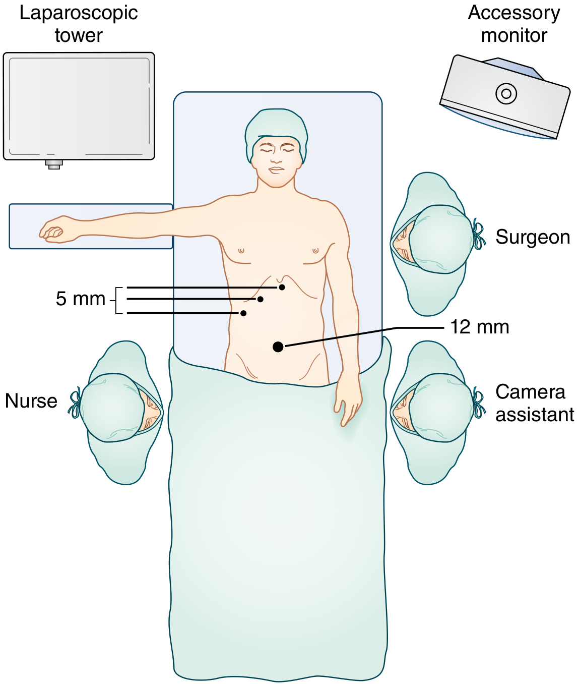
- Surgeon stands to the left of the patient (American technique) or between patient's legs (French technique)
- Two video monitors, one on each side of the table
- After pneumoperitoneum: patient tilted to reverse Trendelenburg 30° + 15° left rotation
- This causes colon and duodenum to fall away from the liver by gravity - crucial for exposure
STEP 3: Establishing Pneumoperitoneum
- Non-combustible (safe with electrocautery)
- Rapidly absorbed from peritoneal cavity
- Soluble in blood
⚠️ Caution: CO₂ causes hypercarbia in patients with significant cardiopulmonary disease. Anaesthetist monitors CO₂ levels throughout.
| Technique | How | When Used |
|---|---|---|
| Veress (Closed) needle technique | Blind insertion of a spring-loaded Veress needle at the umbilicus → insufflate CO₂ → then insert trocar | Most common in uncomplicated cases |
| Hasson (Open) technique | Small umbilical incision → directly visualise peritoneum → insert port under vision → insufflate | Preferred by many; safer in previous surgery, obesity, pregnancy |
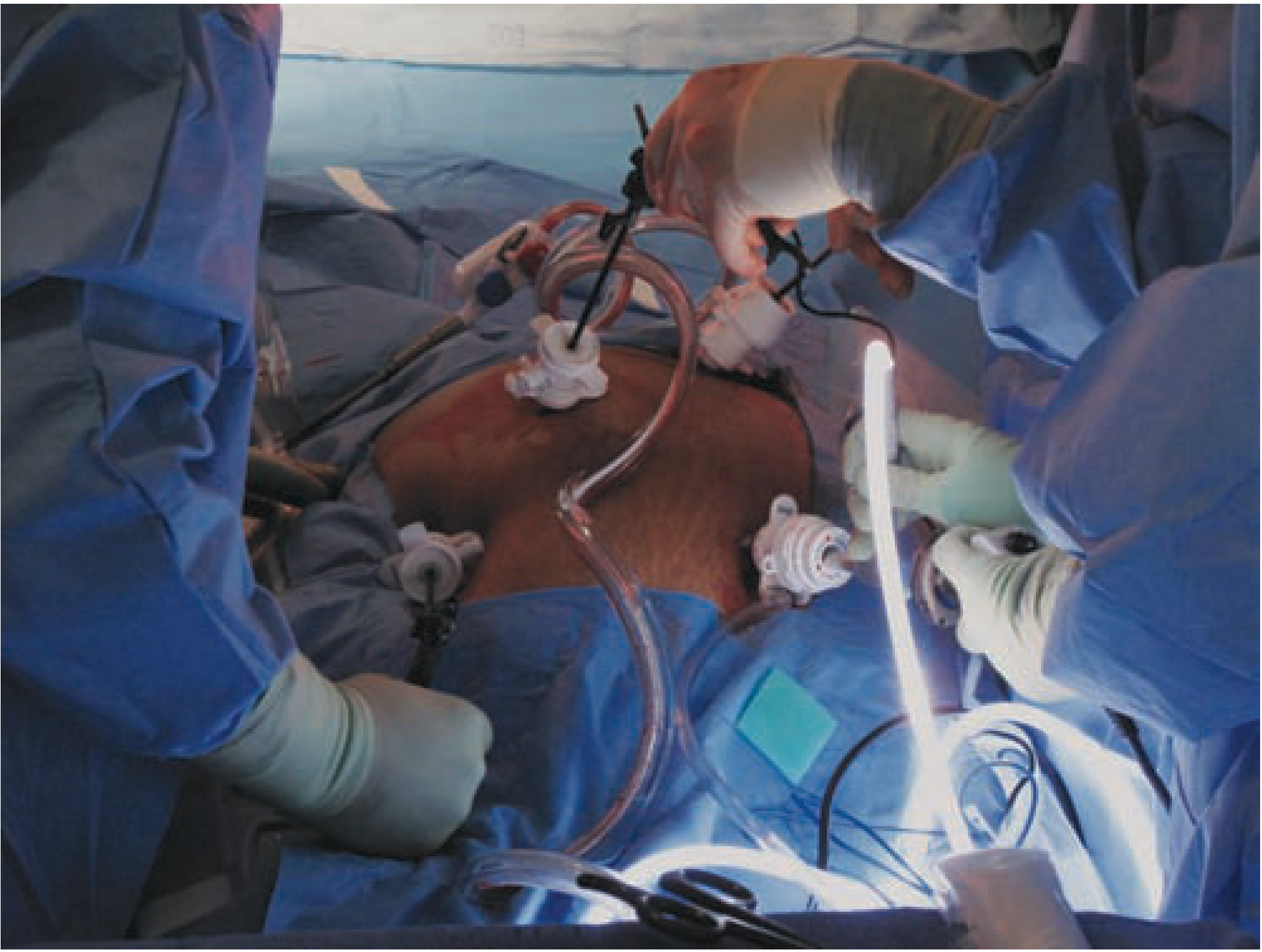
STEP 4: Port (Trocar) Placement - The Standard 4-Port Technique
| Port | Size | Location | Purpose |
|---|---|---|---|
| Port 1 (Camera port) | 10-12 mm | Umbilicus (or just above/below) | Laparoscope camera; also used to extract gallbladder at end |
| Port 2 (Working port) | 5 mm | Subxiphoid / Epigastric (~5 cm below xiphoid) | Surgeon's main dissecting instruments (Maryland forceps, hook cautery) |
| Port 3 (Retracting port) | 5 mm | Right midclavicular line, subcostal | Surgeon's left hand - grasps gallbladder infundibulum, pulls it laterally |
| Port 4 (Fundus port) | 5 mm | Right anterior axillary line, between 12th rib and iliac crest | Assistant retracts gallbladder fundus cephalad (upward) |
STEP 5: Retraction and Exposure
-
Fundus grasped and pushed cephalad (upward toward right shoulder) by the assistant via Port 4 - this rolls the liver up and out of the way
-
Infundibulum (neck/Hartmann's pouch) grasped and retracted inferolaterally (downward and to the right) by the surgeon via Port 3 - this stretches open the Triangle of Calot and pulls the cystic duct AWAY from the CBD (critical for safety)
"A key maneuver is for the surgeon's left operative instrument to distract the infundibulum/Hartmann pouch laterally."
- Sabiston Textbook of Surgery
STEP 6: Dissection of the Hepatocystic Triangle (Triangle of Calot)
- Superior: under-surface of the liver
- Medial: common hepatic duct
- Lateral: cystic duct
- The peritoneum on BOTH anterior AND posterior aspects of the hepatocystic triangle is incised
- Dissection begins on the gallbladder side (known structure) and proceeds distally - never start blindly in an unknown area
- Use fine-tipped dissecting forceps (Maryland dissector) to sweep fibroareolar tissue off the infundibulum
- The dissection starts 4-5 cm proximal to the neck of the gallbladder (modified top-down technique)
- Keep dissecting until the lower third of the gallbladder is separated from the liver bed - so you can see liver substance (cystic plate) through the window
- The Calot's node (lymph node) is a useful landmark - it almost always sits directly on top of the cystic artery
STEP 7: Achieving the Critical View of Safety (CVS)
This is the MOST IMPORTANT safety step in the entire operation. Do NOT clip or cut anything until this is confirmed.
- The hepatocystic triangle is completely cleared of fat and fibrous tissue
- Only TWO structures are seen entering the gallbladder (cystic duct + cystic artery) - nothing else
- The lower third of the gallbladder is separated from the liver bed (cystic plate visible)
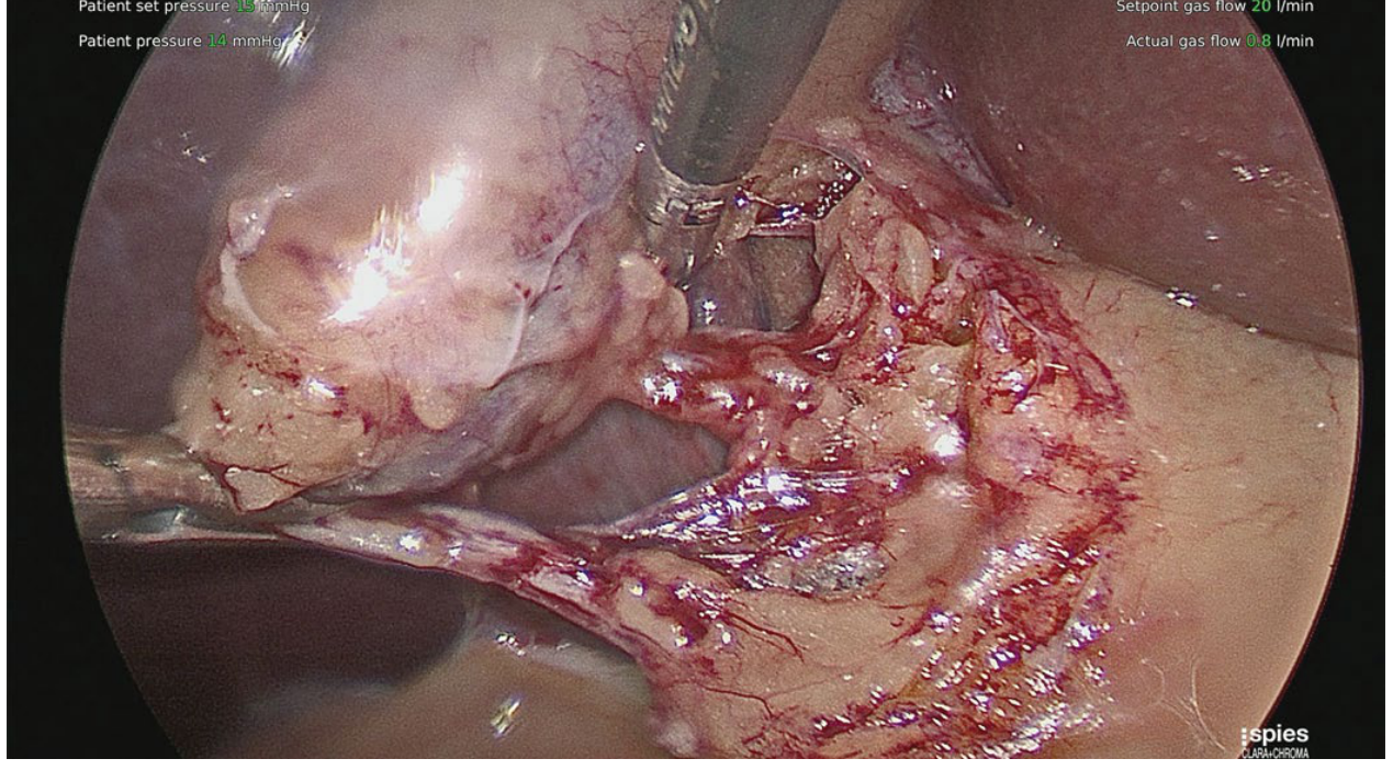
"There should be two, and only two, structures (the cystic duct and artery) crossing this window - this is the 'critical view of safety,' which should be demonstrated prior to clipping or cutting any tubular structures."
- Maingot's Abdominal Operations
STEP 8: Intraoperative Cholangiogram (Selective Use)
- Anatomy is unclear or abnormal
- Suspected CBD stones (gallstone pancreatitis, dilated CBD on USS)
- Abnormal LFTs
- Unclear biliary anatomy during dissection
STEP 9: Clipping and Dividing the Cystic Structures
Cystic ARTERY first:
- 2 clips on the patient side (liver side)
- 1 clip on the gallbladder side
- Divided between clips with scissors
Cystic DUCT second:
- 2 clips on the patient side (CBD side)
- 1 clip on the gallbladder side
- Divided between clips with scissors
- Never clip anything until CVS is confirmed
- Always place clips at right angles to the duct
- Leave adequate length of cystic duct stump (too short → CBD risk; too long → stump chole)
- Inspect clips before and after division
STEP 10: Dissection of Gallbladder from Liver Bed
- Electrocautery (hook cautery) is used to dissect the gallbladder off its hepatic fossa
- Plane: just below the gallbladder serosa, superficial to the liver parenchyma
- Small venules drain directly from the gallbladder into the liver bed → excellent haemostasis needed
- Kept attached by the fundus until complete - the superior traction of the fundus at this stage gives excellent exposure back to the Triangle of Calot for one final check of the clips
"The cystic duct and cystic artery clips are inspected just before completion of the dissection of the fundic attachments because the superior traction of the fundus has provided exposure to the porta and triangle of Calot."
- Sabiston
STEP 11: Specimen Extraction
- Gallbladder is placed in a retrieval bag (reduces port-site wound contamination)
- Bag brought out through the umbilical (12 mm) port
- If the gallbladder is large or contains big stones: the umbilical fascial incision may need to be slightly extended to allow extraction
- If bile spills during extraction: irrigate thoroughly and retrieve all stones - spilled stones can cause late subhepatic abscesses, fistulae, or empyemas years later
STEP 12: Final Inspection, Irrigation, and Port Closure
- Inspect the liver bed for haemostasis and bile leak (watch for 2-3 minutes)
- Inspect the gallbladder clips once more - confirm they are secure
- Irrigate if any soiling occurred
- Deflate CO₂ (full deflation reduces shoulder-tip pain from diaphragmatic irritation)
- Close port sites:
- 12 mm umbilical port: Fascial closure mandatory (risk of port-site hernia)
- 5 mm ports: Skin closure only (fascia does not routinely require closure)
- Subcuticular or interrupted skin sutures at each port site
Conversion to Open Surgery
- Inability to achieve CVS
- Dense adhesions obscuring anatomy
- Uncontrolled bleeding
- Bile duct injury identified intraoperatively
- Bile duct obstruction/malignancy found
- Equipment failure
"Laparoscopic cholecystectomy for acute cholecystitis may carry longer operative times and a higher conversion rate to the open procedure."
- Sabiston
Complications
| Category | Complications |
|---|---|
| Bile duct injury | Most feared (1.5-3x higher than open surgery); often due to misidentification |
| Bile leak | From cystic duct stump, clip slippage, or duct of Luschka |
| Haemorrhage | Cystic artery, liver bed, port-site vessels |
| Retained CBD stones | Missed choledocholithiasis (treat with post-op ERCP) |
| Bowel/visceral injury | During trocar insertion especially |
| Pneumoperitoneum-related | CO₂ embolism, vagal reaction, cardiac arrhythmias, hypercarbia |
| Trocar-related | Vascular injury (aorta, IVC, iliac vessels) - most dangerous during initial blind entry |
| Port-site hernia | Especially at 12 mm umbilical port if fascia not closed |
| Wound infection | From bile/stone spillage |
Key Takeaways for an Intern
Lap Chole in 12 Steps:
1. GA + patient prep (supine, arm tucked)
2. OR setup (reverse Trendelenburg + left tilt after insufflation)
3. Pneumoperitoneum (Veress needle or Hasson open) with CO₂ at 12-15 mmHg
4. 4 ports (12mm umbilical, 5mm subxiphoid, 5mm midclavicular, 5mm axillary)
5. Retract: fundus UP + infundibulum DOWN and LATERAL
6. Lyse adhesions
7. Dissect hepatocystic triangle (both anterior & posterior peritoneum)
8. Achieve CRITICAL VIEW OF SAFETY (most important step!)
9. Cholangiogram if needed
10. Clip + divide cystic ARTERY then cystic DUCT (2+1 clips)
11. Hook cautery to dissect gallbladder from liver bed
12. Bag + extract via umbilical port → inspect → irrigate → close fascia at 12mm port
The cardinal rule: Never clip until Critical View of Safety is confirmed.
The commonest serious complication: CBD injury from misidentification.
Explain courvoisiers law simply
Courvoisier's Law - Simple Explanation for Interns
The Law - One Sentence
"In a jaundiced patient, a palpable, non-tender gallbladder is unlikely to be due to gallstones - it usually means malignant obstruction."
The Diagram (This is All You Need to Remember)
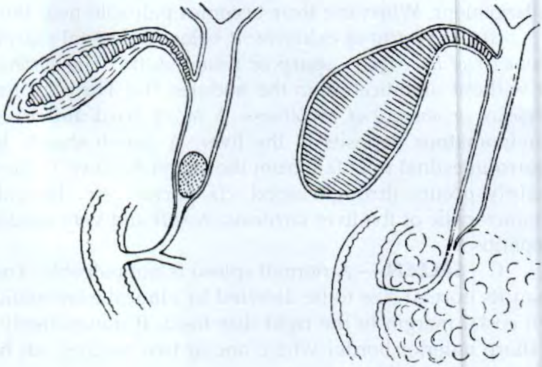
The Logic - WHY Does This Happen?
Scenario 1: Gallstone obstructing the CBD
- Gallstones don't appear suddenly. They form in the gallbladder over years
- During that time, repeated bouts of cholecystitis and inflammation damage the gallbladder wall
- The wall becomes fibrosed, scarred, and thickened - it loses its ability to stretch
- So when a stone finally slips into the CBD and blocks it, the gallbladder cannot distend - it is already stiff from chronic scarring
- Result: gallbladder NOT palpable
Scenario 2: Malignant obstruction (e.g., carcinoma head of pancreas)
- The tumour grows slowly and silently compresses the CBD from outside
- The gallbladder itself has never been inflamed - its wall is normal and healthy
- A healthy gallbladder wall is thin and elastic, like a balloon
- Bile backs up behind the obstruction → gallbladder fills up and distends massively
- Result: gallbladder palpable and non-tender (no inflammation)
"When the common duct is obstructed by a stone, distension of the gallbladder (which is likely to be chronically inflamed) is rare; when the duct is obstructed in some other way, such as a neoplasm, distension of the normal gallbladder is common."
- Bailey & Love's Surgery 28E
Summary Table
| Feature | Gallstone CBD obstruction | Malignant obstruction |
|---|---|---|
| Gallbladder wall | Scarred, fibrotic (from chronic cholecystitis) | Normal, thin, elastic |
| Gallbladder distension | Cannot distend | Distends easily |
| Gallbladder palpable? | No | Yes |
| Tenderness | If acute - tender | Non-tender (no inflammation) |
| Pain | Usually colicky | Often painless jaundice |
| Typical patient | Middle-aged woman, obese, "5 F's" | Older male, progressive jaundice + weight loss |
| Typical cause | Choledocholithiasis | Ca head of pancreas, cholangiocarcinoma, ampullary ca, periampullary malignancy |
Causes of a Courvoisier Gallbladder (Palpable + Non-Tender in Jaundice)
- Carcinoma of the head of pancreas - most common cause
- Carcinoma of the ampulla of Vater (ampullary carcinoma)
- Cholangiocarcinoma (distal bile duct)
- Duodenal carcinoma
- Mirizzi syndrome (rarely - stone impacted in cystic duct compressing CBD)
"Deep jaundice without pain, particularly with a palpable gallbladder (Courvoisier sign), suggests neoplastic obstruction of the bile duct, even when the patient has stones in the gallbladder."
- Sleisenger & Fordtran's Gastrointestinal and Liver Disease
What Investigations Follow?
- Ultrasound abdomen - dilated CBD? Liver metastases? Pancreatic mass?
- Contrast-enhanced CT abdomen - defines the tumour, checks resectability (involvement of SMA, portal vein, coeliac axis)
- MRI/MRCP - maps biliary tree, shows level of obstruction
- ERCP - if diagnostic doubt, cholangitis, or deep jaundice needing stenting (also gets brush cytology/biopsy)
- Tumour markers - CA 19-9 (pancreatic ca), CEA
- LFTs, coagulation - obstructive pattern (↑ALP, ↑bilirubin, prolonged PT from vitamin K malabsorption)
Important Caveat (Exam Trap!)
- A jaundiced patient with gallstones can still have a palpable gallbladder if there is no prior cholecystitis (first-time obstruction, no scarring yet)
- Conversely, a malignant obstruction may not produce a palpable gallbladder if the tumour is too proximal (above the cystic duct insertion)
The law tells you what is unlikely, not what is impossible.
One-Line Exam Answer
Courvoisier's Law: "In obstructive jaundice, a palpable non-tender gallbladder is unlikely due to stones - because stone disease causes chronic cholecystitis and fibrosis preventing distension. It usually indicates periampullary malignancy, most often carcinoma of the head of the pancreas."
How much bile leak is considered dangerous significant
"bile leak" grading classification definition ISGLS
"bile leak" definition "postoperative day 3" bilirubin drain
ISGLS bile leak definition grading grade A B C drain bilirubin postoperative day 3
Bile Leak - How Much is Significant?
The Short Answer
- Biochemical definition - drain fluid bilirubin vs serum bilirubin ratio
- Output volume per day - low vs high output
- Clinical impact - does the patient need more than just a drain?
1. The ISGLS Definition (Gold Standard)
A bile leak is defined as: Drain fluid with a bilirubin concentration at least 3 times the concurrent serum bilirubin, measured on or after postoperative day 3
The need for radiological intervention (percutaneous drainage of a biloma) or relaparotomy for bile peritonitis - even without a surgical drain in place
2. ISGLS Grading - Grade A, B, C
| Grade | Definition | Clinical Impact | Management |
|---|---|---|---|
| Grade A | Bile leak present BUT patient is in good condition; leak adequately controlled by existing drain; output decreasing daily | Minimal - no change in management needed | Conservative: leave drain in, monitor, nothing else required |
| Grade B | Requires active intervention BEYOND simple drain management - BUT does NOT require relaparotomy. Grade A leak that persists >1 week → upgrades to Grade B | Moderate - changes management; prolongs hospital stay; patient may go home with drain | Antibiotics + percutaneous drainage of collections + ERCP with stent/sphincterotomy + percutaneous transhepatic cholangiodrainage (PTCD) |
| Grade C | Requires relaparotomy to control the leak | Severe - life-threatening if not treated | Reoperation: suture closure, bilioenteric anastomosis (Roux-en-Y hepaticojejunostomy) |
Grade A alone = not dangerous. Grade B and C = clinically significant.
3. Volume Thresholds - The Practical Numbers
| Volume | Category | Significance | Action |
|---|---|---|---|
| Low output | <200-300 mL/24h | Usually minor leak (cystic duct stump, duct of Luschka) | Conservative management - drain alone; most close spontaneously |
| High output | >200 mL/24h (Fischer's) or >300 mL/24h (HPB literature) | Significant - suggests major ductal injury or ongoing active leak | ERCP + sphincterotomy/stenting urgently; if haemodynamically unstable → relaparotomy |
"If the output is low, it can be managed with drainage alone as these are usually from the cystic duct stump or a small accessory duct of Luschka and will likely close on their own. If the patient is hemodynamically unstable or there is high output from the drain (>200 mL/24 hours), then the best modality for diagnosis and therapy is an ERCP with sphincterotomy/stenting."
- Fischer's Mastery of Surgery 8E
4. Sources of Bile Leak - Why It Matters
| Source | Common Setting | Severity | Likely to Close Spontaneously? |
|---|---|---|---|
| Cystic duct stump leak | After cholecystectomy (clip slippage, necrosis) | Usually low-grade | Yes - with ERCP stenting |
| Duct of Luschka (small accessory duct between gallbladder bed and liver) | Post-cholecystectomy | Usually minor | Often yes |
| CBD / hepatic duct injury | Mis-clipping, thermal injury during lap chole | Major - Grade B/C | No - needs surgical repair |
| Cut surface leak | After liver resection / hepatectomy | Variable | Depends on size |
| Bilioenteric anastomosis leak | After Whipple's, hepaticojejunostomy | Potentially major | Sometimes with drainage ± stenting |
5. Warning Signs That Make a Leak Immediately Dangerous
- Bile peritonitis - bile irritates the peritoneum severely; causes chemical peritonitis with intense pain, rigidity, fever
- Haemodynamic instability - sepsis from infected biloma/peritonitis
- Rising bilirubin - undrained bile leaking into the cavity
- Failure to improve with conservative management after 48-72 hours
- Biloma enlarging on serial imaging
"If peritonitis is present or develops during management of the leak, then drainage is inadequate and reoperation may be required."
- Fischer's Mastery of Surgery 8E
6. Amsterdam Classification (Post-Cholecystectomy Leaks Specifically)
| Type | Description | Treatability |
|---|---|---|
| A | Cystic duct or aberrant hepatic duct (duct of Luschka) leak | ERCP success ~100% |
| B | Major bile duct leak ± stricture | ERCP + often surgical |
| C | Bile duct stricture without leak | Balloon dilation / stenting |
| D | Complete transection of bile duct | Surgery required |
Quick Summary for the Intern
Bile Leak = Dangerous When:
BIOCHEMICAL DEFINITION (ISGLS):
Drain bilirubin ≥ 3× serum bilirubin ON or AFTER Day 3
VOLUME THRESHOLD:
Low output < 200-300 mL/day → Conservative (drain alone, may close)
High output > 200-300 mL/day → ERCP + stent needed urgently
GRADE (ISGLS):
Grade A → Drain-controlled, patient well → Not dangerous
Grade B → Needs ERCP/PTCD/antibiotics → Significant
Grade C → Needs relaparotomy → Life-threatening
RED FLAGS (act immediately regardless of volume):
✗ Bile peritonitis (pain, rigidity, fever)
✗ Haemodynamic instability / sepsis
✗ Enlarging biloma on imaging
✗ Rising serum bilirubin
✗ Not improving after 48-72h of drainage
Teach me about PAOD and gangrene with standard definitions