
CAD CAM technology in restorative dentistry and endodontics . 10 marks univerisity like answer elaborately
Reading File
Finding Sources
Finding Sources
Finding Sources
Searching the Web
CAD CAM technology in restorative dentistry and endodontics applications
CAD/CAM Technology in Restorative Dentistry and Endodontics
Introduction
CAD/CAM stands for Computer-Aided Design and Computer-Aided Manufacturing. It is a digital technology that allows a clinician or technician to design a dental restoration on a computer using a virtual 3-dimensional model of the prepared tooth, and then fabricate that restoration using automated milling or additive manufacturing (3D printing) equipment. Introduced into dentistry in the early 1980s (Duret, and later Mörmann and Brandestini with the CEREC system), CAD/CAM has since transformed prosthodontic, restorative, and increasingly endodontic practice by replacing time-consuming manual impression, wax-up, and casting techniques with rapid, reproducible, and highly accurate digital workflows.
Components of a CAD/CAM System
A CAD/CAM system has three basic components:
- Data acquisition unit (scanner/digitizer) - captures the 3D surface geometry of the prepared tooth either directly (intraoral scanner - optical/laser scanning of the mouth) or indirectly (scanning of a stone cast or impression).
- Designing unit (software) - the acquired data is displayed as a virtual model on screen; the operator (or automated software) designs the restoration - contours, occlusal anatomy, margins, connectors, and material thickness - using CAD software.
- Manufacturing unit - converts the digital design into a physical restoration by:
- Subtractive manufacturing - milling/grinding a block of ceramic, composite, or metal using diamond burs under water/air coolant.
- Additive manufacturing - 3D printing (stereolithography, digital light processing, fused deposition modelling) used for models, surgical guides, temporary crowns, and increasingly definitive restorations.
Classification of CAD/CAM Systems
| Type | Description | Example |
|---|---|---|
| Chairside systems | Scanning, designing, and milling done in the dental office in a single visit | CEREC (Sirona), E4D |
| Laboratory systems | Impression/cast scanned in lab; design and milling done centrally | 3Shape, Cerec inLab |
| Centralized/industrial systems | Scan data sent to an industrial milling center; restoration returned by mail | Procera (Nobel Biocare), Lava (3M) |
Systems are also described by generation based on integration of scanning-design-milling steps, with later generations offering open architecture (STL file compatibility) allowing interchange between different manufacturers' scanners, software, and mills.
Applications in Restorative Dentistry
CAD/CAM has become the mainstay of modern indirect restorative dentistry:
- Inlays, onlays, and veneers - milled from monolithic ceramic blocks (lithium disilicate, leucite-reinforced glass ceramic) with excellent marginal fit and aesthetics in a single visit.
- Full crowns and fixed partial dentures (bridges) - both anterior and posterior, in ceramic, zirconia, or hybrid ceramic materials.
- Implant-supported restorations - custom abutments and implant crowns/bridges with precise fit to the implant platform.
- Removable prosthodontics - CAD/CAM-milled or printed denture bases and try-in dentures, improving fit and reducing chair-time.
- Diagnostic wax-ups and smile design - virtual mock-ups for aesthetic planning before definitive treatment.
- Materials used: feldspathic and leucite-reinforced ceramics, lithium disilicate glass-ceramic, zirconia (monolithic and layered), hybrid ceramics/resin-ceramic composites, and PMMA for provisional restorations.
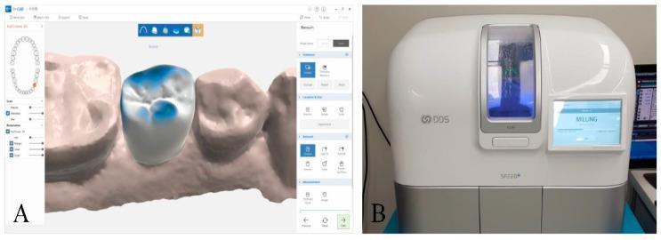
Chairside systems like CEREC allow same-day, single-visit crowns - the tooth is prepared, optically scanned (eliminating conventional impressions), the restoration is designed on-screen, milled from a ceramic block in about 10-20 minutes, and cemented, all in one appointment.
Applications in Endodontics
CAD/CAM and associated digital/3D-printing technology have introduced a distinct discipline sometimes called "Guided Endodontics":
-
3D-printed endodontic access guides - Cone-beam CT (CBCT) data is fused with an intraoral/model scan; software plans the ideal access cavity and canal trajectory, and a physical guide (with a metal sleeve to direct the bur) is 3D-printed. This is invaluable in:
- Locating calcified/obliterated canals
- Conservative access cavity preparation in teeth with extensive restorations
- Avoiding perforation in cases with altered canal anatomy (e.g., dens invaginatus, dilacerated roots)
-
Guided apical surgery - CAD/CAM-fabricated surgical guides help precisely locate the osteotomy site and control the angle/depth of resection during apicoectomy, minimizing damage to adjacent anatomical structures (inferior alveolar nerve, sinus floor).
-
Custom endodontic post and core fabrication - CAD/CAM milling of fiber-reinforced or zirconia posts and cores that are custom-fitted to the canal anatomy, improving retention and reducing risk of root fracture compared to prefabricated posts.
-
CAD/CAM in regenerative and restorative interface - fabrication of customized intracanal medicament carriers, custom post-endodontic restorations (endocrowns) that are milled directly onto the pulp chamber/root canal orifice geometry from a single ceramic block, ideal for badly broken-down endodontically treated posterior teeth as they eliminate the need for a separate post-core and crown.
-
3D-printed models for treatment planning and simulation - printed replicas of complex root canal systems used for case planning, patient education, and endodontic training.
-
Digital workflow integration - CBCT + intraoral scan superimposition (double-scan technique) allows a single digital dataset to plan both the endodontic access/surgery and the final restoration, streamlining interdisciplinary treatment.
Advantages of CAD/CAM Technology
- Eliminates conventional impressions, improving patient comfort and reducing distortion errors
- High precision and reproducibility of fit (marginal and internal adaptation)
- Reduced chair-time; many restorations completed in a single visit
- Reduced laboratory dependency and turnaround time
- Consistent, homogeneous, defect-free industrially processed blocks (fewer porosities than cast/hand-layered restorations)
- Enhanced conservation of tooth structure in guided endodontic access
- Improved case documentation, communication, and archiving of digital records
- Greater accuracy in complex or high-risk cases (calcified canals, surgical guidance)
Disadvantages / Limitations
- High initial cost of equipment (scanners, milling units, software) and per-unit block/material cost
- Steep learning curve for clinicians and technicians
- Limited to certain restoration types/materials (some CAD/CAM ceramics still show lower flexural strength than metal-ceramic in high-stress areas)
- Difficulty capturing subgingival margins or in cases with limited access/moisture control (bleeding, saliva) during intraoral scanning
- Guided endodontic access requires an additional CBCT scan (radiation exposure) and lab/printing time before treatment
- Milling burs wear over time, potentially affecting long-term precision
- Not all clinical situations (e.g., very deep subgingival margins, extensive edentulous ridges) are fully suited to digital scanning
Recent Advances
- Integration of artificial intelligence for automated margin detection and restoration design suggestions
- Hybrid manufacturing combining milling and 3D printing (dual workflows) for both provisional and definitive prostheses
- Intraoral scanners with AI-assisted CBCT fusion enabling one-visit guided endodontic treatment planning
- Development of stronger 3D-printable ceramic-filled resins for permanent restorations, narrowing the gap with milled ceramics
Conclusion
CAD/CAM technology has fundamentally changed the practice of restorative dentistry by enabling precise, efficient, single-visit fabrication of inlays, onlays, veneers, crowns, and bridges from strong, aesthetic materials such as lithium disilicate and zirconia. In endodontics, the same digital principles - CBCT-guided design and 3D-printed guides - have given rise to guided endodontic access and surgery, custom post-core systems, and endocrowns, improving precision while conserving tooth structure in complex cases. Despite cost and learning-curve barriers, continued advances in scanning accuracy, materials science, and AI-assisted design are expected to make CAD/CAM an increasingly indispensable part of everyday clinical dental practice.
Note: The institutional medical textbook library used for verification is focused on general medicine/surgery specialties and does not carry dedicated dental/prosthodontic textbooks, so this answer is compiled from established dental literature and current clinical sources such as the PMC review on CAD-CAM applications in dentistry.
This is a shared conversation. Sign in to Orris to start your own chat.