
Anatomy of spinal cord
Reading File
Reading File
Finding Sources
Finding Sources
Searching Images
labeled cross-sectional anatomy diagram of spinal cord gray and white matter dorsal ventral horns tracts
Searching PubMed
"spinal cord"[MeSH Terms] AND anatomy
Searching the Web
spinal cord anatomy segments enlargements conus medullaris authoritative anatomy
Reading File
Reading File
Reading File
Reading Web Page
https://www.ncbi.nlm.nih.gov/books/NBK537004
Anatomy of the spinal cord
The spinal cord is the caudal continuation of the medulla oblongata. It lies within the vertebral canal, conducts sensory information to the brain and motor commands from it, and contains reflex centers.
1. Extent and termination
- Begins at the foramen magnum, continuous with the medulla.
- In adults it usually ends as the conus medullaris opposite about L1-L2 vertebral level. In neonates it ends lower, near L3.
- The cord is shorter than the vertebral column. Thus, lower spinal nerve roots descend within the vertebral canal before exiting, forming the cauda equina.
- A thin pial strand, the filum terminale, extends from the conus and anchors inferiorly.
2. Spinal segments and nerves
There are 31 spinal cord segments, each giving a pair of spinal nerves:
| Region | Segments / nerve pairs |
|---|---|
| Cervical | 8 (C1-C8) |
| Thoracic | 12 (T1-T12) |
| Lumbar | 5 (L1-L5) |
| Sacral | 5 (S1-S5) |
| Coccygeal | 1 (Co1) |
3. Enlargements
The cord has two widened regions because of the increased number of neurons serving the limbs:
- Cervical enlargement: supplies the upper limb via the brachial plexus.
- Lumbosacral enlargement: supplies the lower limb via the lumbosacral plexus.
Campbell's Operative Orthopaedics, 15th ed., p. 2225.
4. Coverings and spaces
From outside inward:
- Vertebral canal
- Epidural space: contains fat and internal vertebral venous plexus
- Dura mater
- Arachnoid mater
- Subarachnoid space: contains cerebrospinal fluid
- Pia mater: closely adherent to the cord
The pia forms denticulate ligaments, which help stabilize the cord laterally.
5. External features
- Anterior median fissure: deep groove on the ventral surface.
- Posterior median sulcus: shallow dorsal midline groove.
- Posterolateral sulcus: entry point of dorsal sensory rootlets.
- Anterolateral sulcus: exit point of ventral motor rootlets.
6. Internal structure in cross-section
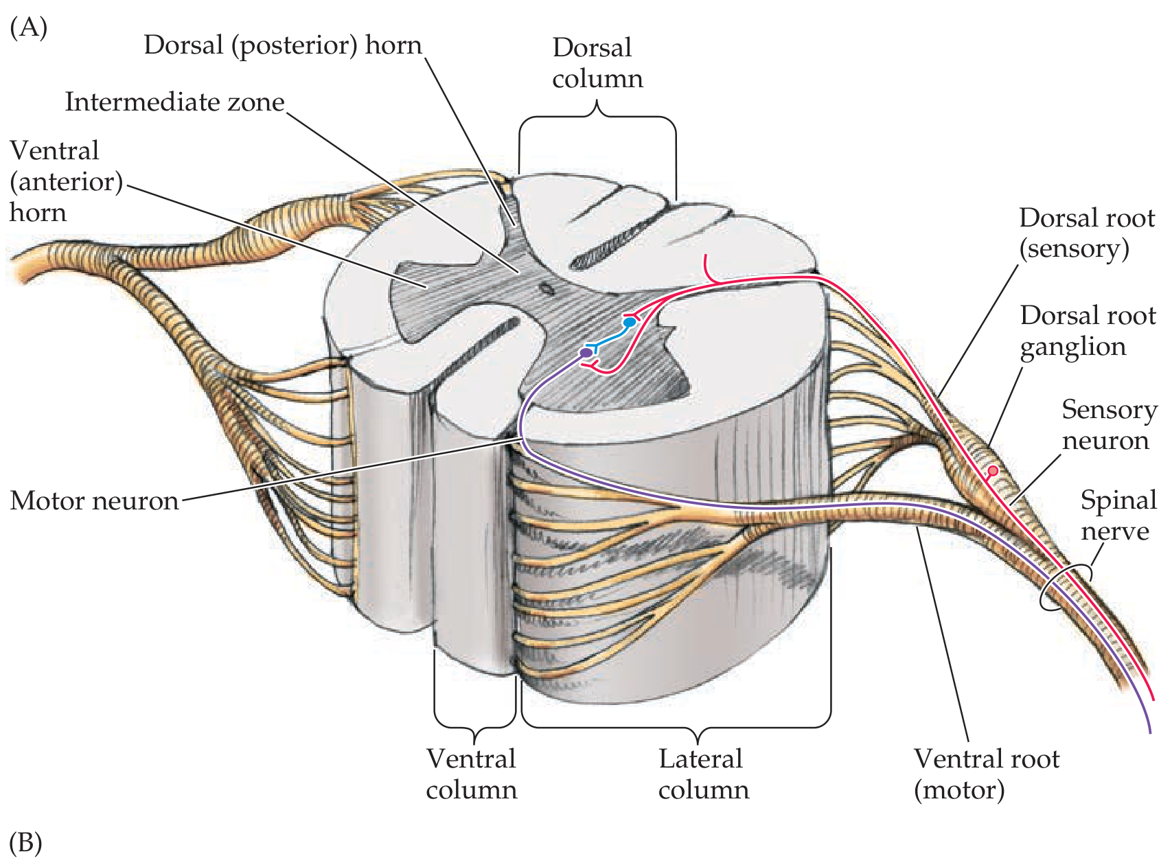
The spinal cord has central gray matter surrounded by peripheral white matter.
Gray matter
It has an H-shaped or butterfly-shaped appearance.
- Posterior (dorsal) horn: receives and processes sensory input.
- Anterior (ventral) horn: contains lower motor neurons supplying skeletal muscle.
- Lateral horn:
- Present prominently at T1-L2: preganglionic sympathetic neurons.
- Sacral parasympathetic outflow arises from S2-S4 intermediate gray matter.
- Central canal: a narrow CSF-containing channel in the center, continuous superiorly with the fourth ventricle.
Gray matter is especially prominent in the cervical and lumbosacral enlargements. Localization in Clinical Neurology, 8e, pp. 242-243.
White matter
White matter consists of myelinated axons organized into three paired columns, called funiculi:
- Posterior (dorsal) funiculus
- Lateral funiculus
- Anterior (ventral) funiculus
They contain:
- Ascending tracts: sensory pathways to the brain
- Descending tracts: motor pathways from the brain
The white matter is greatest at cervical levels, where many ascending fibers have entered but descending fibers have not yet terminated. Neuroanatomy through Clinical Cases, 3rd ed., p. 250.
7. Roots and spinal nerve formation
- Dorsal root: carries sensory, afferent fibers into the cord.
- Has a dorsal root ganglion, containing cell bodies of primary sensory neurons.
- Ventral root: carries motor, efferent fibers from the cord.
- Dorsal and ventral roots unite in the intervertebral foramen to form a short mixed spinal nerve.
8. Major functional tracts
| Pathway | Main function |
|---|---|
| Dorsal columns | Fine touch, vibration, conscious proprioception |
| Spinothalamic tract | Pain, temperature, crude touch |
| Corticospinal tract | Voluntary motor control |
| Spinocerebellar tracts | Unconscious proprioception and coordination |
9. Blood supply
- One anterior spinal artery supplies approximately the anterior two-thirds of the cord.
- Two posterior spinal arteries supply much of the posterior one-third.
- Segmental radiculomedullary arteries reinforce this supply.
- The major thoracolumbar feeder is the artery of Adamkiewicz, usually arising on the left in the lower thoracic or upper lumbar region.
High-yield summary
Dorsal = sensory input; ventral = motor output.
Gray matter = neuronal cell bodies; white matter = ascending and descending tracts.
Cord ends at L1-L2 in adults; nerve roots below form the cauda equina.
Gray matter = neuronal cell bodies; white matter = ascending and descending tracts.
Cord ends at L1-L2 in adults; nerve roots below form the cauda equina.
This is a shared conversation. Sign in to Orris to start your own chat.