
Prinary lesion
- Macule - flat, color change, <1 cm (e.g., freckle)
- Patch - flat, color change, >1 cm (e.g., vitiligo)
- Papule - raised, solid, <1 cm (e.g., wart)
- Plaque - raised, flat-topped, >1 cm (e.g., psoriasis)
- Nodule - raised, solid, >1 cm, deeper (e.g., lipoma)
- Vesicle - fluid-filled blister, <1 cm (e.g., herpes)
- Bulla - fluid-filled blister, >1 cm (e.g., pemphigus)
- Pustule - pus-filled (e.g., acne)
- Wheal - transient, edematous (e.g., urticaria)
- Cyst - encapsulated, fluid or semi-solid
primary skin lesions types macule papule vesicle diagram
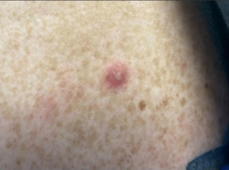
Clinical photograph of a characteristic vesiculopustular skin lesion associated with disseminated gonococcal infection (DGI). The primary lesion is a solitary, erythematous papule, approximately 3-5 mm in diameter, with a centralized greyish-white pustule/vesicle and a dusky, necrotic appearance at the core. The surrounding skin shows generalized solar lentigines (freckling) but no acute inflammatory changes. This visual finding is a hallmark of the arthritis-dermatitis syndrome seen in Neisseria gonorrhoeae dissemination, typically presenting as sparse, painless, non-pruritic lesions on distal extremities. This image serves as an educational reference for identifying cutaneous manifestations of systemic sexually transmitted infections in a clinical setting.
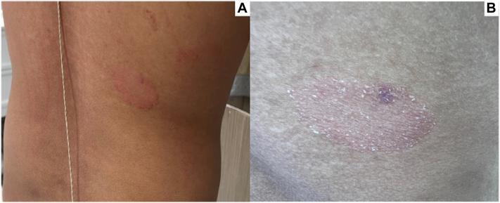
Two-panel clinical photograph displaying primary skin lesions on the mid-right back, characteristic of pityriasis rosea. Panel A shows a 2.5-cm, well-demarcated, annular red plaque on the torso, along with a secondary, smaller, ill-defined erythematous papule located superiorly. Panel B provides a high-detail close-up of the primary plaque, illustrating its morphology: it features a raised, erythematous border and a slightly depressed center with fine, surface scaling. A focal hyperpigmented macule is visible within the center of the lesion, marking the site of a prior punch biopsy. These visual findings—specifically the larger herald-style plaque with centrifugal scaling—are key clinical indicators of spongiotic dermatitis and help differentiate it from other papulosquamous disorders such as tinea corporis or nummular eczema.
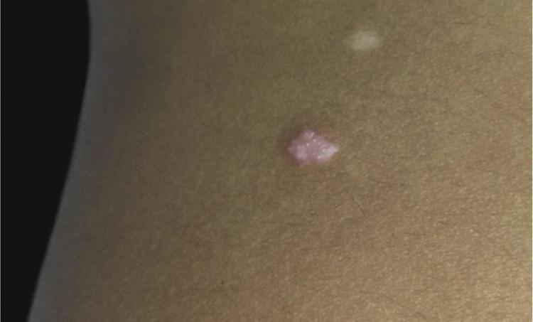
This clinical photograph displays a solitary, well-circumscribed papule on the left flank of a pediatric patient. The primary lesion is approximately 5 mm x 3 mm, exhibiting a rose or pink-erythematous coloration and a smooth, slightly dome-shaped surface. Superior and lateral to the main lesion, a smaller, faint whitish hypopigmented macule is also visible. The surrounding skin appears normal in texture and tone. This image serves as a dermatological example of a solitary adnexal tumor, such as syringocystadenoma papilliferum, characterized by its pink hue and gradual growth. The presentation is relevant for clinical training in identifying benign skin neoplasms and differentiating them from other pediatric papular lesions.
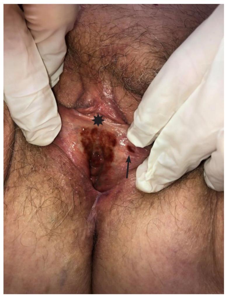
Clinical photograph of the female vulvar region demonstrating pigmented skin lesions. A large, well-demarcated hyperpigmented patch (indicated by an asterisk) is visible at the vulvar vestibule and extending over the clitoral frenulum. This primary macule exhibits variegated brownish to blackish tones and irregular borders. Adjacent to this main lesion on the left labium minus, a smaller, approximately 0.5 cm satellite papule (indicated by an arrow) is present with indistinct margins. The surrounding tissue shows mild erythema but lacks significant atrophy or architectural distortion. This visual evidence is used in a dermatological and gynecological context to differentiate between inflammatory conditions like lichen planus, pigmented purpuric dermatoses, and melanocytic neoplasms such as vulvar melanoma. The image is intended for medical education regarding the clinical presentation of vulvar hyperpigmentation and the importance of identifying satellite lesions.
skin lesion morphology classification primary lesions table
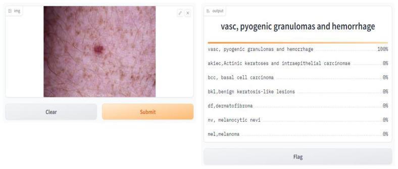
This clinical photograph displays a solitary skin lesion on a pinkish-tan background of skin characterized by scattered solar lentigines or ephelides. The primary lesion is a dark red to purplish-red nodule, indicative of a vascular etiology. Its morphology is slightly raised and circumscribed, consistent with a pyogenic granuloma. The deep coloration suggests focal hemorrhage or high vascular density within the lesion. The surrounding skin shows normal dermatoglyphic patterns and fine hairs, but is also notable for numerous small, light-brown macules reflecting chronic sun exposure. This image is used in the context of dermatological diagnostics and machine learning for automated skin cancer classification, specifically representing the 'VASC' (vascular lesions) category, which includes pyogenic granulomas, angiomas, and related hemorrhagic vascular conditions.

This clinical photograph, presented in grayscale, captures the genital and pubic region of a patient exhibiting a dense eruption of skin lesions. The primary morphology consists of multiple well-demarcated, dome-shaped papules and pustules. A defining characteristic of these lesions is umbilication, where each lesion features a distinct central depression or pit. The lesions are distributed widely across the hair-bearing and adjacent skin areas, appearing both in discrete forms and as clusters. While the grayscale rendering limits color assessment, there is no evidence of gross surrounding tissue necrosis or extensive cellulitis in the visible field. This presentation is highly characteristic of viral dermatological infections, specifically such as Molluscum Contagiosum or infections caused by certain Orthopoxviruses (e.g., Mpox/Monkeypox virus), emphasizing the educational focus on identifying umbilicated genital lesions within the context of sexually transmitted infections (STIs) and public health diagnostic pathways.
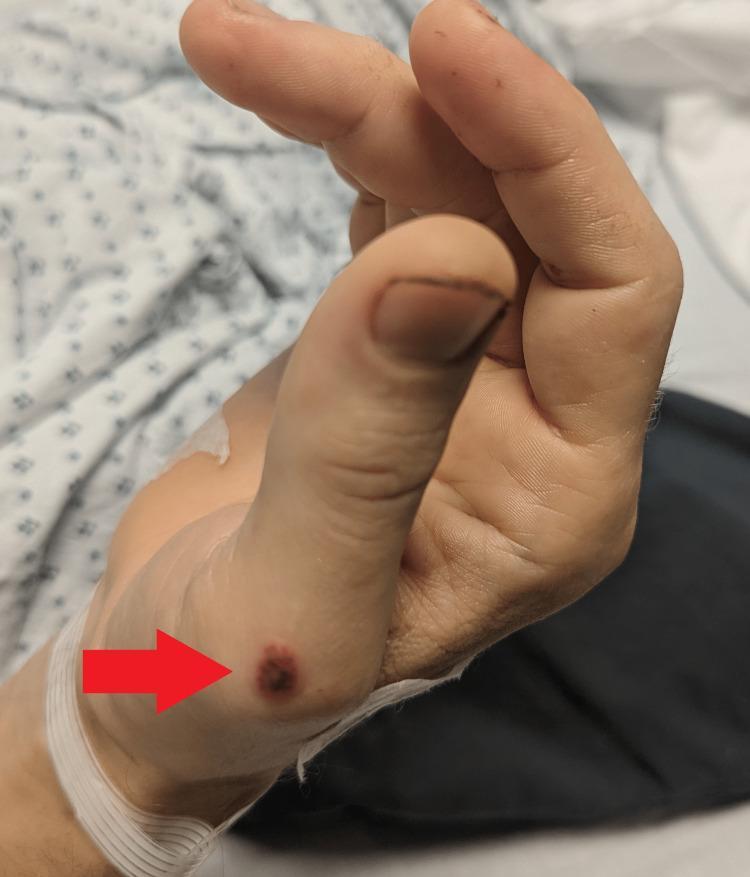
This clinical photograph displays a primary skin lesion on the lateral aspect of a patient's hand near the base of the thumb (thenar eminence). The lesion, highlighted by a red arrow, is a well-demarcated, circular papule approximately 1-2 cm in diameter. Its morphology is characterized by a dark red to violaceous (purpuric) hue with a slightly raised surface and irregular texture. The surrounding skin exhibits a fair tone with faint peripheral erythema. A medical identification band or bandage is visible around the wrist. Clinically, such lesions are associated with systemic conditions, often categorized within vasculitic or rheumatologic frameworks, and may manifest alongside symptoms like arthralgias, fever, and mucosal ulcers. The image serves as an educational tool for identifying discrete purpuric papules in the context of multi-systemic disease.
Primary Lesions - Definition
"The differential diagnosis of a rash should be based on the morphology of the lesion... the physician must first identify the primary lesion, the typical element of the eruption." - Symptom to Diagnosis: An Evidence-Based Guide, 4th Ed.
Classification of Primary Lesions
NON-ELEVATED (Flat) Lesions
| Lesion | Definition | Size | Example |
|---|---|---|---|
| Macule | Flat, circumscribed area of color change, no elevation or depression | < 1 cm (some texts ≤10 mm) | Freckle, petechiae, early rash of roseola |
| Patch | Large macule | ≥ 1 cm (some texts >10 mm) | Vitiligo, Mongolian spot, port-wine stain |
ELEVATED SOLID Lesions
| Lesion | Definition | Size | Example |
|---|---|---|---|
| Papule | Small, solid, elevated "bump" | < 1 cm | Wart, molluscum, lichen planus |
| Plaque | Flat-topped, raised, plateau-like lesion; often a confluence of papules | ≥ 1 cm | Psoriasis, mycosis fungoides |
| Nodule | Palpable, solid lesion, deeper than papule (dermis/subcutaneous fat) | 1-5 cm | Lipoma, rheumatoid nodule, dermatofibroma |
| Tumor / Mass | Large, solid growth | ≥ 5 cm | Malignant neoplasm |
ELEVATED FLUID-FILLED Lesions
| Lesion | Definition | Size | Example |
|---|---|---|---|
| Vesicle | Circumscribed, elevated blister filled with serous fluid | < 1 cm | Herpes simplex, chickenpox (early), contact dermatitis |
| Bulla | Large vesicle | ≥ 1 cm | Pemphigus vulgaris, bullous pemphigoid, large burns |
| Pustule | Elevated blister filled with purulent material (pus) | Any size | Acne, folliculitis, impetigo |
TRANSIENT Lesion
| Lesion | Definition | Example |
|---|---|---|
| Wheal (Hive) | Transient, raised, flat-topped papule or plaque due to superficial dermal edema; erythematous periphery with central pallor | Urticaria (allergic reaction) |
ENCLOSED Lesion
| Lesion | Definition | Example |
|---|---|---|
| Cyst | Encapsulated lesion filled with soft/fluid material, lined by epithelium | Epidermal inclusion cyst, dermoid cyst |
How Primary Lesions Fit into Reaction Patterns
- Papulosquamous - primary lesion is a thin, flat-topped papule or plaque with scale (e.g., psoriasis, lichen planus, pityriasis rosea)
- Eczematous - thin erythematous papules/plaques with epidermal spongiosis
- Vesicular/Bullous - fluid-filled blisters as the main feature
- Pustular - pus-containing lesions (e.g., AGEP, candida, acne)
- Nodular - deeper, firmer elevations
Primary vs. Secondary Lesions
| Secondary Lesion | Arises From |
|---|---|
| Scale | Excessive epidermis shedding |
| Crust | Solidified exudate from broken vesicles/pustules |
| Erosion | Loss of superficial epidermis (heals without scar) |
| Ulcer | Full-thickness skin loss (heals with scar) |
| Fissure | Linear epidermal tear into dermis |
| Excoriation | Scratching-induced loss of epidermis |
| Lichenification | Epidermal thickening from chronic rubbing |
| Scar | New connective tissue after full-thickness injury |
Key Clinical Tip
- Identify the primary lesion (before secondary changes)
- Determine the reaction pattern (papulosquamous? vesicular? nodular?)
- Note distribution (dermatomal? flexural? extensor? sun-exposed?)
- Examine scalp, nails, and mucous membranes
Adhesive capsulitis
adhesive capsulitis frozen shoulder MRI arthrogram glenohumeral joint
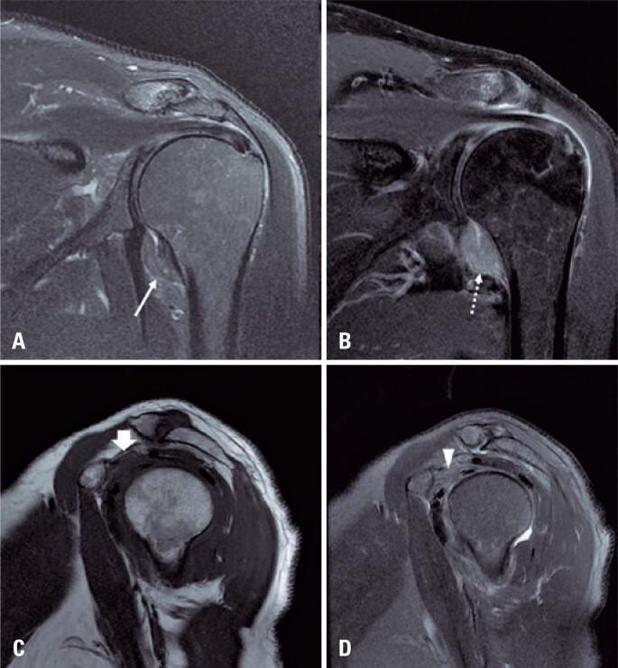
This diagnostic image set consists of four MRI views of a human shoulder illustrating the classic findings of adhesive capsulitis (frozen shoulder). Image A (coronal T2-weighted with fat suppression) and Image B (coronal T1-weighted post-gadolinium with fat suppression) demonstrate significant thickening of the glenohumeral joint capsule and pericapsular edema, particularly within the axillary recess. The post-contrast image (B) shows intense enhancement of the thickened capsule, indicating active synovitis and hypervascularity. Images C (sagittal T1-weighted) and D (sagittal T2-weighted with fat suppression) focus on the rotator interval. They reveal the characteristic obliteration of the normal subcoracoid fat pad by hypointense inflammatory tissue on T1 (arrow in C) and hyperintense edema on T2 (arrowhead in D). These imaging features are essential for confirming adhesive capsulitis in patients presenting with clinical shoulder stiffness and pain, allowing for the differentiation from other pathology such as rotator cuff tears or labral injuries.
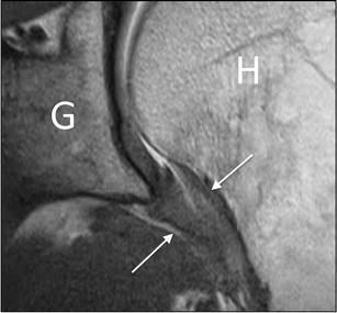
This diagnostic image is a coronal oblique proton density MRI of the glenohumeral joint. Key anatomical landmarks are labeled, including the glenoid (G) and the proximal humerus (H). The focus of the image is the axillary pouch of the joint capsule, indicated by white arrows. Visually, the inferior capsule within the axillary recess demonstrates significant pathological thickening, a hallmark diagnostic feature of adhesive capsulitis (frozen shoulder). Unlike the normally thin and pliable appearance of the capsule, this presentation shows a bulky, intermediate-signal tissue filling the inferior joint space. This imaging modality is utilized in musculoskeletal radiology to assess for capsular inflammation and fibrosis when clinical range of motion is restricted. The image illustrates the relationship between the joint capsule's morphological changes and the surrounding bony structures, providing clear visual evidence of the soft tissue involvement in adhesive capsulitis.
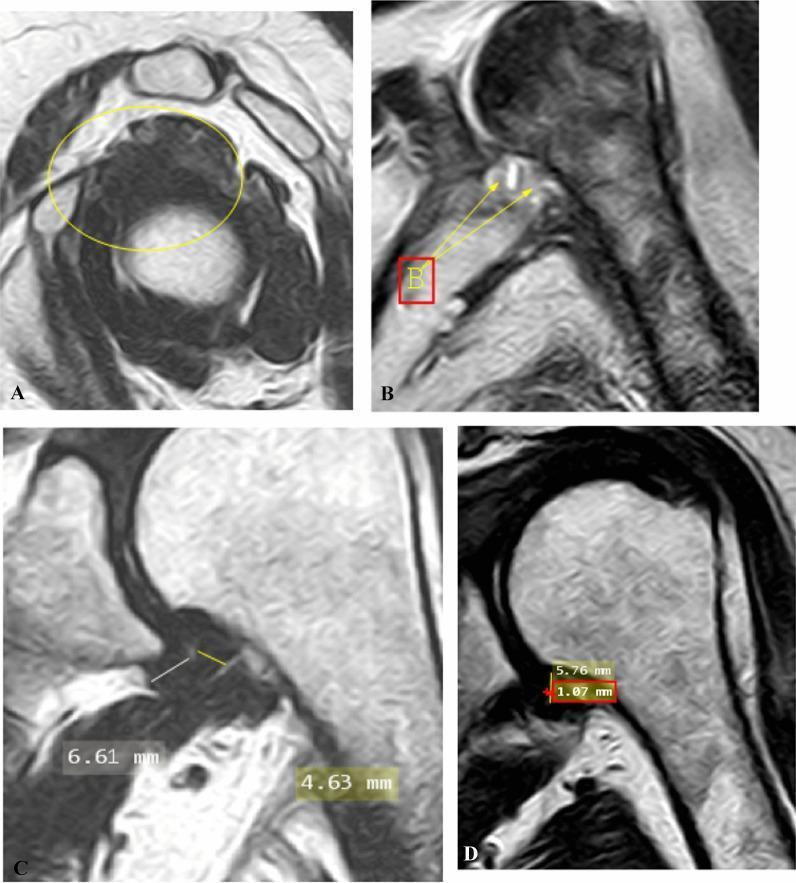
This composite of four magnetic resonance imaging (MRI) panels demonstrates diagnostic features of adhesive capsulitis (frozen shoulder) in the left shoulder. (A) Oblique sagittal T2-weighted MRI shows complete obliteration of the subcoracoid fat triangle (yellow circle), with an absence of normal fat planes and the coracohumeral ligament. (B) Oblique coronal fat-suppressed proton density (PD)-weighted image highlights hyperintense signal (arrows) indicating edema at both the glenoid and humeral insertions of the axillary recess. (C) Oblique coronal T2-weighted image demonstrates quantitative assessment of joint capsule thickening, measuring 6.61 mm at the glenoid portion and 4.63 mm at the humeral portion of the axillary recess. (D) Oblique coronal T2-weighted image shows the measurement of the maximal height (5.76 mm) and width (1.07 mm) of the axillary recess. These findings collectively illustrate key radiological markers used to stage and diagnose inflammatory and fibrotic changes in the glenohumeral joint capsule and surrounding rotator interval.
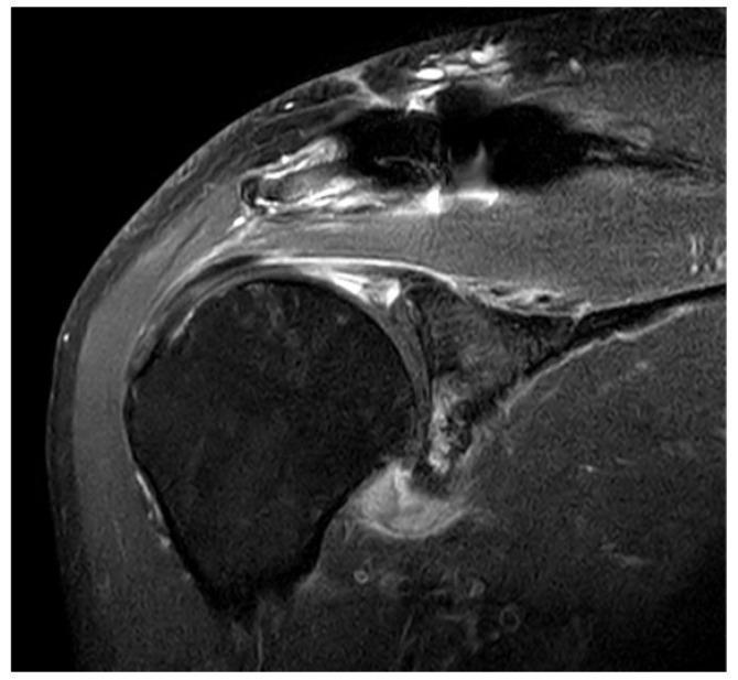
This diagnostic image is a coronal T2-weighted fat-suppressed MRI of the right shoulder joint. The primary finding is a significantly thickened glenohumeral joint capsule demonstrating increased signal intensity, most prominent at the axillary capsular pouch. These findings are characteristic of adhesive capsulitis (frozen shoulder), indicating inflammatory changes and edema within the joint capsule. The image clearly shows the relationship between the humeral head, glenoid, and the inferior redundant fold of the capsule (axillary pouch). The surrounding soft tissues, including the deltoid muscle and subacromial space, are visible with high contrast. The increased signal intensity within the inferior capsule suggests active inflammation or synovitis, which is a hallmark finding in patients presenting with painful shoulder stiffness. The MRI provides sufficient resolution to distinguish these pathological capsular changes from normal thin, low-signal ligamentous structures.
"adhesive capsulitis" treatment management
Adhesive Capsulitis (Frozen Shoulder)
Definition & Terminology
Synovial inflammation → Reactive capsular fibrosis → Contracted joint capsule
Epidemiology
- Incidence: ~2% of the general population
- Most common in ages 40-70 years
- ~70% are women
- 20-30% develop contralateral frozen shoulder
- Diabetic patients have bilateral disease 50% of the time
- Insulin-dependent diabetics are 5x more likely to develop frozen shoulder
- Rarely recurs in the same shoulder
Risk Factors
| Risk Factor | Notes |
|---|---|
| Female sex | 70% of cases |
| Age >49 years | Peak 40-70 |
| Diabetes mellitus | 5x increased risk; insulin-dependent |
| Prolonged immobilization | Most significant contributing factor |
| Cervical disc disease | |
| Hyperthyroidism | |
| Stroke / Myocardial infarction | |
| Autoimmune diseases | |
| Trauma / Post-surgical | |
| Hyperlipidemia | Proposed but needs further study |
Stages of Adhesive Capsulitis
| Stage | Arthroscopic/Clinical Features | Symptoms |
|---|---|---|
| 1 - Pre-adhesive | Fibrinous inflammatory synovitis, early adhesion formation | Gradual onset of diffuse pain, especially nocturnal; pain lying on affected side |
| 2 - Acute adhesive synovitis | Synovial proliferation; early inferior capsular fold adhesions | Progressive pain; patient restricts movement, stiffness begins |
| 3 - Maturation | Less synovitis, more fibrosis; axillary fold obliterated | Significantly restricted motion (4-12 months); pain less severe |
| 4 - Chronic/Frozen | Fully mature adhesions | Motion severely reduced; pain only at end ranges or sudden movement |
| 5 - Thawing | Resolution phase | Motion gradually returns; pain diminishes; may last weeks to months |
Clinical Features
- Gradual onset of diffuse shoulder pain, worst at night
- Pain exacerbated by lying on the affected side
- Progressively restricted motion in all planes
- Global loss of both active AND passive movement (distinguishes from rotator cuff tear)
- Pathognomonic sign: loss of external rotation - earliest and most consistent finding
- Internal rotation limited to sacral level
- ~50% loss of external rotation
- Abduction < 90 degrees
- No redness/local warmth (distinguishes from septic arthritis)
Investigations
| Investigation | Finding |
|---|---|
| X-ray (plain film) | Usually normal - rules out OA, fracture, calcific tendinitis |
| Arthrography | Reduced joint volume (<10 mL); lack of filling of axillary fold; irregular margins - most specific |
| MRI | Thickened inferior capsule; increased synovial blood flow ("burning sign"); useful to exclude other pathology |
| Bone scan | May be positive; positive scan predicts good response to steroid injection |
Differential Diagnosis
- Rotator cuff tear - loss of active but NOT passive motion
- Osteoarthritis - X-ray changes present
- Calcific tendinitis - calcium visible on X-ray
- Septic arthritis - fever, redness, raised inflammatory markers
- Fracture/dislocation
- Inflammatory arthritis (RA, etc.)
Treatment
Conservative (First-line)
| Modality | Notes |
|---|---|
| NSAIDs | Pain and inflammation control |
| Intra-articular corticosteroid injection | 40 mg/mL methylprednisolone acetate into glenohumeral joint + subacromial bursa; most effective in painful/freezing stage |
| Oral corticosteroids | Short-term improvement in pain and ROM |
| Physiotherapy | Pendulum exercises → wall-climbing → active ROM → strengthening; ice packs, ultrasound, TENS |
| Distension arthrography (hydrodilatation) | 20-30 mL saline + local anaesthetic into joint; disrupts adhesions and facilitates rehab |
"Avoid abduction initially to prevent impingement until joint motion becomes suppler." - Campbell's Operative Orthopaedics, 15th Ed.
Interventional / Surgical (if conservative fails)
| Option | Indication |
|---|---|
| Manipulation Under Anaesthesia (MUA) | Prolonged stiffness (>6 months); 70% improvement; worse results in diabetics (71% vs 90% full ROM) |
| Arthroscopic capsular release | When MUA fails; release of coracohumeral ligament and rotator interval |
| Open surgical release | Last resort when arthroscopic release fails |
Prognosis
- Most resolve spontaneously over 1-2 years (longer in diabetics)
- Only ~50% are completely pain-free even after resolution
- Dominant shoulder involvement = better prognosis
- Prevention is the best treatment for secondary frozen shoulder (e.g., post-surgical, post-MI)
Recent Evidence (2024-2025)
- A 2024 network meta-analysis in Rheumatology (PMID 38538951) evaluated pharmacological interventions for early-stage frozen shoulder - corticosteroid injections remain the most supported early intervention.
- A 2024 systematic review (PMID 39420459) confirmed that muscle energy techniques and joint mobilization significantly improve ROM and pain in frozen shoulder.