
Cholesteatoma of attic. Its management Types. Mastoid obliteration procedure and precention of discharging cavity. In detail for long question answer from standard ENT textbook in simplified manner
Reading File
Now let me search the medical library for comprehensive content on cholesteatoma, attic, and mastoid obliteration simultaneously.
Finding Sources
Finding Sources
Finding Sources
Now let me read all key sections in parallel:
Reading File
Reading File
Reading File
Excellent content so far. Let me now read the detailed Scott-Brown's section on cholesteatoma management and the Cummings section on mastoid obliteration details:
Reading File
Reading File
Finding Sources
Reading File
Finding Sources
Finding Sources
Reading File
Reading File
Excellent! Now let me get the Shambaugh section and look for prevention/discharging cavity, plus search for relevant images:
Reading File
Finding Sources
Searching Images
cholesteatoma attic ear CT scan diagram
I now have comprehensive content from Cummings Otolaryngology, Scott-Brown's, and Shambaugh's Surgery of the Ear. Let me also fetch the CT image of cholesteatoma that was found earlier:
Loading Image
Finding Sources
I now have all the essential content from the major ENT textbooks. Let me compile a comprehensive, well-structured answer.
Cholesteatoma of the Attic - Comprehensive Review
1. Definition and Nature
A cholesteatoma is a collection of keratinizing squamous epithelium (skin cells) in the middle ear cleft, which actively erodes adjacent bony structures. The term is a misnomer - it is neither a true tumor nor does it contain cholesterol. It behaves like a slowly expanding, locally destructive lesion.
Attic (Epitympanic) Cholesteatoma is the most common acquired type. It arises from the pars flaccida region of the tympanic membrane, with keratin accumulating in the lateral epitympanic (Prussak's) space.
- Accounts for ~98% of all cholesteatomas
- Most common presentation: a retraction pocket in the pars flaccida with accumulation of keratin debris
- The Prussak space is the lateral epitympanic recess, bounded by:
- Laterally: pars flaccida of the tympanic membrane
- Medially: neck of the malleus
- Superiorly: lateral malleal ligament
- Inferiorly: lateral malleal fold
2. Pathogenesis
Four main theories explain how an attic cholesteatoma forms:
| Theory | Mechanism |
|---|---|
| Retraction pocket theory (most accepted) | Eustachian tube dysfunction → negative middle ear pressure → retraction of pars flaccida → accumulation of desquamated keratin |
| Squamous metaplasia theory | Chronic inflammation causes middle ear mucosa to undergo squamous metaplasia |
| Basal cell hyperplasia | Proliferating basal cells of the tympanic membrane extend into the middle ear |
| Implantation theory | Iatrogenic or traumatic implantation of squamous epithelium |
Bone erosion mechanism: Cholesteatoma produces cytokines (IL-1, IL-6, TNF-α), prostaglandins, and activates osteoclasts via the RANKL pathway, causing progressive bone resorption. This is NOT simple pressure necrosis but an active enzymatic and cytokine-driven process.
3. Clinical Features
Symptoms:
- Foul-smelling, scanty, painless otorrhoea (classic presentation)
- Conductive hearing loss (due to ossicular erosion)
- Ear fullness or blocked sensation
- Complications when advanced: facial nerve palsy, vertigo, labyrinthine fistula, intracranial spread
Otoscopic signs:
- Marginal perforation or retraction pocket in the pars flaccida (attic region)
- White, pearly, cheese-like debris in the attic
- Destruction of the scutum (bony spur at the lateral attic wall)
- An attic crust is a hallmark - when removed, reveals squamous debris underneath
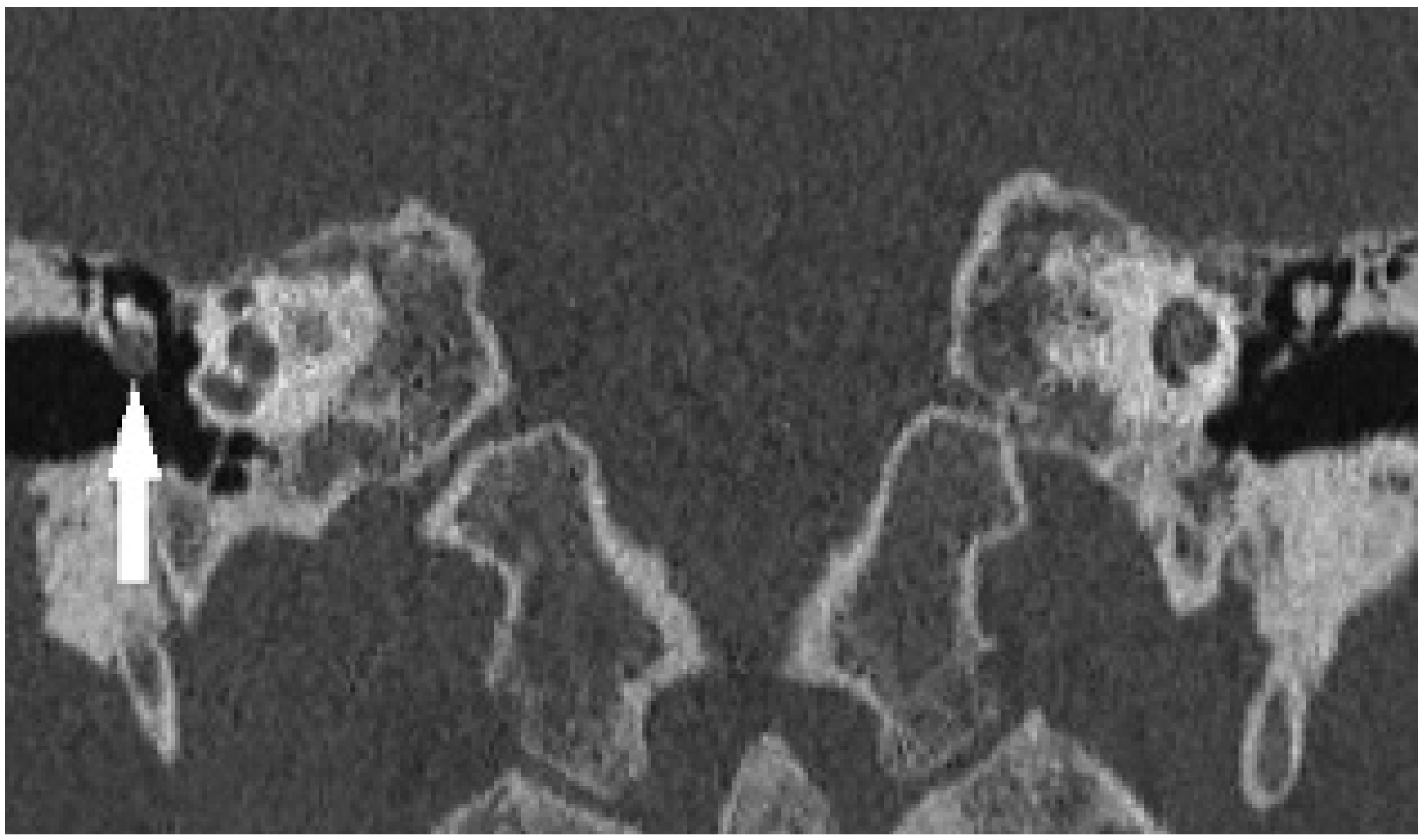
CT scan findings:
- Soft tissue density mass in the epitympanum
- Scutum erosion (the bony lateral wall of the attic)
- Ossicular chain erosion (especially long process of incus)
- Lateral epitympanic (Prussak space) opacification
- CT has high negative predictive value for excluding cholesteatoma
Below: Coronal CT scan showing right lateral epitympanic pars flaccida cholesteatoma (arrow) with early ossicular erosion
4. Complications of Untreated Cholesteatoma
Intratemporal:
- Ossicular chain erosion (most common) → conductive hearing loss
- Labyrinthine (horizontal semicircular canal) fistula → vertigo, sensorineural hearing loss
- Facial nerve dehiscence/palsy
- Mastoiditis
Intracranial:
- Extradural (epidural) abscess
- Subdural abscess
- Meningitis
- Brain abscess (temporal lobe / cerebellum)
- Lateral sinus thrombophlebitis
- Otitic hydrocephalus
5. Management of Attic Cholesteatoma
Key principle: Cholesteatoma can ONLY be eradicated by surgical resection. Medical treatment has no role except pre-operative optimization. The primary goal is disease eradication; hearing reconstruction is secondary.
Pre-operative Assessment
- Pure tone audiogram + tympanogram
- CT temporal bone (mandatory) - identifies extent of erosion, tegmen and sigmoid sinus position, facial nerve canal
- MRI with diffusion-weighted imaging (non-EPI DWI) - detects residual/recurrent disease post-operatively
- Thorough otoscopy (remove any crust to assess depth of retraction)
Surgical Approaches: Classification
Surgical approaches divide into two fundamental groups:
Group 1: Resection and Reconstruction (Closed / Canal Wall-Up techniques)
Group 2: Exteriorization (Open / Canal Wall-Down techniques)
TYPE 1: Closed Techniques (Canal Wall-Up = CWU)
The posterior bony canal wall is preserved. Disease is removed through a mastoidectomy approach while the external auditory canal remains intact.
A. Atticotomy (Limited Attic Approach)
- Best for: Small, early attic cholesteatoma confined to Prussak's space without extension into the mastoid antrum
- Removes the bony lateral attic wall (scutum) to expose the Prussak space
- Can be done trancanally or via a postauricular approach
- Defect in the scutum is repaired with cartilage (from tragus or conchal bowl)
- Advantage: Minimally invasive, preserves canal wall, no mastoid cavity
- Disadvantage: Limited exposure; not suitable for extensive disease
B. Combined Approach Tympanoplasty (CAT) / Intact Canal Wall Mastoidectomy
- The standard CWU procedure for moderate-to-extensive cholesteatoma
- Steps:
- Postauricular incision
- Cortical mastoidectomy - opens the mastoid antrum and air cells
- Posterior tympanotomy (facial recess approach) - opening between the facial nerve, chorda tympani, and posterior canal wall
- Cholesteatoma matrix carefully dissected and removed under microscope
- Tympanoplasty (myringoplasty) to repair the tympanic membrane
- Ossicular chain reconstruction (if needed) with PORP/TORP
- Second-look operation: Usually planned at 9-12 months to check for residual disease (because hidden areas under the preserved canal wall can harbor residual keratin)
Advantages of CWU:
- Normal anatomy preserved
- No mastoid bowl to maintain
- Water tolerance better
- Hearing aid fitting easier
Disadvantages of CWU:
- Residual disease rate: 11-27%
- Recurrent disease rate: 5-13%
- Usually requires second-look surgery
- Hidden recesses (sinus tympani, facial recess) may conceal residual disease
- New retraction pockets can form behind the preserved canal wall
TYPE 2: Open Techniques (Canal Wall-Down = CWD)
The posterior bony canal wall is removed, creating a large open mastoid cavity that communicates with the external auditory canal. Disease is exteriorized rather than enclosed.
A. Modified Radical Mastoidectomy (Bondy Procedure)
- Specifically for: Attic cholesteatoma limited to the epitympanum, where disease is lateral to the ossicular chain (between the ossicles and the lateral attic wall)
- The Bondy approach: removes the scutum and adjacent superior/posterior canal wall to exteriorize the epitympanum
- The uninvolved middle ear is NOT entered
- The cholesteatoma matrix is maintained in place as a lining over the ossicular heads
- The tympanic membrane and ossicular chain are left intact
- Result: Creates a small, shallow cavity opening into the ear canal
- Key distinction from radical mastoidectomy: Middle ear is undisturbed, ossicles are preserved, hearing is maintained
B. Canal Wall-Down Mastoidectomy (Radical / Modified Radical)
- Used for more extensive cholesteatoma involving mastoid antrum and air cells
- Creates a large mastoid bowl communicating with the external ear canal
- Requires a meatoplasty (enlarging the external auditory meatus) to allow the cavity to self-clean and be examined
Advantages of CWD:
- Better exposure of epitympanum and mesotympanum
- Lower residual disease rate: 2-10%
- Disease can be monitored by direct visualization in clinic
- No need for routine second-look surgery
Disadvantages of CWD:
- Creates a permanent mastoid cavity requiring regular aural toilet/debridement
- Slow epithelialization of cavity (months)
- Risk of chronic discharging cavity (the dreaded "problem cavity")
- Difficulties with hearing aid fitting (infection risk from mold occlusion)
- Meatoplasty cosmetically and functionally challenging
- Caloric vertigo from wind/water exposure to exposed semicircular canals
Comparing CWU vs CWD at a Glance
| Feature | Canal Wall-Up (CWU) | Canal Wall-Down (CWD) |
|---|---|---|
| Canal wall | Preserved | Removed |
| Cavity created | No (normal anatomy) | Yes (mastoid bowl) |
| Residual disease | 11-27% | 2-10% |
| Recurrent disease | 5-13% | 6-20% |
| Second-look needed | Usually yes | Usually not |
| Cavity maintenance | Not needed | Regular aural toilet |
| Water exposure | Well tolerated | Problematic |
| Hearing aid | Well tolerated | Risky (infection) |
| Preferred in children | Yes | Rarely |
Indications favoring CWD over CWU:
- Only hearing ear (to avoid second surgery under anesthesia)
- Poor anesthetic risk / cannot return for second-look
- Unreliable follow-up
- Very extensive disease (poorly pneumatized mastoid)
- Intraoperative: labyrinthine fistula, low-lying tegmen, anteriorly placed sigmoid sinus
TYPE 3: Endoscopic Ear Surgery (EES)
- Increasingly used as primary or adjunct approach
- Transcanal endoscopic atticotomy for limited attic disease
- Endoscopic assistance during CWU mastoidectomy to inspect hidden recesses (sinus tympani, facial recess, anterior epitympanum)
- Significantly reduces residual disease rates in CWU surgery
- Reduces need for second-look procedures
6. Mastoid Obliteration
What is it?
Mastoid obliteration means filling in the mastoid cavity (created after CWD mastoidectomy) with biological or synthetic materials to eliminate dead space, prevent chronic discharge, and create a trouble-free ear.
Historical Background
- Originally advanced by Palva in the 1970s as a modification of Popper's flap
- Involved a postauricular musculoperiosteal flap rotated inward to fill the mastoid bowl
- Later combined with bone chips and bone pâté
Why Obliterate?
The persistent discharging cavity is a major problem after CWD mastoidectomy:
- Large dead space accumulates moisture and keratin debris
- Poorly epithelialized recesses become chronically infected
- Patients require frequent hospital attendance for aural toilet
- Caloric vertigo from exposed semicircular canals
- Hearing aid fitting problems
Mastoid obliteration addresses all these by:
- Reducing cavity volume
- Filling recesses at risk for debris accumulation
- Restoring a more anatomical contour
- Reducing future need for regular bowl cleanings
- Reducing cholesteatoma recidivism rates (systematic review of 13 studies, 1534 patients showed recurrent/residual rates of only 5.8%/5.9% in obliterated cavities)
When to Obliterate?
Primary obliteration - Done at the same time as the CWD mastoidectomy
- Goal: reduce bowl size, fill recesses, minimize future maintenance
- Requires confidence that all cholesteatoma has been removed
Secondary (Rehabilitation) obliteration - Done as a later procedure in a chronically discharging open cavity
- Goal: achieve a dry ear in a troublesome bowl
- Results: ~90% achieve a well-healed, dry cavity at 1-year follow-up (Bernardeschi et al.)
- Also used to treat vertiginous symptoms from the exposed semicircular canal
Materials Used for Obliteration
Autologous (from the patient):
| Material | Source | Notes |
|---|---|---|
| Bone pâté | Cortical bone dust from burr | Most commonly used; packed into dead space |
| Bone chips | Cortical bone fragments | Combined with pâté |
| Cartilage | Tragus or conchal bowl | Good structural support for scutum reconstruction |
| Temporalis muscle/fascia flap | Postauricular region | Palva flap - vascularized pedicled flap, excellent healing |
| Abdominal fat | Abdominal wall | Free fat graft; bulky; used for large cavities |
| Periosteal flap | Postauricular periosteum | Combined with bone |
Alloplastic / Biomaterials:
| Material | Notes |
|---|---|
| Hydroxyapatite (HA) | Biocompatible ceramic; integrates well; widely used |
| Tricalcium phosphate (TCP) | Bioresorbable ceramic; good biocompatibility |
| Biosynthetic ceramic | 90% healed cavities at 1 year in secondary obliteration series |
| Titanium canal wall prosthesis | For canal wall reconstruction |
The Palva Flap (Musculoperiosteal Flap) - Technique
This is the most widely used obliteration technique:
- Postauricular incision placed 1 cm behind the postauricular crease
- A musculoperiosteal flap based on the postauricular tissue is elevated - includes skin, subcutaneous tissue, periosteum, and muscle
- After completing the CWD mastoidectomy and removing all disease, the mastoid cavity is saucerized (smoothed and lowered using burrs to eliminate overhangs)
- The mastoid bowl is then partially filled with bone pâté/chips (from the cortical drilling)
- The Palva flap is rotated inward to lie on top of the bone pâté, obliterating the remaining dead space
- The flap is secured and the meatoplasty is created
- A wide meatoplasty (enlargement of the ear canal opening) ensures good ventilation and visual access for follow-up
7. Prevention of a Discharging (Problem) Cavity
A chronically discharging mastoid cavity is one of the most troublesome complications of CWD mastoidectomy. Key principles of prevention:
Surgical Technique Principles (Intraoperative Prevention)
-
Adequate saucerization - Lower the facial ridge to the level of the facial nerve. A high facial ridge creates a hidden recess that cannot self-clean and traps debris.
-
Smooth, rounded cavity - Eliminate all bony overhangs and sharp corners. The cavity should be smooth-walled so epithelium can migrate across it and debris does not accumulate.
-
Wide meatoplasty - The meatus must be wide enough to allow:
- Adequate air circulation (prevents moisture trapping)
- Adequate drainage
- Direct visualization of the entire cavity without equipment
- Easy clinic-based debridement when needed
- Rule of thumb: the meatus should be at least as wide as the mastoid bowl
-
Obliteration of dead space - Use bone pâté, cartilage, or musculoperiosteal flap to fill the mastoid cavity and reduce its volume (as described above)
-
Lowered tegmen plate - Eliminates the overhanging tegmen which can hide residual disease
-
Eustachian tube function - Sealing or packing the eustachian tube orifice is sometimes done to prevent middle ear secretions from contaminating the cavity
Post-operative Management
-
Regular aural toilet - Gentle microsuction cleaning (at 6-8 week intervals initially, then every 3-6 months once healed) to remove debris and keep the cavity clean
-
Antibiotic/antifungal eardrops - For episodes of infection; avoid prolonged use of aminoglycoside drops
-
Acetic acid 2% drops - Acidifies the canal, prevents bacterial overgrowth (particularly Pseudomonas), can be used for regular maintenance
-
Water precautions - Protect the cavity from water entry (custom earmolds or cotton wool with Vaseline for bathing/swimming)
-
Avoiding hearing aid molds - Standard behind-the-ear hearing aids with custom molds occlude the meatoplasty, trap moisture, and frequently cause chronic discharge. Alternatives:
- Bone-anchored hearing aids (BAHA)
- Contralateral routing of signal
- Open-fit hearing aids
-
Secondary obliteration - If the cavity remains chronically discharging despite conservative measures, secondary obliteration with a vascularized flap or biosynthetic material is highly effective (~90% success)
Causes of a Discharging/Problem Cavity and Their Solutions
| Cause | Solution |
|---|---|
| High facial ridge with dependent sump | Revision surgery to lower the ridge |
| Inadequate meatoplasty | Revision meatoplasty |
| Residual/recurrent cholesteatoma | Second-look / revision surgery |
| Granulation tissue | Topical steroid drops, microsuction; silver nitrate cautery |
| Fungal infection (Aspergillus, Candida) | Antifungal eardrops (clotrimazole); microsuction |
| Chronic bacterial infection | Culture-specific antibiotic eardrops |
| Exposed bone (osteitis) | Microsuction, topical agents; may need revision |
| Hearing aid occlusion | Change to BAHA or open-fit hearing aid |
8. Summary Flow for Attic Cholesteatoma
ATTIC CHOLESTEATOMA
        |
        ├── Small / Limited to Prussak space
        │        └── ATTICOTOMY ± scutum reconstruction
        │
        ├── Moderate / Involves antrum / mastoid
        │        └── CANAL WALL-UP (CWU) mastoidectomy
        │             + Second-look at 9-12 months
        │             ± Endoscopic-assisted inspection
        │
        └── Extensive / Poor pneumatization / Unreliable follow-up
                 └── CANAL WALL-DOWN (CWD) mastoidectomy
                      + Wide meatoplasty
                      ± Mastoid obliteration (Palva flap + bone pâté)

 Special case - Epitympanic only, lateral to ossicles:
                 └── BONDY PROCEDURE (modified radical mastoidectomy)
                      - Middle ear NOT entered
                      - Ossicles preserved
                      - Hearing preserved
9. Post-operative Surveillance
- Non-EPI DWI MRI (diffusion-weighted MRI) has become the gold standard for detecting residual and recurrent cholesteatoma post-operatively
- Has high positive predictive value for cholesteatoma (can detect lesions as small as 3 mm)
- Increasingly replacing routine second-look surgery in CWU cases
- First scan typically at 12-18 months post-op
Key Points for Exam
- Attic (pars flaccida) cholesteatoma originates in Prussak's space and erodes the scutum first
- ONLY surgical treatment is definitive - no medical cure
- Two groups of surgery: Resection + Reconstruction (CWU) vs Exteriorization (CWD)
- Bondy procedure = specifically for disease lateral to ossicles in the epitympanum only; middle ear NOT entered; hearing preserved
- CWU has higher recidivism (11-27%) but better quality of life; CWD has lower recidivism (2-10%) but requires cavity maintenance
- Mastoid obliteration - Palva musculoperiosteal flap + bone pâté - reduces cavity problems and recidivism
- For discharging cavity: lower the facial ridge, widen the meatoplasty, obliterate dead space
- DWI MRI is now the surveillance tool of choice for detecting residual/recurrent disease
- Complications (mnemonic LIMFADES): Labyrinthine fistula, Intracranial spread, Mastoiditis, Facial palsy, Abscess, Deafness (SNHL), sinus thrombophlebitis
Sources: Cummings Otolaryngology Head and Neck Surgery (7th ed), Scott-Brown's Otorhinolaryngology Head & Neck Surgery Vol 2, Shambaugh Surgery of the Ear
This is a shared conversation. Sign in to Orris to start your own chat.