Jugular vein mbbs level
Reading File
Finding Sources
Finding Sources
Reading File
Reading File
Finding Sources
Loading Image
Finding Sources
Reading File
Reading File
Finding Sources
Finding Sources
Loading Image
Reading File
I now have comprehensive content. Let me compile the complete MBBS-level notes on the jugular veins.
Jugular Veins - MBBS Level Notes
Sources: Gray's Anatomy for Students | Color Atlas of Human Anatomy Vol. 2 | Goldman-Cecil Medicine | Medical Physiology
Overview
There are three jugular veins on each side of the neck:
- Internal jugular vein (IJV) - the main venous drainage of the neck and head
- External jugular vein (EJV) - superficial trunk
- Anterior jugular vein (AJV) - smallest, midline region
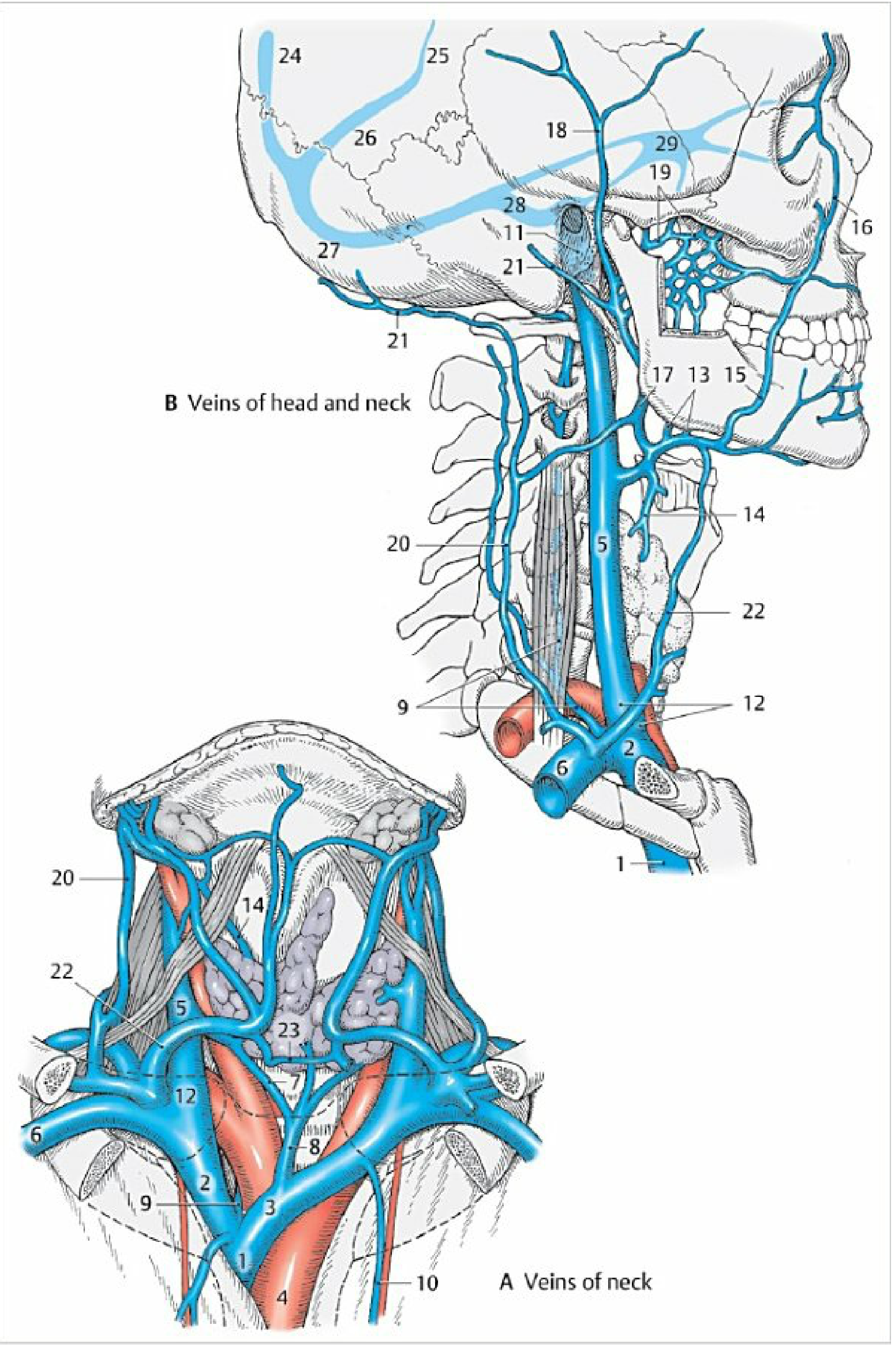
Fig: Veins of the neck (A) and head and neck (B). The IJV (5) descends within the carotid sheath alongside the common carotid artery. The EJV (20) crosses the sternocleidomastoid superficially. The anterior jugular vein (22) connects via the jugular venous arch (23). - Color Atlas of Human Anatomy Vol. 2
1. Internal Jugular Vein (IJV)
Origin
- Begins at the jugular foramen as a direct continuation of the sigmoid sinus (a dural venous sinus)
- The initial dilated portion is called the superior bulb of the jugular vein
- It also receives the inferior petrosal sinus soon after formation
Course
- Exits the skull through the jugular foramen alongside CN IX (glossopharyngeal), CN X (vagus), and CN XI (accessory)
- Enters the carotid sheath and descends the neck
- Initially lies posterior to the internal carotid artery, then becomes lateral to the common carotid artery farther down
- The vagus nerve (CN X) lies posteriorly between the IJV and the carotid artery throughout
- Before joining the subclavian vein, it presents another dilatation - the inferior bulb of the jugular vein
Termination
- Joins the subclavian vein posterior to the sternal end of the clavicle
- Forms the brachiocephalic vein (right and left)
- This junction is called the venous angle (angle of Pirogoff)
Carotid Sheath Contents (mnemonic: "VAN")
| Structure | Position |
|---|---|
| IJV | Lateral |
| Common carotid artery | Medial |
| Vagus nerve (CN X) | Posterior (between the two) |
Tributaries of IJV
From the cranial cavity (via dural sinuses):
- Inferior petrosal sinus
Extracranial tributaries:
- Facial vein (begins as angular vein at medial canthus, anastomoses with ophthalmic vein)
- Lingual vein
- Pharyngeal veins (from pharyngeal plexus)
- Superior thyroid vein (with superior laryngeal vein)
- Middle thyroid veins
- Occipital vein
- Sternocleidomastoid vein
- Meningeal veins
Note: The retromandibular vein receives the superficial temporal veins and pterygoid plexus, then joins the facial vein - Color Atlas of Human Anatomy
2. External Jugular Vein (EJV)
Formation
- Formed near the angle of the mandible by the union of:
- Posterior branch of the retromandibular vein +
- Posterior auricular vein
Course
- Descends in the superficial fascia of the neck
- Crosses superficially over the sternocleidomastoid muscle
- Enters the posterior triangle and descends vertically
Termination
- Empties into the internal jugular vein or subclavian vein near the venous angle
Tributaries
- Transverse cervical veins
- Suprascapular vein
- Anterior jugular vein (frequently)
The EJV is used clinically to assess mean right atrial pressure. Central venous pressure (CVP) = height of jugular distension above the sternal angle of Louis + 5 cm. Normal CVP = 5-10 cm H2O. - Goldman-Cecil Medicine
3. Anterior Jugular Vein (AJV)
- Begins at the level of the hyoid bone
- Descends near the midline
- The two AJVs may be connected just above the sternum by a transverse vessel called the jugular venous arch
- Usually drains into the EJV
4. Dural Venous Sinuses (IJV Connections)
The IJV ultimately collects blood from all the dural venous sinuses:
| Sinus | Drains Into |
|---|---|
| Superior sagittal sinus | Transverse sinus |
| Inferior sagittal sinus | Straight sinus |
| Straight sinus | Transverse sinus |
| Transverse sinus | Sigmoid sinus |
| Sigmoid sinus | IJV (at jugular foramen) |
| Cavernous sinus | Connects via inferior petrosal sinus to IJV |
5. Jugular Venous Pulse (JVP) - MBBS Clinical Importance
The IJV transmits pressure changes from the right atrium and is used to assess right heart function.
How to Examine
- Patient at 45 degrees incline
- Look for pulsations in the neck (EJV for mean pressure, IJV for waveform)
- Height of distension above the sternal angle + 5 cm = CVP
JVP Waveform
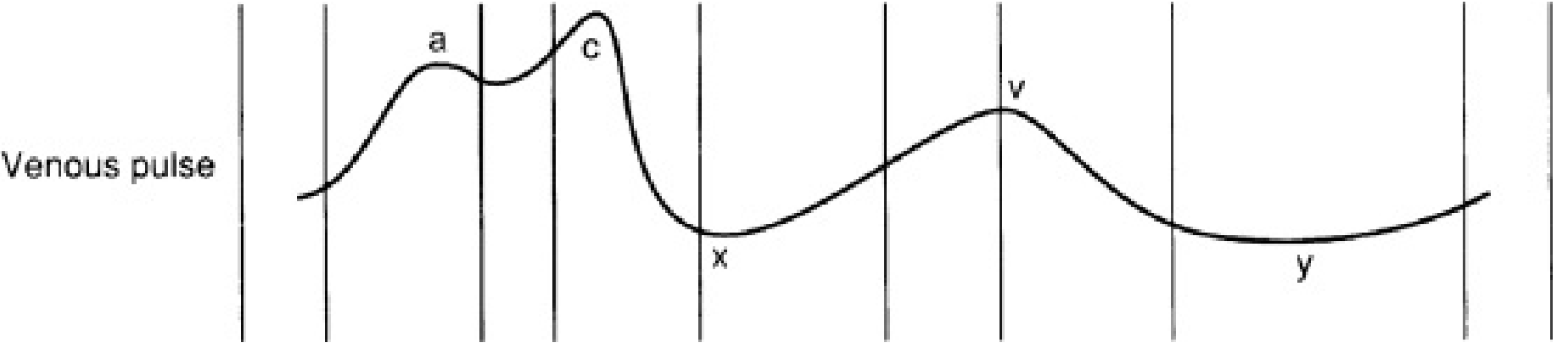
| Wave/Descent | Cause | Condition if Abnormal |
|---|---|---|
| a wave | Right atrial contraction | Absent in AF; giant in tricuspid stenosis, pulmonary hypertension |
| c wave | Tricuspid valve closure + carotid artery pulsation | Small, often not visible |
| x descent | Atrial relaxation + downward displacement of tricuspid | Absent in AF |
| v wave | Venous filling while tricuspid is closed (isovolumetric RV contraction) | Giant in tricuspid regurgitation |
| y descent | Tricuspid valve opens, ventricular filling begins | Slow in tricuspid stenosis; steep in constrictive pericarditis |
Clinical Significance of JVP Changes
- Raised JVP - right heart failure, SVC obstruction, cardiac tamponade, constrictive pericarditis
- Absent a wave - atrial fibrillation (no atrial contraction)
- Kussmaul's sign - JVP rises on inspiration (paradoxical); seen in constrictive pericarditis and cardiac tamponade
- Normal - JVP falls on inspiration
6. Clinical Relevance at MBBS Level
Central Venous Cannulation (IJV approach)
- Right IJV preferred - larger, more direct course to the superior vena cava
- Lower part of IJV lies in the triangle between the sternal and clavicular heads of the SCM
- Landmark: apex of the triangle formed by the two SCM heads
- Complications: pneumothorax, arterial puncture (carotid), air embolism, thoracic duct injury (left side), arrhythmias
Neck Dissection
- Radical neck dissection may sacrifice the IJV
- Bilateral ligation → dangerous (raised intracranial pressure)
Jugular Foramen Syndrome (Vernet's Syndrome)
- Lesion at jugular foramen affects CN IX, X, XI all exiting together
- Dysphagia, hoarse voice, weakness of trapezius/SCM
Facial Vein Danger Zone
- Facial vein communicates with cavernous sinus via the angular vein and ophthalmic veins
- Thrombophlebitis of the face ("danger triangle") can spread to the cavernous sinus
Key Points Summary
| Feature | IJV | EJV |
|---|---|---|
| Origin | Sigmoid sinus at jugular foramen | Retromandibular + posterior auricular veins |
| Location | Deep, inside carotid sheath | Superficial, in superficial fascia |
| Terminates | Joins subclavian → brachiocephalic | Subclavian or IJV |
| Valves | Has valves (inferior bulb) | Has valves |
| Used for JVP | Waveform assessment | Mean pressure assessment |
| Central line access | Yes (preferred) | Possible but less preferred |
- Gray's Anatomy for Students, p. 1151-1153
- Color Atlas of Human Anatomy Vol. 2, p. 131-132
- Goldman-Cecil Medicine, p. 391
- Medical Physiology (Boron & Boulpaep)
This is a shared conversation. Sign in to Orris to start your own chat.