
Features of contralateral and ipsilateral hemiplegia
corticospinal tract pyramidal decussation hemiplegia diagram
Contralateral vs. Ipsilateral Hemiplegia
Anatomical Basis
- Above the decussation (cerebral cortex, corona radiata, internal capsule, midbrain, upper pons, lower pons down to the upper medulla) - the lesion produces contralateral hemiplegia.
- Below the decussation (cervical spinal cord) - the lesion produces ipsilateral hemiplegia.
CONTRALATERAL HEMIPLEGIA
General Features (UMN pattern)
- Weakness/paralysis of the face, arm, and leg on the opposite side of the lesion
- Spasticity (clasp-knife rigidity) - develops after initial flaccid phase
- Hyperreflexia (exaggerated deep tendon reflexes)
- Babinski sign (extensor plantar response)
- No significant muscle atrophy (unless disuse, or if anterior horn cells are also damaged at the cord level)
- Hemiplegic gait (circumduction of the leg)
By Level of Lesion
| Location | Weakness Pattern | Distinguishing Features |
|---|---|---|
| Cerebral cortex | Contralateral face + arm + leg (all together only if extensive) | Focal deficits possible (just leg or arm/face); seizures; aphasia (dominant hemisphere); anosognosia; homonymous hemianopia; astereognosis |
| Internal capsule | Contralateral face + arm + leg (dense, all equal) | "Pure motor hemiplegia" (capsular lacune); no cortical signs |
| Midbrain | Contralateral arm + leg + lower face | Weber syndrome: ipsilateral CN III palsy + contralateral hemiplegia (crossed!) |
| Upper pons | Contralateral arm + leg + lower face | Associated CN V, VI, VII palsies ipsilateral |
| Lower pons | Contralateral arm + leg | Millard-Gubler syndrome: ipsilateral CN VI + VII palsy + contralateral hemiplegia |
| Medulla (medial) | Contralateral arm + leg, spares face | Dejerine (medial medullary) syndrome: ipsilateral tongue weakness + contralateral hemiplegia + contralateral hemisensory loss |
IPSILATERAL HEMIPLEGIA
1. Cervical Spinal Cord Lesion (below decussation)
- Ipsilateral hemiplegia (corticospinal tract already crossed)
- Spares the face entirely
- UMN signs below the level of the lesion on the ipsilateral side
- Brown-Sequard syndrome (cord hemisection): ipsilateral hemiplegia + ipsilateral loss of vibration and proprioception (posterior column) + contralateral loss of pain and temperature (spinothalamic tract)
2. Kernohan's Phenomenon (False Localizing Sign)
- Caused by transtentorial (uncal) herniation
- The herniated uncus pushes the midbrain so far contralaterally that the opposite cerebral peduncle is compressed against the tentorial notch
- This damages the contralateral (to the original lesion) corticospinal tract - producing hemiplegia on the same side as the original lesion (ipsilateral)
- This is a "false localizing sign" - the hemiplegia incorrectly suggests a lesion on the same side as the brain mass
Crossed (Alternating) Hemiplegia - The Brainstem Signature
- Ipsilateral cranial nerve palsy (at the level of the lesion)
- Contralateral hemiplegia (corticospinal tract involvement below the cranial nerve nucleus)
| Syndrome | Level | Ipsilateral CN | Contralateral Body |
|---|---|---|---|
| Weber | Midbrain | CN III palsy (ptosis, fixed dilated pupil, down-and-out eye) | Hemiplegia |
| Claude | Midbrain tegmentum | CN III palsy + ataxia | Hemiplegia |
| Millard-Gubler | Lower pons | CN VI + VII palsy | Hemiplegia |
| Foville | Pons | CN VI + ipsilateral conjugate gaze palsy + CN VII | Contralateral hemiplegia |
| Dejerine (medial medullary) | Medulla | Hypoglossal (CN XII) tongue weakness | Hemiplegia + hemisensory loss |
| Raymond | Pons | CN VI palsy | Contralateral hemiplegia |
Quick Summary Table
| Type | Side of Lesion vs. Weakness | Level | Key Clue |
|---|---|---|---|
| Contralateral (cortex/capsule) | Opposite | Supratentorial | Cortical signs, aphasia, seizures |
| Contralateral (brainstem) | Opposite body, same-side CN palsy | Brainstem | Crossed signs |
| Ipsilateral (cervical cord) | Same side, no face | Below decussation | Brown-Sequard pattern |
| Ipsilateral (Kernohan's) | Same side as mass | Uncal herniation | False localizing sign |
- Adams and Victor's Principles of Neurology, 12th Ed. - p. 1724-1732 (Hemiplegia chapter)
- Bradley and Daroff's Neurology in Clinical Practice - p. 468-469 (Cerebral and subcortical lesions)
- Neuroanatomy through Clinical Cases, 3rd Ed. - p. 171 (Herniation syndromes, Kernohan's phenomenon)
- Localization in Clinical Neurology, 8e - Crossed syndromes and brainstem localization
Brown Sequard syndrome
Brown Sequard syndrome spinal cord hemisection diagram ipsilateral contralateral deficits
Brown-Séquard Syndrome (Spinal Cord Hemisection)
Definition
Anatomical Basis
| Tract | Location in cord | Crosses at | Effect of hemisection |
|---|---|---|---|
| Lateral corticospinal tract | Lateral funiculus (ipsilateral) | Already crossed at pyramidal decussation | Ipsilateral UMN weakness below lesion |
| Posterior (dorsal) columns | Posterior funiculus (ipsilateral) | Crosses in medulla (nucleus gracilis/cuneatus) | Ipsilateral loss of proprioception, vibration, 2-point discrimination, tactile localization |
| Spinothalamic tract (anterolateral system) | Anterior/lateral funiculus (contralateral - already crossed) | Crosses at the same spinal cord level (ventral commissure), 2-3 segments above entry | Contralateral loss of pain and temperature, 2-6 segments below lesion |
Classic Clinical Features
IPSILATERAL (same side as lesion) - below the level:
- UMN hemiplegia/hemiparesis: spasticity, hyperreflexia, Babinski sign
- Loss of proprioception (joint position sense)
- Loss of vibration sense
- Loss of 2-point discrimination and tactile localization (fine/discriminative touch)
- Loss of crude light touch partially (dorsal column contribution)
CONTRALATERAL (opposite side) - 1-2 dermatomes below the lesion:
- Loss of pain sensation
- Loss of temperature sensation
- Crude touch is largely preserved (spinothalamic tract partially preserves it)
AT THE LEVEL of the lesion (ipsilateral):
- LMN signs: flaccid weakness, wasting, areflexia (anterior horn damage)
- Ipsilateral band of pain/temperature loss (1-2 segments) - from damage to the posterior horn cells before their axons have crossed
- A narrow zone of hyperalgesia just above the lesion (irritation of dorsal root entry zone)
Important Nuances
- The contralateral spinothalamic loss begins 1-2 dermatomes below the lesion (because the entering fibers ascend 2-3 segments before crossing in the ventral commissure)
- Crude "light touch" is partially preserved bilaterally - it travels in both dorsal columns and spinothalamic tract; the intact contralateral spinothalamic tract compensates partially
- At cervical levels (C8-T2): if the ciliospinal center of Budge is involved, there may be an ipsilateral Horner syndrome (ptosis, miosis, anhidrosis)
Causes
- Penetrating injury (stab wounds) - most common overall cause
- Blunt trauma (hyperextension > flexion injuries)
- Herniated cervical disc (lateral)
- Vertebral fracture-dislocation
- Multiple sclerosis (demyelinating plaques)
- Spinal cord tumors (primary or metastatic) - lateral compression
- Spinal cord ischemia / epidural hematoma
- Spinal subdural hematoma
- Hematomyelia
- Posttraumatic or infectious arachnoiditis
- Cervical manipulation
- Spinal cord herniation
Brown-Séquard Plus Syndrome
- Asymmetrical hemiplegia rather than complete unilateral paralysis
- Hypoalgesia more prominent on the less paretic side
- A mixed or incomplete pattern
Prognosis
- Recover ambulatory function
- Regain bowel and bladder continence
- Those with more upper > lower extremity weakness have especially favorable ambulation outcomes
Comparison with Other Cord Syndromes
| Feature | Brown-Séquard | Central Cord | Anterior Cord |
|---|---|---|---|
| Motor loss | Ipsilateral UMN | Arms > legs bilaterally | Bilateral (UMN) |
| Pain/temp loss | Contralateral | Bilateral "suspended" (cape/vest) | Bilateral below lesion |
| Proprioception/vibration | Ipsilateral loss | Preserved | Preserved |
| Cause | Penetrating trauma, MS | Hyperextension in elderly | Anterior spinal artery infarct |
- Bradley and Daroff's Neurology in Clinical Practice - Brown-Séquard section, p. 1306
- Neuroanatomy through Clinical Cases, 3rd Ed. - Hemicord lesions, Figure 7.10B
- Guyton and Hall Medical Physiology - Spinal cord transection, p. 615
- Localization in Clinical Neurology, 8e - Causes of BSS
- Tintinalli's Emergency Medicine - BSS overview
Simple diagram to explain Brown Sequard syndrome
A clean medical educational diagram explaining Brown-Séquard Syndrome (spinal cord hemisection). The diagram shows: 1. At the top: a cross-section of the spinal cord with the LEFT half shaded/lesioned (labeled "Lesion - Left hemisection"). Three tracts are labeled inside: "Lateral Corticospinal Tract (ipsilateral)", "Posterior Columns (ipsilateral)", and "Spinothalamic Tract (already crossed - contralateral)". 2. In the middle: a simple human body outline (front view), split down the middle, with color-coded deficit zones: - LEFT side of body: Red shading labeled "Ipsilateral (LEFT) side: UMN weakness (spasticity, hyperreflexia, Babinski), Loss of proprioception & vibration" - RIGHT side of body: Blue shading labeled "Contralateral (RIGHT) side: Loss of pain & temperature (1-2 dermatomes below lesion)" 3. At the level of lesion: a small yellow band across both sides labeled "At lesion level: LMN signs ipsilaterally + narrow band of ipsilateral pain/temp loss" 4. A small legend box in the corner: Red = Ipsilateral motor + dorsal column loss, Blue = Contralateral spinothalamic loss 5. Title at top: "Brown-Séquard Syndrome" with subtitle "Spinal Cord Hemisection" Clean white background, medical textbook style, clear labels, simple arrows showing tract pathways.
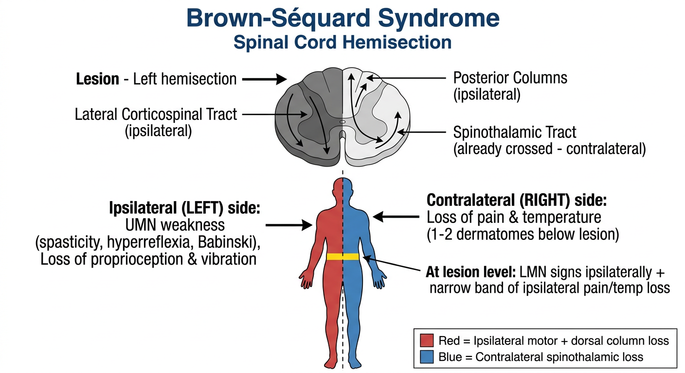
- Left half shaded = the side of the cord lesion
- Red (left body) = ipsilateral deficits: UMN weakness (spasticity, hyperreflexia, Babinski) + loss of proprioception and vibration - because the corticospinal tract and posterior columns have already crossed above the cord
- Blue (right body) = contralateral deficits: loss of pain and temperature, starting 1-2 dermatomes below the lesion - because the spinothalamic fibers cross within the cord before ascending
- Yellow band = at the lesion level itself: a narrow ipsilateral strip of pain/temp loss (posterior horn damage before fibres cross) + LMN signs (anterior horn damage)
Create a downloadable PDF summary of Brown-Séquard Syndrome for reference
ls -la /tmp/workspace/ 2>/dev/null || mkdir -p /tmp/workspace && echo "workspace ready"
mkdir -p /tmp/workspace/bss-pdf && python3 << 'EOF' from reportlab.lib.pagesizes import A4 from reportlab.lib import colors from reportlab.lib.styles import getSampleStyleSheet, ParagraphStyle from reportlab.lib.units import cm, mm from reportlab.platypus import SimpleDocTemplate, Paragraph, Spacer, Table, TableStyle, HRFlowable, KeepTogether from reportlab.lib.enums import TA_CENTER, TA_LEFT, TA_JUSTIFY from reportlab.platypus import Image import urllib.request, os # Download the diagram image img_url = "https://cdn.orris.care/image-gen/d72a506e8ccb4f67a9d37049f9922a8a.png" img_path = "/tmp/workspace/bss-pdf/bss_diagram.png" urllib.request.urlretrieve(img_url, img_path) print("Image downloaded:", os.path.exists(img_path)) EOF
~/bss-pdf/generate_bss_pdf.py
from reportlab.lib.pagesizes import A4
from reportlab.lib import colors
from reportlab.lib.styles import getSampleStyleSheet, ParagraphStyle
from reportlab.lib.units import cm, mm
from reportlab.platypus import (SimpleDocTemplate, Paragraph, Spacer, Table,
TableStyle, HRFlowable, KeepTogether, Image)
from reportlab.lib.enums import TA_CENTER, TA_LEFT, TA_JUSTIFY
from reportlab.pdfbase import pdfmetrics
from reportlab.pdfbase.ttfonts import TTFont
OUTPUT = "/tmp/workspace/bss-pdf/Brown_Sequard_Syndrome.pdf"
IMG_PATH = "/tmp/workspace/bss-pdf/bss_diagram.png"
# ── Colours ──────────────────────────────────────────────────────────────────
NAVY = colors.HexColor("#1a3a5c")
TEAL = colors.HexColor("#0f7173")
RED = colors.HexColor("#c0392b")
BLUE = colors.HexColor("#2471a3")
ORANGE = colors.HexColor("#d35400")
LBLUE = colors.HexColor("#d6eaf8")
LRED = colors.HexColor("#fadbd8")
LYELLOW = colors.HexColor("#fef9e7")
LGREY = colors.HexColor("#f2f3f4")
WHITE = colors.white
BLACK = colors.black
doc = SimpleDocTemplate(
OUTPUT,
pagesize=A4,
leftMargin=1.8*cm, rightMargin=1.8*cm,
topMargin=1.5*cm, bottomMargin=1.8*cm,
title="Brown-Séquard Syndrome – Clinical Reference",
author="Orris Medical AI"
)
W, H = A4
CW = W - 3.6*cm # content width
styles = getSampleStyleSheet()
# ── Custom styles ─────────────────────────────────────────────────────────────
def S(name, **kw):
return ParagraphStyle(name, **kw)
title_style = S("Title2", fontSize=20, textColor=WHITE,
alignment=TA_CENTER, fontName="Helvetica-Bold",
spaceAfter=2, leading=24)
subtitle_style = S("Sub", fontSize=11, textColor=colors.HexColor("#d0e8f5"),
alignment=TA_CENTER, fontName="Helvetica", spaceAfter=0)
h1 = S("H1", fontSize=13, textColor=WHITE, fontName="Helvetica-Bold",
spaceAfter=3, leading=16, leftIndent=6)
h2 = S("H2", fontSize=11, textColor=NAVY, fontName="Helvetica-Bold",
spaceBefore=8, spaceAfter=3, leading=14)
body = S("Body2", fontSize=9.5, textColor=BLACK, fontName="Helvetica",
leading=14, spaceAfter=3, alignment=TA_JUSTIFY)
bullet = S("Bullet2", fontSize=9.5, textColor=BLACK, fontName="Helvetica",
leading=14, spaceAfter=2, leftIndent=14, firstLineIndent=-10)
small = S("Small", fontSize=8.5, textColor=colors.HexColor("#555555"),
fontName="Helvetica-Oblique", leading=12, alignment=TA_CENTER)
note_style = S("Note", fontSize=8.5, textColor=colors.HexColor("#444444"),
fontName="Helvetica-Oblique", leading=12, spaceAfter=2)
def bull(text, color=BLACK):
return Paragraph(f"• {text}", bullet)
def section_header(title, bg=TEAL):
data = [[Paragraph(title, h1)]]
t = Table(data, colWidths=[CW])
t.setStyle(TableStyle([
("BACKGROUND", (0,0), (-1,-1), bg),
("TOPPADDING", (0,0), (-1,-1), 5),
("BOTTOMPADDING", (0,0), (-1,-1), 5),
("LEFTPADDING", (0,0), (-1,-1), 8),
("ROUNDEDCORNERS", [4]),
]))
return t
# ── Build story ───────────────────────────────────────────────────────────────
story = []
# ── Title block ───────────────────────────────────────────────────────────────
title_data = [[Paragraph("Brown-Séquard Syndrome", title_style)],
[Paragraph("Spinal Cord Hemisection | Clinical Reference Summary", subtitle_style)]]
title_tbl = Table(title_data, colWidths=[CW])
title_tbl.setStyle(TableStyle([
("BACKGROUND", (0,0), (-1,-1), NAVY),
("TOPPADDING", (0,0), (-1,-1), 12),
("BOTTOMPADDING", (0,0), (-1,-1), 12),
("ROUNDEDCORNERS", [6]),
]))
story.append(title_tbl)
story.append(Spacer(1, 10))
# ── Definition ────────────────────────────────────────────────────────────────
story.append(section_header("Definition"))
story.append(Spacer(1, 4))
story.append(Paragraph(
"Brown-Séquard Syndrome (BSS) results from <b>hemisection (damage to one lateral half) of the "
"spinal cord</b>. It produces a characteristic <b>dissociated sensorimotor deficit</b>: "
"motor loss and one class of sensory loss <b>ipsilateral</b> to the lesion, "
"and a different sensory modality lost <b>contralaterally</b>.",
body))
story.append(Paragraph(
"BSS accounts for <b>1–4%</b> of all traumatic spinal cord injuries and most commonly "
"occurs at the <b>cervical spine</b>.",
body))
story.append(Spacer(1, 6))
# ── Diagram ───────────────────────────────────────────────────────────────────
story.append(section_header("Diagram – Tract Deficits in BSS"))
story.append(Spacer(1, 4))
img = Image(IMG_PATH, width=CW*0.88, height=CW*0.62)
img.hAlign = "CENTER"
story.append(img)
story.append(Paragraph("Figure: Cross-section of cord (left hemisection) showing ipsilateral vs. contralateral deficits.", small))
story.append(Spacer(1, 6))
# ── Anatomical basis ──────────────────────────────────────────────────────────
story.append(section_header("Anatomical Basis – Three Key Tracts"))
story.append(Spacer(1, 4))
tract_data = [
[Paragraph("<b>Tract</b>", S("th", fontSize=9, textColor=WHITE, fontName="Helvetica-Bold", leading=12)),
Paragraph("<b>Location</b>", S("th", fontSize=9, textColor=WHITE, fontName="Helvetica-Bold", leading=12)),
Paragraph("<b>Where it crosses</b>", S("th", fontSize=9, textColor=WHITE, fontName="Helvetica-Bold", leading=12)),
Paragraph("<b>Deficit after hemisection</b>", S("th", fontSize=9, textColor=WHITE, fontName="Helvetica-Bold", leading=12))],
[Paragraph("Lateral Corticospinal", S("tc", fontSize=8.5, fontName="Helvetica", leading=12)),
Paragraph("Lateral funiculus (ipsilateral)", S("tc", fontSize=8.5, fontName="Helvetica", leading=12)),
Paragraph("Pyramidal decussation (medulla) – already crossed", S("tc", fontSize=8.5, fontName="Helvetica", leading=12)),
Paragraph("<b>Ipsilateral</b> UMN weakness", S("tc", fontSize=8.5, fontName="Helvetica-Bold", textColor=RED, leading=12))],
[Paragraph("Posterior Columns", S("tc", fontSize=8.5, fontName="Helvetica", leading=12)),
Paragraph("Posterior funiculus (ipsilateral)", S("tc", fontSize=8.5, fontName="Helvetica", leading=12)),
Paragraph("Nucleus gracilis/cuneatus in medulla – already crossed", S("tc", fontSize=8.5, fontName="Helvetica", leading=12)),
Paragraph("<b>Ipsilateral</b> loss of proprioception, vibration, 2-point discrimination", S("tc", fontSize=8.5, fontName="Helvetica-Bold", textColor=RED, leading=12))],
[Paragraph("Spinothalamic Tract", S("tc", fontSize=8.5, fontName="Helvetica", leading=12)),
Paragraph("Anterolateral funiculus (contralateral – already crossed)", S("tc", fontSize=8.5, fontName="Helvetica", leading=12)),
Paragraph("Crosses within cord at same/adjacent level via ventral commissure", S("tc", fontSize=8.5, fontName="Helvetica", leading=12)),
Paragraph("<b>Contralateral</b> loss of pain & temperature (1–2 dermatomes below)", S("tc", fontSize=8.5, fontName="Helvetica-Bold", textColor=BLUE, leading=12))],
]
col_w = [CW*0.18, CW*0.22, CW*0.32, CW*0.28]
tract_tbl = Table(tract_data, colWidths=col_w, repeatRows=1)
tract_tbl.setStyle(TableStyle([
("BACKGROUND", (0,0), (-1,0), TEAL),
("BACKGROUND", (0,1), (-1,1), LRED),
("BACKGROUND", (0,2), (-1,2), LRED),
("BACKGROUND", (0,3), (-1,3), LBLUE),
("GRID", (0,0), (-1,-1), 0.4, colors.HexColor("#cccccc")),
("VALIGN", (0,0), (-1,-1), "TOP"),
("TOPPADDING", (0,0), (-1,-1), 5),
("BOTTOMPADDING", (0,0), (-1,-1), 5),
("LEFTPADDING", (0,0), (-1,-1), 5),
("RIGHTPADDING", (0,0), (-1,-1), 5),
]))
story.append(tract_tbl)
story.append(Spacer(1, 6))
# ── Clinical features two-column ──────────────────────────────────────────────
story.append(section_header("Clinical Features"))
story.append(Spacer(1, 4))
ipsi_items = [
"<b>UMN hemiplegia/hemiparesis</b> – spasticity, hyperreflexia, Babinski sign",
"Loss of <b>proprioception</b> (joint position sense)",
"Loss of <b>vibration sense</b>",
"Loss of <b>2-point discrimination</b> and tactile localisation",
"Partial loss of crude light touch",
"(At C8–T2) <b>Ipsilateral Horner syndrome</b> – ptosis, miosis, anhidrosis",
]
contra_items = [
"Loss of <b>pain sensation</b>",
"Loss of <b>temperature sensation</b>",
"Onset <b>1–2 dermatomes below</b> the lesion level",
"Crude touch largely <b>preserved</b>",
]
at_level_items = [
"<b>LMN signs ipsilaterally</b> – flaccid weakness, wasting, areflexia (anterior horn)",
"Narrow ipsilateral band of <b>pain/temp loss</b> (posterior horn damage before fibres cross)",
"Zone of <b>hyperalgesia</b> just above lesion (dorsal root zone irritation)",
]
def feature_box(title, items, bg, tc):
content = [Paragraph(f"<b>{title}</b>", S("bh", fontSize=9.5, textColor=WHITE,
fontName="Helvetica-Bold", leading=13, alignment=TA_CENTER))]
for i in items:
content.append(Paragraph(f"• {i}", S("bi", fontSize=8.5, textColor=BLACK,
fontName="Helvetica", leading=13, leftIndent=6, spaceAfter=2)))
tbl = Table([[c] for c in content], colWidths=[(CW-10)/2])
tbl.setStyle(TableStyle([
("BACKGROUND", (0,0), (-1,0), tc),
("BACKGROUND", (0,1), (-1,-1), bg),
("TOPPADDING", (0,0), (-1,-1), 5),
("BOTTOMPADDING", (0,0), (-1,-1), 4),
("LEFTPADDING", (0,0), (-1,-1), 7),
("RIGHTPADDING", (0,0), (-1,-1), 7),
("BOX", (0,0), (-1,-1), 0.6, tc),
]))
return tbl
ipsi_box = feature_box("IPSILATERAL (same side as lesion) – below lesion", ipsi_items, LRED, RED)
contra_box = feature_box("CONTRALATERAL (opposite side) – below lesion", contra_items, LBLUE, BLUE)
side_tbl = Table([[ipsi_box, Spacer(10, 1), contra_box]],
colWidths=[(CW-10)/2, 10, (CW-10)/2])
side_tbl.setStyle(TableStyle([
("VALIGN", (0,0), (-1,-1), "TOP"),
("TOPPADDING", (0,0), (-1,-1), 0),
("BOTTOMPADDING", (0,0), (-1,-1), 0),
("LEFTPADDING", (0,0), (-1,-1), 0),
("RIGHTPADDING", (0,0), (-1,-1), 0),
]))
story.append(side_tbl)
story.append(Spacer(1, 5))
# At-level box
at_level_box = feature_box("AT THE LEVEL OF LESION (ipsilateral)", at_level_items,
LYELLOW, ORANGE)
story.append(at_level_box)
story.append(Spacer(1, 6))
# ── Causes ────────────────────────────────────────────────────────────────────
story.append(section_header("Causes / Aetiology"))
story.append(Spacer(1, 4))
cause_data = [
[Paragraph("<b>Traumatic (most common)</b>", S("ch", fontSize=9.5, textColor=WHITE,
fontName="Helvetica-Bold", leading=12)),
Paragraph("<b>Non-traumatic</b>", S("ch", fontSize=9.5, textColor=WHITE,
fontName="Helvetica-Bold", leading=12))],
[Paragraph(
"• Penetrating injury – stab wounds (most common overall)\n"
"• Blunt trauma – hyperextension > flexion injuries\n"
"• Herniated cervical disc (lateral)\n"
"• Vertebral fracture-dislocation\n"
"• Compression injuries",
S("cb", fontSize=8.5, fontName="Helvetica", leading=14)),
Paragraph(
"• Multiple sclerosis (demyelinating plaques)\n"
"• Spinal cord tumours – primary or metastatic\n"
"• Spinal cord ischaemia / epidural haematoma\n"
"• Hematomyelia\n"
"• Posttraumatic / infectious arachnoiditis\n"
"• Spinal subdural haematoma\n"
"• Spinal cord herniation",
S("cb", fontSize=8.5, fontName="Helvetica", leading=14))],
]
cause_tbl = Table(cause_data, colWidths=[CW/2, CW/2])
cause_tbl.setStyle(TableStyle([
("BACKGROUND", (0,0), (-1,0), NAVY),
("BACKGROUND", (0,1), (0,1), LGREY),
("BACKGROUND", (1,1), (1,1), colors.HexColor("#eafaf1")),
("GRID", (0,0), (-1,-1), 0.4, colors.HexColor("#aaaaaa")),
("VALIGN", (0,0), (-1,-1), "TOP"),
("TOPPADDING", (0,0), (-1,-1), 6),
("BOTTOMPADDING", (0,0), (-1,-1), 6),
("LEFTPADDING", (0,0), (-1,-1), 8),
]))
story.append(cause_tbl)
story.append(Spacer(1, 6))
# ── BSS Plus ──────────────────────────────────────────────────────────────────
story.append(section_header("Brown-Séquard Plus Syndrome", bg=colors.HexColor("#6c3483")))
story.append(Spacer(1, 4))
story.append(Paragraph(
"The <b>pure classic</b> form is rarely encountered clinically. More commonly, "
"patients present with <b>Brown-Séquard Plus</b> (Taylor & Gleave, 1957):",
body))
story.append(bull("Asymmetrical hemiplegia rather than complete unilateral paralysis"))
story.append(bull("Hypoalgesia more prominent on the <i>less</i> paretic side"))
story.append(bull("Mixed/incomplete pattern of deficits"))
story.append(Paragraph(
"<b>Prognosis note:</b> Paradoxically, pure BSS carries a <i>worse prognosis</i> than "
"BSS Plus. Overall, BSS has the <b>best functional motor recovery</b> of all incomplete "
"spinal cord syndromes. Most patients regain ambulatory function and bowel/bladder "
"continence.",
body))
story.append(Spacer(1, 6))
# ── Comparison table ──────────────────────────────────────────────────────────
story.append(section_header("Comparison with Other Incomplete Cord Syndromes"))
story.append(Spacer(1, 4))
comp_data = [
[Paragraph("<b>Feature</b>", S("t", fontSize=9, textColor=WHITE, fontName="Helvetica-Bold")),
Paragraph("<b>Brown-Séquard</b>", S("t", fontSize=9, textColor=WHITE, fontName="Helvetica-Bold")),
Paragraph("<b>Central Cord</b>", S("t", fontSize=9, textColor=WHITE, fontName="Helvetica-Bold")),
Paragraph("<b>Anterior Cord</b>", S("t", fontSize=9, textColor=WHITE, fontName="Helvetica-Bold")),
Paragraph("<b>Posterior Cord</b>", S("t", fontSize=9, textColor=WHITE, fontName="Helvetica-Bold"))],
["Motor loss",
"Ipsilateral UMN",
"Arms > legs bilaterally",
"Bilateral UMN (below)",
"Mild or none"],
["Pain/Temp",
"Contralateral loss",
"Bilateral suspended 'cape'",
"Bilateral loss below",
"Preserved"],
["Proprioception/Vibration",
"Ipsilateral loss",
"Preserved",
"Preserved",
"Bilateral loss"],
["Common cause",
"Penetrating trauma, MS",
"Hyperextension (elderly)",
"Anterior spinal artery infarct",
"Posterior cord ischaemia"],
["Prognosis",
"Best of all incomplete",
"Good – arms worse than legs",
"Variable",
"Good"],
]
cw2 = [CW*0.16, CW*0.20, CW*0.20, CW*0.22, CW*0.22]
comp_tbl = Table(comp_data, colWidths=cw2)
comp_tbl.setStyle(TableStyle([
("BACKGROUND", (0,0), (-1,0), TEAL),
("ROWBACKGROUNDS",(0,1), (-1,-1), [WHITE, LGREY]),
("GRID", (0,0), (-1,-1), 0.4, colors.HexColor("#bbbbbb")),
("FONTNAME", (0,0), (-1,0), "Helvetica-Bold"),
("FONTSIZE", (0,0), (-1,-1), 8.5),
("VALIGN", (0,0), (-1,-1), "TOP"),
("TOPPADDING", (0,0), (-1,-1), 5),
("BOTTOMPADDING", (0,0), (-1,-1), 5),
("LEFTPADDING", (0,0), (-1,-1), 5),
("FONTNAME", (0,1), (0,-1), "Helvetica-Bold"),
]))
story.append(comp_tbl)
story.append(Spacer(1, 8))
# ── Memory Aid ────────────────────────────────────────────────────────────────
story.append(section_header("Memory Aid", bg=colors.HexColor("#1a6b3a")))
story.append(Spacer(1, 4))
mem_data = [[
Paragraph(
"<b>\"Same side – Motor and Posterior column loss\"</b><br/>"
"Motor weakness + proprioception/vibration → <b>IPSILATERAL</b>",
S("m", fontSize=9.5, textColor=BLACK, fontName="Helvetica", leading=14)),
Paragraph(
"<b>\"Opposite side – Pain and Temperature\"</b><br/>"
"Pain and temperature loss → <b>CONTRALATERAL</b> (1–2 levels below)",
S("m", fontSize=9.5, textColor=BLACK, fontName="Helvetica", leading=14)),
]]
mem_tbl = Table(mem_data, colWidths=[CW/2, CW/2])
mem_tbl.setStyle(TableStyle([
("BACKGROUND", (0,0), (0,0), colors.HexColor("#d5f5e3")),
("BACKGROUND", (1,0), (1,0), colors.HexColor("#d6eaf8")),
("BOX", (0,0), (-1,-1), 0.6, colors.HexColor("#1a6b3a")),
("GRID", (0,0), (-1,-1), 0.4, colors.HexColor("#aaaaaa")),
("TOPPADDING", (0,0), (-1,-1), 8),
("BOTTOMPADDING", (0,0), (-1,-1), 8),
("LEFTPADDING", (0,0), (-1,-1), 10),
]))
story.append(mem_tbl)
story.append(Spacer(1, 8))
# ── Footer ────────────────────────────────────────────────────────────────────
story.append(HRFlowable(width=CW, thickness=0.5, color=colors.HexColor("#aaaaaa")))
story.append(Spacer(1, 3))
story.append(Paragraph(
"Sources: Adams & Victor's Principles of Neurology 12e · Bradley and Daroff's Neurology in Clinical Practice · "
"Neuroanatomy through Clinical Cases 3e · Guyton & Hall Medical Physiology · "
"Localization in Clinical Neurology 8e · Tintinalli's Emergency Medicine | "
"Generated by Orris Medical AI · August 2026",
S("foot", fontSize=7, textColor=colors.HexColor("#888888"), fontName="Helvetica-Oblique",
alignment=TA_CENTER, leading=10)))
# ── Build ─────────────────────────────────────────────────────────────────────
doc.build(story)
print("PDF created successfully at", OUTPUT)
python3 /tmp/workspace/bss-pdf/generate_bss_pdf.py
ls -lh /tmp/workspace/bss-pdf/Brown_Sequard_Syndrome.pdf
Brown Sequard Syndrome
PDF Document · PDF
- Title block with full heading
- Diagram (the one generated earlier showing ipsilateral vs. contralateral deficits)
- Anatomical basis table - all three tracts, where they cross, and what deficit each produces
- Clinical features - colour-coded boxes: red for ipsilateral deficits, blue for contralateral, yellow for at-level findings
- Causes - traumatic vs. non-traumatic, side by side
- Brown-Séquard Plus explanation with prognosis
- Comparison table vs. Central Cord, Anterior Cord, and Posterior Cord syndromes
- Memory aid - a quick two-box reminder for the exam
- Source citations at the footer