
Aortic stenosis
"aortic stenosis"[MeSH Terms] AND management
"aortic stenosis" AND "transcatheter aortic valve"
aortic stenosis pathophysiology diagnosis management guidelines 2024 2025
https://123sonography.com/blog/news/highlights-updates-escea…
Aortic Stenosis
Overview
Etiology
| Type | Features |
|---|---|
| Calcific/Degenerative | Most common in adults >65. Calcium deposits on normal tricuspid cusps, causing progressive obstruction |
| Congenital Bicuspid Valve | 1-2% of population; affects 66% of patients. Severe AS develops 1-2 decades earlier than in tricuspid valves. Often associated with aortic root dilation - risk of dissection/rupture |
| Rheumatic | Rare in Western countries. Results from fusion of commissures. Usually coexists with MS |
- Braunwald's Heart Disease, p. 1036
- Grainger & Allison's Diagnostic Radiology, p. 338
Pathophysiology
- Maintains systolic function initially
- Increases LV end-diastolic pressure -> diastolic dysfunction
- Makes the LV noncompliant and highly dependent on atrial kick for filling (hence AF is very poorly tolerated)
- Eventually reduces LV compliance and stroke volume
| Symptom | Mechanism | Mortality if untreated |
|---|---|---|
| Angina | Increased O₂ demand (hypertrophied LV) + decreased coronary perfusion time | 50% 5-year mortality |
| Syncope | Fixed CO unable to meet demands on exertion; also baroreceptor reflex miscommunication | 50% 3-year mortality |
| Dyspnea/HF | Elevated LVEDP -> pulmonary congestion | 50% 2-year mortality |
- Symptom to Diagnosis, p. 9340
Clinical Features
- Atrial fibrillation (poorly tolerated - LV relies on atrial kick)
- Acquired von Willebrand deficiency (large vWF multimers disrupted by turbulent flow across stenotic valve) - presents as GI bleeding (Heyde syndrome)
Diagnosis
Echocardiography (first-line)
| Parameter | Mild | Moderate | Severe |
|---|---|---|---|
| Peak velocity (m/s) | <3.0 | 3.0-4.0 | ≥4.0 |
| Mean gradient (mmHg) | <20 | 20-40 | ≥40 |
| AVA (cm²) | >1.5 | 1.0-1.5 | <1.0 |
| AVA indexed (cm²/m²) | - | - | <0.6 |
EOA = (SV of LVOT) / (VTI across aortic valve)
- Valve area <1 cm² but mean gradient <40 mmHg
- Occurs when LV stroke volume index is <35 mL/m²
- More common in women with small LV cavities and in AF
- Must distinguish true severe (low flow due to poor LV function) from pseudo-severe (moderate AS that appears severe at low flow)
- Use dobutamine stress echo OR cardiac CT calcium scoring: an Agatston score ≥1600 in men / ≥1200 in women confirms severe AS without needing stress echo (2025 ESC/EACTS update)
Chest X-ray
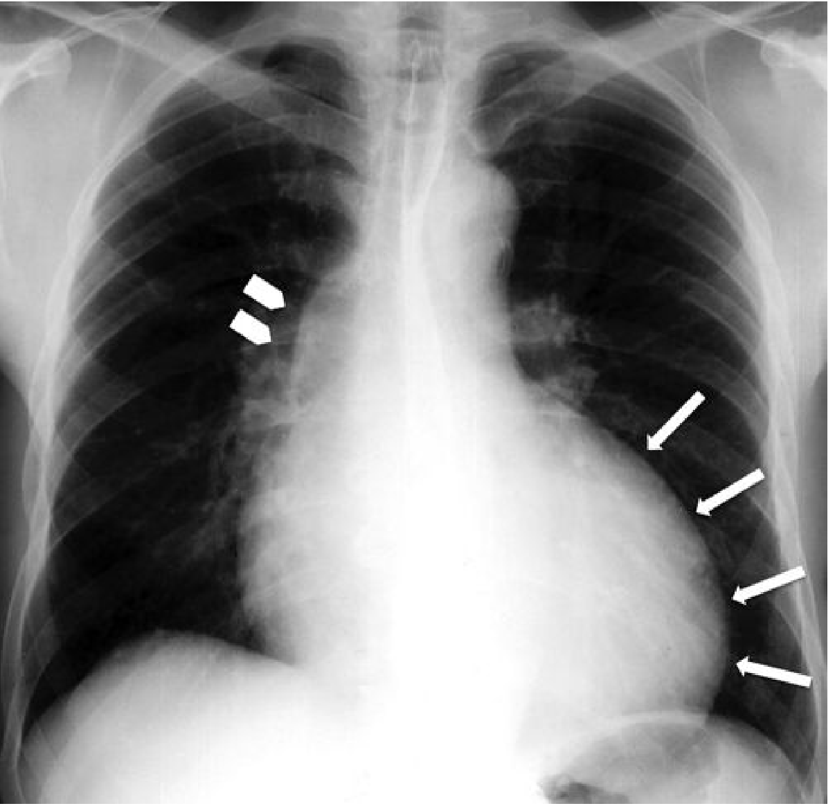
- Rounding of LV apex (LVH)
- Poststenotic dilation of the ascending aorta
- Aortic valve calcification visible on lateral view
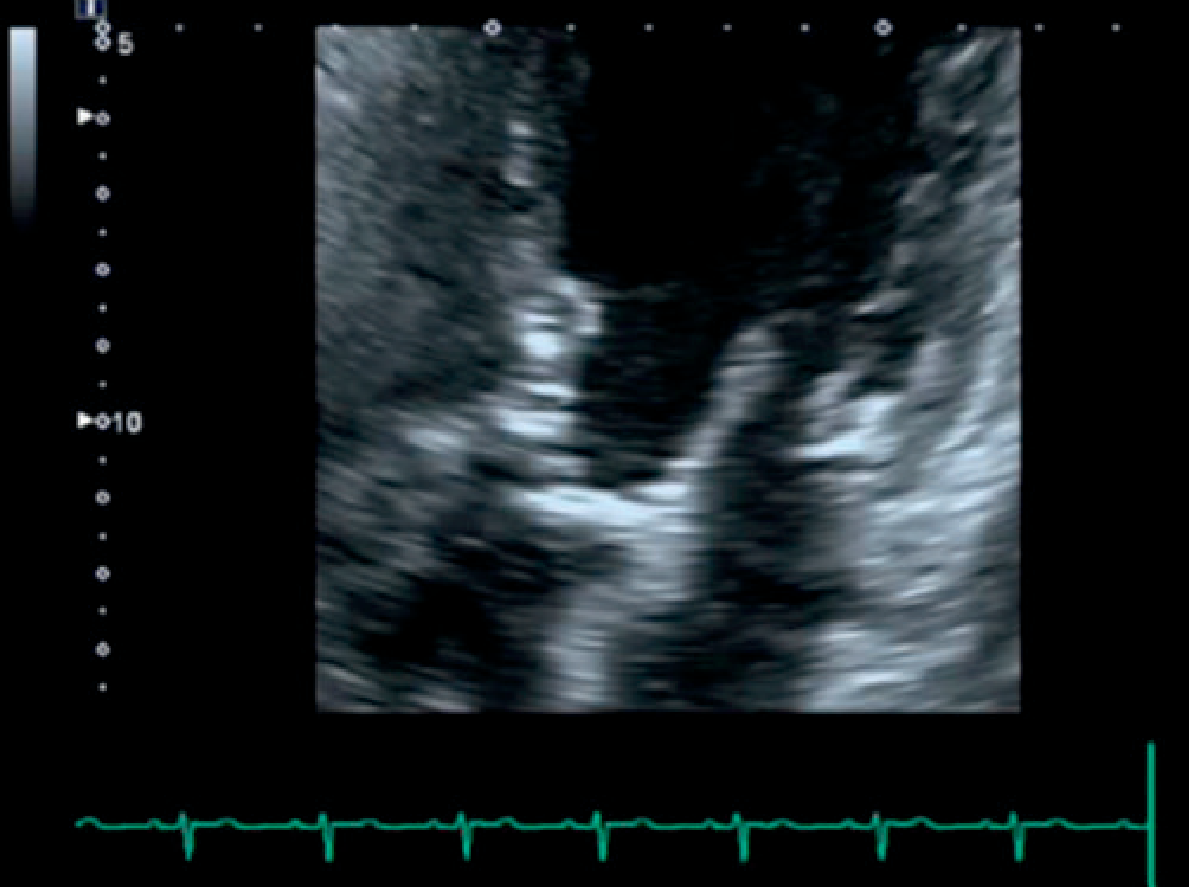
Echocardiogram (B-mode)
Cardiac CT
- Excellent for calcium scoring (Agatston score)
- Allows direct planimetry of the valve orifice
- Essential for pre-TAVR anatomy assessment (annulus sizing, coronary ostia height, access)
- Agatston score of 150 is 100% sensitive for discriminating velocities <2.5 m/s from >2.5 m/s
CMR
- Direct planimetry of valve orifice possible
- Less sensitive than CT for calcium detection
- Useful adjunct when echo is suboptimal
Management
Medical Therapy
- Diuretics cautiously for HF symptoms (avoid over-diuresis given diastolic dysfunction)
- ACE inhibitors/ARBs - can be used cautiously; avoid vasodilators that precipitate hypotension
- Rate control for AF (these patients are preload-dependent)
Intervention Indications
- Symptomatic severe AS (dyspnea, angina, syncope) - this is the primary indication regardless of age or LV function
- Asymptomatic severe AS with LVEF <50%
- Asymptomatic severe AS undergoing cardiac surgery for another reason
- Early intervention is a reasonable option vs. watchful waiting in asymptomatic severe AS with normal LVEF
- Asymptomatic patients with peak velocity ≥5.5 m/s or very rapid progression (velocity increase >0.3 m/s per year) - intervention recommended even without symptoms
TAVR vs. SAVR
| Patient Profile | Preferred Approach |
|---|---|
| High/prohibitive surgical risk | TAVR (any age) |
| Age >80 or life expectancy <10 years | TAVR |
| Age 65-80, low-intermediate surgical risk | Shared decision-making; TAVR or SAVR both acceptable |
| Age <65-70, low surgical risk | SAVR preferred (concern for long-term valve durability with TAVR) |
| Symptomatic, any age, high surgical risk + expected survival >12 months with acceptable QoL | TAVR |
- PARTNER trial (inoperable patients, mean age 83): 1-year mortality 30% TAVR vs. 50% medical therapy
- PARTNER low-risk trial: composite of death, stroke, or hospitalization at 1 year 8.5% (TAVR) vs. 15.1% (SAVR)
- Registry data: 30-day TAVR mortality has declined from 4% to 2% over time
- Braunwald's Heart Disease, p. 1036
2025 ESC/EACTS Guideline Update (Key Changes)
- CT calcium scoring elevated to same level as dobutamine stress echo for LFLG-AS workup
- Stronger recommendation for early intervention in asymptomatic patients (2A)
- Continued 2A recommendation for high-risk asymptomatic patients (peak velocity ≥5 m/s)
- TAVR expanded for intermediate-risk patients (previously required high risk)
- For patients <70 at low surgical risk, SAVR remains preferred
Prognosis
Hand drawn diagrams for examination
Which diagrams would you like for aortic stenosis?
Hand drawn diagrams
Hand-drawn medical diagram for exam revision on aortic stenosis pathophysiology. Sketch style with pencil-like lines on white background. Flow chart showing: "Normal AV" arrow to "Calcification / Bicuspid / Rheumatic" arrow to "Valve Orifice Narrows" arrow to "LV Outflow Obstruction" which branches into two paths: Path 1: "LV Pressure Overload" → "Concentric LVH" → "Increased O2 demand" → "Angina" AND "Diastolic Dysfunction" → "↑ LVEDP" → "Pulmonary Congestion" → "Dyspnea / HF". Path 2: "Fixed Cardiac Output" → "Failure to ↑ CO on exertion" → "Syncope". Title at top: "AS Pathophysiology - Cardinal Symptoms". Annotations with small arrows and boxes, hand-written style labels, slight imperfect lines to look genuinely hand drawn.
Hand-drawn medical examination diagram showing auscultation findings in aortic stenosis. Pencil sketch style on white paper. Central drawing of chest outline with stethoscope positions marked. Key finding: Diamond-shaped crescendo-decrescendo systolic murmur drawn as a waveform between S1 and S2 heart sounds, with annotation "Ejection Systolic Murmur" and "Peaks in mid-systole". Radiation arrow going from aortic area (right 2nd ICS) up to neck/carotids labeled "Radiates to carotids". Additional labeled boxes: "S2 Soft or absent (calcified valve)", "S4 gallop (LVH)", "Ejection Click (bicuspid AV, non-calcified)", "Slow rising pulse - Pulsus parvus et tardus". Heart sound timing diagram showing S1----murmur----S2 with diamond shape. Title: "Aortic Stenosis - Clinical Findings". Hand-written style, slight imperfect sketch lines.
Hand-drawn medical exam revision diagram for aortic stenosis echocardiographic grading and severity criteria. Pencil sketch style on white background. Table drawn with hand-drawn lines showing three columns: Mild, Moderate, Severe. Rows: Peak Velocity (less than 3 m/s, 3-4 m/s, 4 m/s or more), Mean Gradient (less than 20 mmHg, 20-40 mmHg, 40 mmHg or more), Valve Area (more than 1.5 cm2, 1.0-1.5 cm2, less than 1.0 cm2), Indexed AVA (-, -, less than 0.6 cm2 per m2). Below table: Small sketch of Doppler waveform showing high velocity jet in severe AS. Box labeled "Continuity Equation: AVA = CSA(LVOT) x VTI(LVOT) / VTI(AV)". Another box: "Low-Flow Low-Gradient AS: AVA less than 1cm2 BUT gradient less than 40mmHg - check stroke volume index less than 35mL/m2 - Use Dobutamine stress echo OR CT calcium score". Title: "Echo Grading of Aortic Stenosis". Hand-written annotations and arrows, slightly imperfect lines.
Hand-drawn medical exam diagram showing aortic stenosis management algorithm. Pencil sketch flowchart style on white paper. Start box: "Severe AS diagnosed (AVA less than 1cm2, Vmax 4m/s, gradient 40mmHg)". Arrow down to diamond decision box: "Symptomatic? (Angina / Syncope / Dyspnea)". YES branch goes right to box: "INTERVENTION INDICATED (Class I)". NO branch goes down to another diamond: "LVEF less than 50% OR undergoing other cardiac surgery?". YES → "INTERVENTION (Class I)". NO → "Asymptomatic Severe AS" box → two sub-boxes: "Vmax more than 5.5 m/s OR rapid progression → Intervention (Class IIa)" and "Otherwise → Watchful waiting + Echo surveillance every 6-12 months". From INTERVENTION box, arrow down to diamond: "Surgical Risk?". Branches: "High/Prohibitive or Age more than 80 → TAVR" and "Low risk, Age less than 70 → SAVR preferred" and "Intermediate / Age 65-80 → Heart Team Decision: TAVR or SAVR". Title: "AS Management Algorithm". Hand-written style, annotated boxes, slightly uneven lines like real exam notes.
400, message: Got more than 4194304 bytes when reading: b'data: {"candidates": [{"content": {"role": "model","parts": [{"inlineData": {"mimeType": "image/png"...'.
We encountered an error while processing your request. Please try again or reach out to us for help.