
Lynphomas
I'll start by reading the medical library skill, then search for comprehensive information on lymphomas.
Reading File
Now let me search the medical library for lymphoma content in parallel:
Finding Sources
Finding Sources
Excellent! Let me now read the key textbook sections on lymphomas in parallel - from Robbins Pathology and Goldman-Cecil Medicine:
Reading File
Reading File
Let me get the core lymphoma pathology content from Robbins and the clinical classification/staging from Goldman-Cecil in parallel:
Reading File
Reading File
Now let me read further in Goldman-Cecil for Hodgkin lymphoma, staging, and treatment, and read more Robbins content on key lymphoma subtypes:
Reading File
Reading File
Let me now read the Hodgkin lymphoma section from Robbins and the staging/treatment section from Goldman-Cecil:
Reading File
Reading File
Let me get the Hodgkin lymphoma section from Robbins and the NHL treatment/DLBCL section from Goldman-Cecil:
Finding Sources
Reading File
Now let me get the Hodgkin lymphoma section from Robbins Basic Pathology (very detailed):
Reading File
Reading File
Let me fetch key images and also read the Hodgkin lymphoma HL chapter in Goldman-Cecil for staging/treatment:
Finding Sources
Loading Image
Let me get the Hodgkin lymphoma chapter in Goldman-Cecil and also fetch the follicular lymphoma histology image:
Finding Sources
Loading Image
Let me get the Hodgkin lymphoma treatment chapter from Goldman-Cecil and also the Reed-Sternberg cell image:
Finding Sources
Finding Sources
Let me now read the critical Hodgkin lymphoma morphology (RS cells) from Robbins and the HL treatment from Goldman-Cecil:
Reading File
Reading File
Let me now get the HL staging/treatment plan from Goldman-Cecil and fetch the RS cell image:
Reading File
Loading Image
I now have enough comprehensive information from authoritative textbooks to write a thorough response on lymphomas. Let me compile the full answer.
Lymphomas - Comprehensive Overview
Lymphomas are malignant neoplasms of lymphoid cells. They are broadly divided into two major groups: Hodgkin Lymphoma (HL) and Non-Hodgkin Lymphoma (NHL). The distinction matters clinically because behavior, staging, and treatment differ significantly.
Normal Counterparts - Origin of Lymphomas
Understanding where in lymphoid differentiation a lymphoma arises explains its biology:
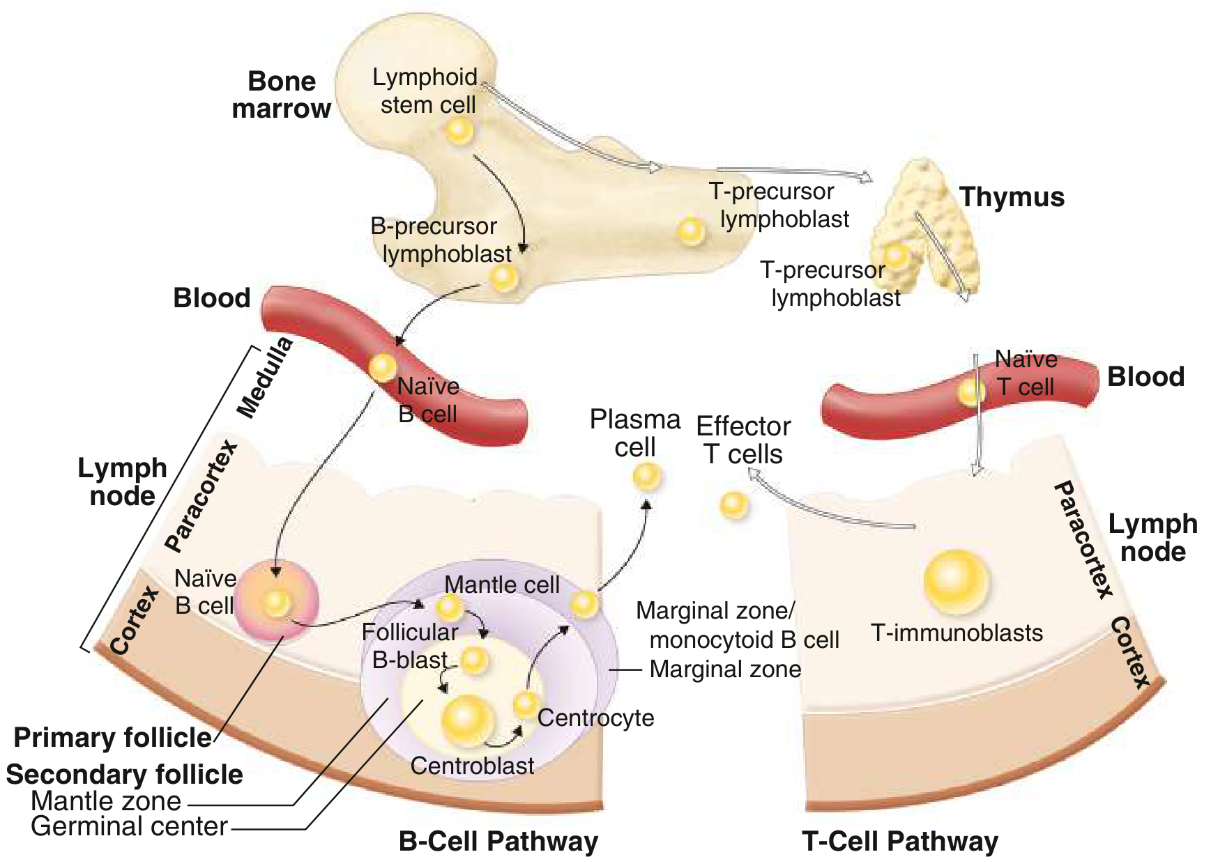
Postulated normal counterparts of B- and T-cell malignancies. Lymphoid stem cells in bone marrow differentiate into B-cell precursors (cortex/germinal center) or T-cell precursors (thymus), giving rise to specific lymphoma subtypes at each stage.
I. CLASSIFICATION
WHO Classification (2016/Updated)
The WHO classification integrates morphology, immunophenotype, cytogenetics, and clinical behavior. Lymphomas are divided by:
- Cell of origin: B-cell vs. T/NK-cell
- Maturity: Precursor (immature) vs. peripheral (mature/post-thymic)
-
In the US and Europe, 85-90% of NHLs are B-cell in origin
-
Most frequent NHL worldwide: Diffuse Large B-Cell Lymphoma (DLBCL) ~30%
-
Second most frequent: Follicular Lymphoma ~20%
-
Less common types (~5-10% each): Extranodal marginal zone/MALT, Peripheral T-cell, Small Lymphocytic Lymphoma (SLL), Mantle Cell Lymphoma
-
Goldman-Cecil Medicine, p. 1951
II. EPIDEMIOLOGY & RISK FACTORS
Autoimmune and Inflammatory Conditions
- Rheumatoid arthritis: 2-fold increased NHL risk
- Sjögren syndrome: 30-40 fold increased marginal zone lymphoma risk
- Hashimoto thyroiditis: increased thyroid lymphoma risk
- Celiac disease: associated with enteropathy-type T-cell lymphoma
Infectious Agents
| Agent | Associated Lymphoma |
|---|---|
| EBV | Burkitt lymphoma (>95% endemic form), post-transplant lymphoproliferative disorders, DLBCL of elderly, NK/T-cell lymphoma |
| HTLV-1 | Adult T-cell leukemia/lymphoma (virtually all cases) |
| HHV-8 | Primary effusion lymphoma (immunocompromised) |
| Hepatitis C virus | Lymphoplasmacytic lymphoma, marginal zone lymphoma |
| H. pylori | Gastric MALT lymphoma |
| Borrelia burgdorferi | Marginal zone B-cell lymphoma of skin |
Environmental Factors
-
Agricultural chemicals (phenoxy herbicides/Agent Orange), ionizing radiation, organic solvents, high-fat diet, ultraviolet radiation
-
Prior treatment for Hodgkin lymphoma: 20-fold increased NHL risk
-
Heavy smoking: increased follicular lymphoma risk
-
Goldman-Cecil Medicine, pp. 845-864
III. NON-HODGKIN LYMPHOMAS (NHL)
A. B-Cell NHLs
1. Follicular Lymphoma
- ~20% of all NHLs; more common in North America and Western Europe
- Cell of origin: Germinal center B cells (centrocytes + centroblasts)
- Key genetic lesion: t(14;18) translocation → BCL2 overexpression → blocks apoptosis
- Morphology: Nodular (follicular) growth pattern; mixture of small centrocytes (cleaved nuclei) and larger centroblasts
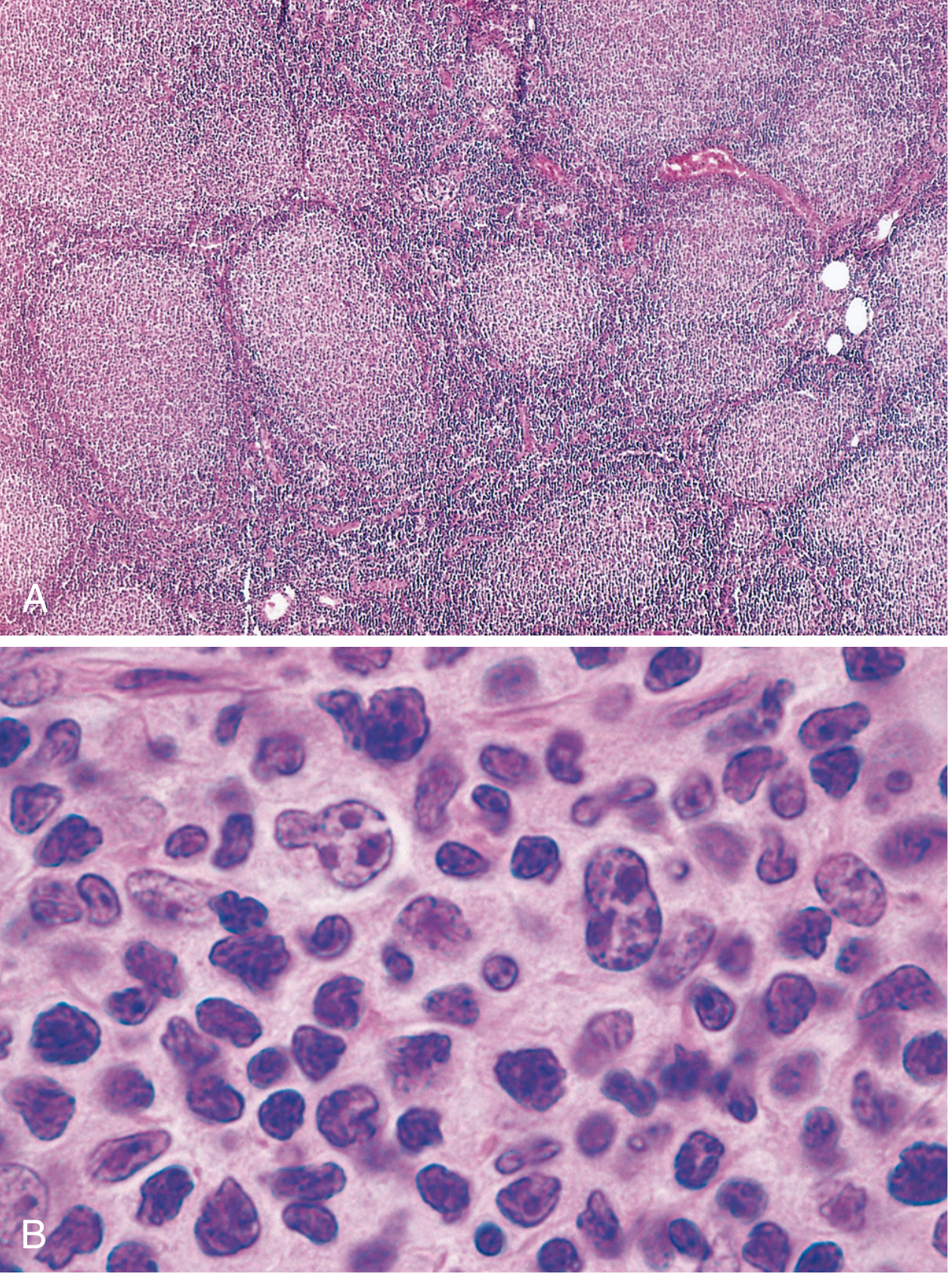
Fig. A - Low power: nodular aggregates throughout the lymph node. Fig. B - High power: small centrocytes with cleaved nuclei mixed with larger centroblasts with nucleoli.
- Immunophenotype: CD20+, BCL2+ (unlike reactive germinal centers), CD10+
- BCL2 immunostain: positive in neoplastic follicles (negative in reactive germinal centers) - key diagnostic feature
- Clinical behavior: Indolent, waxing and waning; median survival 7-9 years; NOT improved by aggressive therapy - palliated with anti-CD20 (rituximab) + gentle chemotherapy
- Histologic transformation: 30-50% transform to DLBCL; if MYC is acquired, prognosis is particularly poor (median survival 3-5 years)
- EZH2 inhibitors are active in the subset with EZH2 gain-of-function mutations
2. Diffuse Large B-Cell Lymphoma (DLBCL)
- Most common NHL (~30%)
- Aggressive but potentially curable
- Includes several distinct subtypes: primary CNS lymphoma, primary mediastinal (thymic) large B-cell lymphoma, leg-type cutaneous DLBCL, plasmablastic lymphoma, double/triple-hit lymphomas
- Double-hit lymphoma: MYC rearrangement + BCL2 and/or BCL6 rearrangements - now classified as "high-grade B-cell lymphoma with MYC and BCL2/BCL6 rearrangements"; requires aggressive regimens (DA-R-EPOCH)
- Primary CNS lymphoma: very sensitive to corticosteroids; treated with high-dose methotrexate ± rituximab; whole-brain RT adds toxicity without benefit
- Plasmablastic lymphoma: Most common in HIV+ patients; CD20 negative, so does NOT benefit from rituximab
3. Mantle Cell Lymphoma
- Cell of origin: Naive B cells in the mantle zone
- Moderately aggressive course
- Key genetic lesion: t(11;14) → cyclin D1 overexpression → confirmed by FISH; CD5+
- Distinct from CLL/SLL by cyclin D1
4. Marginal Zone Lymphoma / MALT Lymphoma
- Indolent tumors of antigen-primed B cells at sites of chronic immune stimulation
- Gastric MALT lymphoma: driven by H. pylori; eradication of H. pylori can induce regression
- Often remain localized for long periods
5. Burkitt Lymphoma
- t(8;14) - MYC translocation (or variants t(2;8) or t(8;22))
- Endemic form: African children, jaw tumors, nearly all EBV+
- Sporadic form: abdominal/ileocecal mass, ~15-35% EBV+
- AIDS-associated form: aggressive, often involves CNS
- "Starry sky" pattern on histology (macrophages phagocytosing apoptotic cells)
6. Small Lymphocytic Lymphoma (SLL) / CLL
- Tissue manifestation of CLL; indolent
- CD5+, CD23+, weak surface Ig
7. Lymphoplasmacytic Lymphoma (Waldenström's Macroglobulinemia)
- B-cell neoplasm with terminal differentiation to plasma cells
- Secretes monoclonal IgM → hyperviscosity syndrome
- Nearly all cases have MYD88 mutations
- No bone destruction (unlike myeloma)
B. T- and NK-Cell NHLs
Represent 5-10% of NHLs in the US/Europe; more common in East Asia.
- Peripheral T-cell lymphoma, NOS: Pleomorphic mixture of malignant T cells; reactive eosinophils and macrophages (cytokine-driven); express CD2, CD3, CD5; significantly worse prognosis than DLBCL
- Anaplastic Large Cell Lymphoma (ALCL): CD30+; ALK+ form has better prognosis; ALK- form has poor prognosis; distinct from Hodgkin lymphoma (CD15-negative)
- Adult T-cell leukemia/lymphoma: caused by HTLV-1; skin lesions, hypercalcemia, leukemia picture
- Mycosis fungoides / Sézary syndrome: Primary cutaneous T-cell lymphoma; starts as skin patches → plaques → tumors (Sézary syndrome = erythroderma + circulating atypical cells)
- Extranodal NK/T-cell lymphoma (nasal type): EBV-associated; midline destructive lesions of the nasopharynx; more common in Asia and Latin America
- Enteropathy-associated T-cell lymphoma: associated with celiac disease; intestinal involvement
C. Lymphoblastic Lymphoma / ALL
- Precursor B or T lymphoblasts
- T-cell LBL: mediastinal mass in young males; CNS involvement common
- B-cell LBL: solid tumors of skin and bones (~85% of ALL has B-cell phenotype)
- Treated with ALL-type regimens (cytarabine, high-dose methotrexate, maintenance therapy, CNS prophylaxis)
IV. HODGKIN LYMPHOMA (HL)
What Makes HL Distinct
- Reed-Sternberg (RS) cells - the neoplastic element
- Robust but ineffective host immune response; RS cells are only a small fraction of the tumor mass
- Arises in a single node/chain and spreads in stepwise contiguous fashion - important for radiotherapy planning
- Robbins Basic Pathology, p. 417
Reed-Sternberg Cells
- Large cells (~45 µm diameter) with bilobed or multilobed nuclei, each with a large "owl-eye" nucleolus (~5-7 µm, size of a small lymphocyte)
- Diagnostic RS cells must have at least two nuclear lobes/nuclei each with a nucleolus
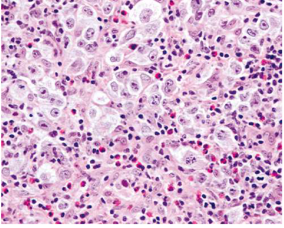
Nodular sclerosing Hodgkin lymphoma: large lacunar cells (RS variants) with abundant pale cytoplasm in a background of lymphocytes and eosinophils. Note the prominent large "owl-eye" nucleoli characteristic of Reed-Sternberg cells.
Pathogenesis of HL
- RS cells originate from germinal center B cells (proven by clonal IGH rearrangements + somatic hypermutation in microdissected RS cells)
- Classic HL RS cells paradoxically fail to express immunoglobulin genes - due to widespread epigenetic reprogramming
- NF-κB activation is the central molecular event:
- EBV LMP-1 (in ~70% of mixed-cellularity subtype) activates NF-κB
- EBV- tumors: loss-of-function mutations in IκB or TNFAIP3
- Immune evasion: RS cells express high levels of PD-L1 and PD-L2 (chromosome 9p amplification) → antagonizes cytotoxic T-cell responses
- Reactive background: RS cells secrete IL-5 (eosinophil chemoattractant), TGF-β (fibrogenic), IL-13 (autocrine growth)
WHO Classification of HL Subtypes
| Subtype | Frequency |
|---|---|
| Nodular sclerosis | 60% (most common) |
| Lymphocyte-rich | 3% |
| Mixed cellularity | 9% |
| Lymphocyte-depleted | 1% (worst prognosis) |
| Nodular Lymphocyte-Predominant HL (NLPHL) | 8% |
Data: Goldman-Cecil Medicine, Table 172-1
Key HL Subtypes
Nodular Sclerosis (most common):
- Young adults; mediastinal involvement common
- Collagen bands divide lymph node into nodules
- Lacunar cells (RS variants with retracted cytoplasm due to formalin fixation)
- Background: lymphocytes, eosinophils, plasma cells, neutrophils
Mixed Cellularity:
- Older adults; EBV present in ~70%
- Diagnostic RS cells easily found
- Background: lymphocytes, eosinophils, plasma cells, histiocytes
Lymphocyte-Depleted:
- Rarest; most aggressive; older/HIV patients
- Few lymphocytes; sheets of RS cells
Nodular Lymphocyte-Predominant HL (NLPHL):
- Distinct entity; neoplastic cells are "popcorn cells" (LP cells/lymphocytic and histiocytic cells)
- CD20+, CD79a+, CD45+ (unlike classic HL)
- CD30-, CD15- (unlike classic HL)
- More indolent course; associated with late relapse
Immunophenotype of Classic HL RS Cells
| Marker | Classic HL | NLPHL | Reactive/ALCL |
|---|---|---|---|
| CD30 | + (90-100%) | - | + (ALCL) |
| CD15 | + (75-85%) | - | - |
| CD20 | +/- (~40%, weak) | strongly + | - |
| PAX5/BSAP | + (>90%) | + | - |
| CD45 | - | + | - |
V. STAGING - ANN ARBOR SYSTEM (Modified Cotswolds)
| Stage | Definition |
|---|---|
| I | Single lymph node region OR one extralymphatic site (Ie) |
| II | Two or more nodal regions, same side of diaphragm (IIe = + local extralymphatic extension) |
| III | Nodal regions on both sides of diaphragm (IIIe = + local extralymphatic) |
| IV | Diffuse extranodal involvement (bone marrow, lung, bone, liver) |
| A | No B symptoms |
| B | ≥1 of: weight loss >10% in 6 months, unexplained fever >38°C, night sweats |
- Goldman-Cecil Medicine, Table 172-3
VI. WORKUP & STAGING INVESTIGATIONS
- Biopsy: Excisional lymph node biopsy (gold standard); fine needle aspiration is inadequate
- Laboratory: CBC, chemistry including LDH, uric acid
- Imaging: CT chest/abdomen/pelvis; FDG-PET/CT is now mandatory for HL and most aggressive NHLs - more sensitive than CT for staging and residual disease assessment; PET can replace bone marrow biopsy in HL
- Bone marrow biopsy: In NHL if results will change management (not needed if PET positive)
- Lumbar puncture: If CNS involvement suspected (DLBCL with high IPI, Burkitt, etc.)
VII. INTERNATIONAL PROGNOSTIC INDEX (IPI) - for aggressive NHLs
5 adverse factors (1 point each):
- Age > 60 years
- Elevated LDH
- Performance status 2-4
- More than 1 extranodal site
- Ann Arbor stage III or IV
| Score | Risk Group |
|---|---|
| 0-1 | Low |
| 2 | Low-intermediate |
| 3 | High-intermediate |
| 4-5 | High |
- Goldman-Cecil Medicine, Table 171-8
VIII. TREATMENT OVERVIEW
Hodgkin Lymphoma
- Limited stage (I-II, favorable): 2-4 cycles ABVD + involved-site radiotherapy
- Advanced stage (III-IV): 6-8 cycles ABVD or escalated BEACOPP
- BEACOPP: superior response rates but more toxicity (infertility with alkylating agents; not seen with ABVD)
- Brentuximab vedotin (anti-CD30 ADC): used in refractory/relapsed HL and upfront with AVD in stage III-IV
- PD-1 checkpoint inhibitors (nivolumab, pembrolizumab): active in relapsed/refractory HL (exploiting PD-L1 overexpression on RS cells)
- Relapsed/refractory HL: Second-line chemo → autologous hematopoietic stem cell transplant (HSCT) for chemosensitive disease
Non-Hodgkin Lymphoma - Key Treatments
| NHL Type | First-line Treatment |
|---|---|
| DLBCL | R-CHOP (rituximab + cyclophosphamide, doxorubicin, vincristine, prednisone) |
| Double/triple-hit | DA-R-EPOCH |
| Follicular (symptomatic) | R-CHOP, R-bendamustine + maintenance rituximab; EZH2 inhibitors (tazemetostat) for EZH2-mutant |
| Mantle cell | Rituximab-based + BTK inhibitors (ibrutinib, acalabrutinib); autologous HSCT in young fit patients |
| Burkitt | Intensive ALL-like regimens (CODOX-M/IVAC); CNS prophylaxis essential |
| MALT/Gastric | H. pylori eradication; radiotherapy for localized; rituximab |
| Primary CNS lymphoma | High-dose methotrexate ± rituximab ± cytarabine; avoid whole-brain RT as initial therapy |
| T-cell lymphomas | CHOP-based; worse prognosis than comparable B-cell lymphomas |
| Lymphoblastic lymphoma | ALL-type regimens with CNS prophylaxis |
Special Situations
- AIDS-associated NHL: Treat same as non-immunocompromised; add CNS prophylaxis; prognosis has improved with HAART
- Post-transplant lymphoproliferative disorder (PTLD): Reduce immunosuppression first; rituximab; chemotherapy for refractory cases
- Elderly patients: Dose-adjusted regimens; careful assessment of cardiac function for anthracyclines
IX. KEY GENETIC LESIONS SUMMARY
| Lymphoma | Translocation/Mutation | Effect |
|---|---|---|
| Follicular lymphoma | t(14;18) | BCL2 overexpression → blocked apoptosis |
| Mantle cell lymphoma | t(11;14) | Cyclin D1 overexpression → uncontrolled cell cycle |
| Burkitt lymphoma | t(8;14) [or t(2;8), t(8;22)] | MYC overexpression → uncontrolled proliferation |
| DLBCL - double hit | MYC + BCL2/BCL6 rearrangements | Highly aggressive |
| Classic Hodgkin HL | 9p amplification (PD-L1/L2); NF-κB activation | Immune evasion; survival signaling |
| NLPHL | - | Germinal center B-cell origin; CD20+ |
| Lymphoplasmacytic lymphoma | MYD88 mutation | NF-κB activation |
| Hairy cell leukemia | BRAF V600E mutation | Constitutive MAPK signaling |
| ALCL | ALK translocation (subset) | ALK+ has better prognosis |
X. CLINICAL PEARLS
- Lymphoma spreads stepwise and contiguously in HL but skips nodes more often in NHL
- "B symptoms" (fever >38°C, night sweats, >10% weight loss) are adverse prognostic signs and affect staging (A vs. B designation)
- RS cells in classic HL: CD15+, CD30+ - the hallmark immunoprofile
- RS-like cells can appear in infectious mononucleosis, solid tumors, and some NHLs - diagnosis requires clinicopathologic correlation
- Follicular lymphoma is incurable with standard therapy but follows an indolent course; transformation to DLBCL occurs in 30-50%
- Gastric MALT lymphoma often regresses with H. pylori eradication alone
- High macrophage infiltrate in HL is a predictor of treatment resistance
- PET/CT has replaced gallium scanning and is now the gold standard for staging and response assessment in HL and aggressive NHLs
Sources: Robbins, Cotran & Kumar - Pathologic Basis of Disease; Robbins & Kumar Basic Pathology; Goldman-Cecil Medicine International Edition (2-volume set)
This is a shared conversation. Sign in to Orris to start your own chat.