
Hypertension
What aspect of hypertension are you interested in? · What is your purpose?
hypertension pathophysiology diagram blood pressure regulation
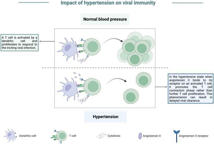
A pathophysiology diagram titled 'Impact of hypertension on viral immunity' comparing cellular immune responses under normal and hypertensive conditions. The top panel, 'Normal blood pressure,' illustrates a dendritic cell presenting antigens to a T cell via receptor-ligand interactions and cytokine release. This stimulation leads to robust T-cell activation and proliferation, resulting in a large cluster of effector T cells essential for viral clearance. The bottom panel, 'Hypertension,' depicts the same initial dendritic cell-T cell interaction but highlights the presence of elevated Angiotensin II. Angiotensin II binds to Angiotensin II receptors expressed on the activated T cell. This signaling inhibits sustained proliferation and instead promotes a T-cell contraction phase, resulting in a significantly reduced population of T cells. The diagram indicates that this hypertensive state leads to an impaired effector response and delayed viral clearance. Key components labeled include dendritic cells, T cells, cytokines, Angiotensin II, and Angiotensin II receptors.
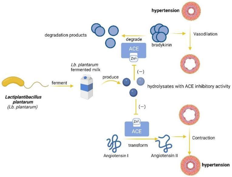
This pathophysiology diagram illustrates the anti-hypertensive mechanism of Lactiplantibacillus plantarum (Lb. plantarum) in functional foods. The process begins with Lb. plantarum fermenting milk, which produces bioactive hydrolysates. These hydrolysates function as Angiotensin-Converting Enzyme (ACE) inhibitors, targeting the zinc-binding (Zn2+) site of the ACE protein. The diagram depicts two physiological pathways influenced by this inhibition: 1) The Bradykinin Pathway: Typically, ACE degrades the vasodilator bradykinin into inactive products. By inhibiting ACE, hydrolysates prevent bradykinin degradation, promoting vasodilation and relieving hypertension. 2) The Renin-Angiotensin Pathway: Normally, ACE transforms Angiotensin I into the potent vasoconstrictor Angiotensin II, leading to vascular contraction and hypertension. The hydrolysates inhibit this transformation, preventing pathological vasoconstriction. The visual contrast emphasizes the transition from constricted, hypertensive blood vessel states to relaxed, dilated states through the modulation of enzymatic activity. This diagram serves as a model for how probiotic-derived peptides can mitigate cardiovascular risk factors by interfering with the biochemical pathways responsible for systemic blood pressure regulation.
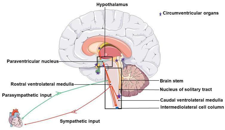
An anatomical and pathophysiology diagram illustrating the brain regions and neural pathways involved in neurogenic hypertension. The illustration features a sagittal view of the human brain with key regulatory centers labeled, including the hypothalamus, paraventricular nucleus (PVN), and components of the brainstem: the rostral ventrolateral medulla (RVLM), nucleus of the solitary tract (NTS), caudal ventrolateral medulla (CVLM), and the intermediolateral cell column (IML). Purple stars denote the locations of circumventricular organs (CVOs) distributed near the hypothalamus and brainstem. The diagram maps the functional connectivity between these regions and the heart. A green line represents the parasympathetic input originating from the RVLM/brainstem area, while a red line represents the sympathetic input originating from the IML, both descending to the cardiac target. This illustration highlights the neuroregulatory network responsible for blood pressure control and the autonomic imbalance (increased sympathetic and decreased parasympathetic tone) characteristic of neurogenic hypertension.
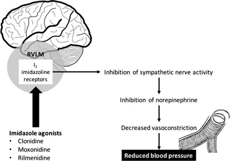
This pathophysiology diagram illustrates the mechanism of action of imidazoline receptor agonists on blood pressure regulation. The visual is divided into a sagittal brain anatomical diagram and a secondary physiological flowchart. On the left, the diagram highlights the 'I1 imidazoline receptors' located within the Rostral Ventrolateral Medulla (RVLM) of the brainstem, situated inferior to the cerebrum and anterior to the cerebellum. Below this, three pharmacological agents—Clonidine, Moxonidine, and Rilmenidine—are listed as imidazole agonists that target these receptors. To the right, a downward flowchart depicts the systemic consequences of receptor activation: stimulation of the RVLM leads to the 'Inhibition of sympathetic nerve activity,' followed by the 'Inhibition of norepinephrine' release. This progression results in 'Decreased vasoconstriction,' visually represented by an illustration of a dilated blood vessel with reduced smooth muscle tension. The final clinical outcome is identified as 'Reduced blood pressure.' The content is designed for intermediate medical education, focusing on neuropharmacology and cardiovascular physiology.
hypertension end organ damage complications retinopathy heart kidney
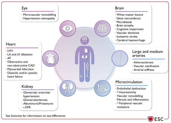
Summary : This figure illustrates the organ-specific and vascular effects of hypertension, detailing pathological changes in the eye, heart, kidney, brain, large and medium arteries, and microcirculation. Each organ system is listed with its associated hypertensive complications. diagram: # Organ Systems and Hypertensive Effects : ## Eye : • Microvascular remodelling • Hypertensive retinopathy ## Heart : • LVH (left ventricular hypertrophy) • LA and LV dilatation (left atrial and left ventricular) • AF (atrial fibrillation) • Obstructive and non-obstructive CAD (coronary artery disease) • Myocardial infarction • Diastolic and/or systolic heart failure ## Kidney : • Glomerular arteriolar hypertension • Glomerulosclerosis • Albuminuria/Proteinuria • ↓ GFR (glomerular filtration rate) ## Brain : • White matter lesions • Silent microinfarcts • Microbleeds • Brain atrophy • Cognitive impairment • Vascular dementia • Ischaemic stroke • Cerebral haemorrhage ## Large and Medium Arteries : • Atherosclerosis • Vascular calcification • Arterial stiffness ## Microcirculation : • Endothelial dysfunction • ↑ Vasoreactivity • Vascular remodelling • Fibrosis and inflammation • ↑ Peripheral vascular resistance # Layout : • Central human figure with icons representing each organ system. • Each organ system is connected to a text box listing its hypertensive complications. • Colour-coded circles and boxes for each organ/vascular system. # Additional Information : • Footnote mentions sex-differences (not detailed in the figure). • ESC (European Society of Cardiology) logo at the bottom right. # Analysis : • The figure demonstrates that hypertension affects multiple organ systems, with distinct pathological changes in each. • Both macrovascular (large arteries) and microvascular (small vessels, microcirculation) complications are highlighted. • The heart, brain, and kidney are shown to be particularly vulnerable, with a wide range of structural and functional consequences. • The diagram visually emphasises the systemic nature of hypertensive disease and its multi-organ impact.
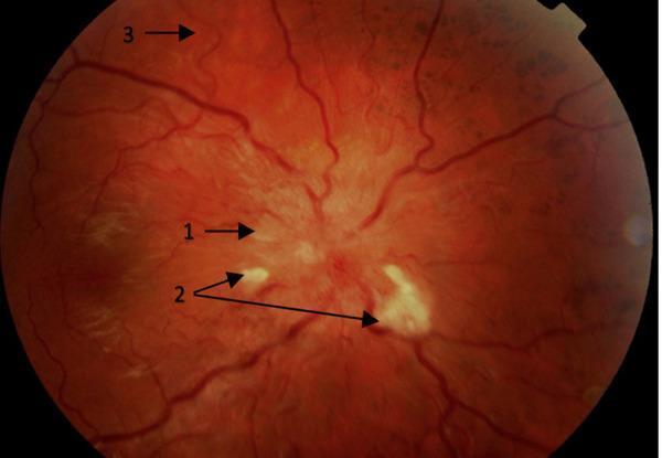
This clinical photograph displays a retinal fundus image highlighting hypertension-mediated end-organ damage (EOD), specifically hypertensive retinopathy. The image is annotated with numerical markers indicating key diagnostic features: (1) points to blurred optic disc margins and papilloedema, signaling severe intracranial pressure or grade IV hypertensive changes; (2) identifies multiple cotton wool spots, which are small, fluffy, whitish lesions representing focal retinal ischemia; and (3) demonstrates generalized arteriolar attenuation and increased vascular tortuosity, where the retinal vessels appear narrowed and more winding than normal. The anatomical focus includes the optic nerve head, the retinal nerve fiber layer, and the retinal vasculature. This material is pedagogically relevant for ophthalmology and internal medicine, illustrating the ocular manifestations of malignant or accelerated hypertension and the importance of fundoscopic examination in assessing systemic cardiovascular risk and subclinical target organ damage.
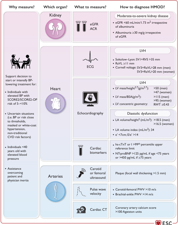
Summary : This figure provides a structured overview of how to measure and diagnose hypertension-mediated organ damage (HMOD) in the kidney, heart, and arteries, including what to measure, diagnostic criteria, and clinical indications for assessment. flowchart/table hybrid: # Why measure? : • Support decision to start or intensify blood pressure (BP)-lowering treatment for: – Individuals with elevated BP and SCORE2/SCORE2-OP risk of 5–<10% – Uncertain situations (BP/risk close to thresholds, masked/white-coat hypertension, non-traditional CVD risk factors) – Individuals <40 years old with elevated BP – Assistance overcoming patient/physician inertia # Which organ? : • Kidney • Heart • Arteries # What to measure? : ## Kidney : • eGFR (estimated glomerular filtration rate) • ACR (albumin-to-creatinine ratio) ## Heart : • ECG • Echocardiography • Cardiac biomarkers ## Arteries : • Carotid or femoral ultrasound • Pulse wave velocity • Cardiac CT # How to diagnose HMOD? : ## Kidney : • Moderate-to-severe kidney disease: – eGFR <60 mL/min/1.73 m² (irrespective of albuminuria) – Albuminuria ≥30 mg/g (irrespective of eGFR) ## Heart : • LVH (left ventricular hypertrophy) by ECG: – Sokolow–Lyon: SV1+RV5 >35 mm – RaVL ≥11 mm – Cornell voltage: SV3+RaVL >28 mm (men), >20 mm (women) • LVH by echocardiography: – LV mass/height².⁷ (g/m².⁷): >50 (men), >47 (women) – LV mass/BSA (g/m²): >115 (men), >95 (women) – LV concentric geometry: RWT ≥0.43 • Diastolic dysfunction: – LA volume/height² (mL/m²): >18.5 (men), >16.5 (women) – LA volume index (mL/m²): 34 – e’ <7cm; E/e’ >14 • Cardiac biomarkers: – hs-cTnT or >99th percentile upper reference limit – NT-proBNP >125 pg/mL (<75 years) or >450 pg/mL (≥75 years) ## Arteries : • Carotid/femoral ultrasound: Plaque (focal wall thickening >1.5 mm) • Pulse wave velocity: – Carotid-femoral PWV >10 m/s – Brachial-ankle PWV >14 m/s • Cardiac CT: Coronary artery calcium score >100 Agatston units # Design Encodings : • Organ sections are colour-coded: kidney (purple), heart (light purple), arteries (blue). • Icons represent each organ and measurement modality. • Diagnostic criteria are listed in shaded boxes for each organ. # Layout : • Four main columns: Why measure?, Which organ?, What to measure?, How to diagnose HMOD? • Rows for kidney, heart, and arteries, each with corresponding measurements and criteria. # Analysis : • The figure provides a comprehensive, organ-based approach to diagnosing HMOD, with clear measurement modalities and diagnostic thresholds for kidney, heart, and arterial damage. • It highlights the importance of tailored assessment based on clinical context and risk, and provides actionable criteria for each organ system. • The use of colour and icons aids rapid identification of organ systems and measurement types.
Hypertension - Comprehensive Medical Exam Guide
1. Definition & Classification
JNC / ESC Classification (ESH/ISH)
| Category | Systolic (mmHg) | Diastolic (mmHg) | |
|---|---|---|---|
| Optimal | < 120 | and | < 80 |
| Normal | 120-129 | and/or | 80-84 |
| High-Normal | 130-139 | and/or | 85-89 |
| Grade 1 HTN | 140-159 | and/or | 90-99 |
| Grade 2 HTN | 160-179 | and/or | 100-109 |
| Grade 3 HTN | ≥ 180 | and/or | ≥ 110 |
| Isolated Systolic HTN | ≥ 140 | and | < 90 |
Rule: When systolic and diastolic fall into different grades, use the higher category.
-
Isolated Systolic HTN: Common in the elderly due to reduced aortic compliance.
-
White-coat HTN: Elevated in clinic only; confirmed by ambulatory BP monitoring (ABPM).
-
Masked HTN: Normal in clinic but elevated on ABPM - carries cardiovascular risk.
-
Park's Textbook of Preventive and Social Medicine
2. Epidemiology
-
Global prevalence: ~1.13 billion adults (2015 data); ~30-40% of all adults worldwide.
-
Age-standardized prevalence: 24% in men, 20% in women globally.
-
Prevalence >60% in those aged >60 years.
-
"Rule of Halves": ~half are unaware, half of those aware are treated, half of those treated are adequately controlled - making it an "iceberg disease."
-
Elevated BP is responsible for ~10 million deaths/year and >200 million DALYs.
-
Projected to increase 15-20% by 2025 due to aging, sedentary lifestyles, and obesity.
-
Park's Textbook of Preventive and Social Medicine
3. Pathophysiology
Key Mechanisms
- Renin (from juxtaglomerular cells) cleaves angiotensinogen → Angiotensin I
- ACE converts Ang I → Angiotensin II (potent vasoconstrictor)
- Ang II → aldosterone secretion → sodium and water retention → increased CO
- Ang II also causes direct vascular smooth muscle contraction → increased TPR
- Increased adrenergic tone → vasoconstriction (α1), tachycardia/increased contractility (β1)
- The rostral ventrolateral medulla (RVLM) is the key brainstem center
- Neurogenic hypertension involves increased sympathetic / decreased parasympathetic tone
- Reduced nitric oxide (NO) bioavailability → impaired vasodilation
- Increased endothelin-1 → vasoconstriction
- Excess dietary sodium → increased plasma volume → increased CO
- Impaired renal sodium excretion is central to essential hypertension
- Chronic high pressure → medial hypertrophy of arterioles → increased TPR (self-perpetuating)
4. Primary vs. Secondary Hypertension
| Feature | Primary (Essential) | Secondary |
|---|---|---|
| Proportion | ~95% | ~5% |
| Cause | Multifactorial/genetic | Identifiable underlying disease |
| Age of onset | Usually middle age | Often younger (<30) or sudden onset |
| Clue to workup | Family history, gradual | Refractory HTN, specific signs/symptoms |
Causes of Secondary Hypertension (Organ Framework)
- Primary hyperaldosteronism (Conn syndrome): hypokalemia, suppressed renin, elevated aldosterone; most common endocrine cause
- Pheochromocytoma: episodic headache, palpitations, sweating (the "triad"); catecholamine excess - screen with plasma/urine metanephrines
- Cushing syndrome: truncal obesity, striae, buffalo hump; cortisol excess → mineralocorticoid effect
- Hypothyroidism (diastolic) / Hyperthyroidism (systolic)
- Hyperparathyroidism: hypercalcemia → vasoconstriction
- Chronic kidney disease (CKD) - most common secondary cause overall
- Acute kidney injury
- Renovascular HTN (renal artery stenosis): atherosclerosis (older men) or fibromuscular dysplasia (young women); abdominal bruit; refractory HTN; use captopril renal scan or Doppler
- Coarctation of the aorta: upper limb > lower limb BP; radio-femoral delay; rib notching on CXR
- Obstructive sleep apnea (OSA): most common cause of resistant hypertension
-
NSAIDs, COX-2 inhibitors, corticosteroids, OCPs, cocaine, alcohol, sympathomimetics (decongestants), cyclosporine/tacrolimus, erythropoietin, amphetamines/modafinil
-
Symptom to Diagnosis, 4th Edition; Robbins & Cotran Pathologic Basis of Disease
Exam Tip: Suspect secondary HTN if: onset < 30 years, resistant to 3+ drugs, sudden onset, hypokalemia without diuretics, abdominal bruit, or features of an endocrine disorder.
5. Clinical Features & Diagnosis
Symptoms
- Often asymptomatic ("silent killer") until complications develop
- Headache (occipital, morning) - actually not specific; usually absent in mild-moderate HTN
- Epistaxis, tinnitus, dizziness (non-specific)
BP Measurement
- Seated, rest 5 min, arm at heart level, 2+ readings, 2+ occasions
- Cuff size matters: too small → falsely high; too large → falsely low
- Methods: office, home (HBPM), ambulatory (ABPM) - ABPM is gold standard
Workup at Diagnosis (Baseline)
- Urinalysis + urine ACR (proteinuria, hematuria)
- Serum creatinine + eGFR (renal function)
- Serum electrolytes (hypokalemia → hyperaldosteronism)
- Fasting glucose + HbA1c (diabetes co-morbidity)
- Lipid panel (cardiovascular risk)
- ECG (LVH: Sokolow-Lyon index SV1 + RV5 >35 mm; Cornell voltage SV3 + RaVL >28 mm in men)
- Echocardiogram (if ECG LVH or symptoms)
- Fundoscopy (retinopathy grading)
- Chest X-ray (cardiomegaly, coarctation rib notching)
6. Target Organ Damage (HMOD - Hypertension-Mediated Organ Damage)
Heart
- Left Ventricular Hypertrophy (LVH) - earliest sign; diastolic dysfunction
- Left atrial dilatation → Atrial Fibrillation
- Coronary artery disease, Myocardial infarction
- Diastolic and/or systolic heart failure
Kidney
- Glomerular arteriolar hypertension → glomerulosclerosis
- Albuminuria/proteinuria (first sign of renal involvement)
- Reduced eGFR, CKD
Brain
- Lacunar infarcts, white matter lesions, microbleeds
- Ischaemic stroke and intracerebral haemorrhage
- Hypertensive encephalopathy (severe HTN)
- Vascular dementia and cognitive impairment
Eyes (Keith-Wagener-Barker Grading)
| Grade | Features |
|---|---|
| I | Arterial narrowing, increased arteriovenous (AV) ratio |
| II | AV nipping/nicking |
| III | Flame haemorrhages, cotton-wool spots, hard exudates |
| IV | Papilloedema (= malignant HTN) |
Large Arteries
- Atherosclerosis, arterial stiffness, vascular calcification, aortic aneurysm
HMOD Assessment Thresholds
| Organ | Parameter | Threshold for HMOD |
|---|---|---|
| Kidney | eGFR | < 60 mL/min/1.73 m² |
| Kidney | ACR | ≥ 30 mg/g |
| Heart (ECG) | Sokolow-Lyon | SV1 + RV5 > 35 mm |
| Heart (Echo) | LV mass/BSA | > 115 g/m² (men), > 95 g/m² (women) |
| Arteries | Carotid-femoral PWV | > 10 m/s |
- Goldman-Cecil Medicine; Comprehensive Clinical Nephrology, 7th Edition
7. Management
Step 1 - Lifestyle Modifications (Non-pharmacological)
| Intervention | Recommendation | SBP Reduction |
|---|---|---|
| Weight loss | Maintain BMI 18.5-24.9 | 5-20 mmHg per 10 kg lost |
| DASH diet | Rich in fruits, vegetables, low-fat dairy; low saturated fat | 8-14 mmHg |
| Sodium restriction | < 2.3 g Na/day (< 6 g NaCl) | 2-8 mmHg |
| Aerobic exercise | ≥ 30 min/day, most days | 5-8 mmHg |
| Alcohol restriction | ≤ 2 drinks/day (men), ≤ 1/day (women) | 2-4 mmHg |
The DASH diet alone reduced SBP by 7.6 mmHg and DBP by 4.2 mmHg in RCTs.
- National Kidney Foundation Primer on Kidney Diseases, 8th Edition
Step 2 - Pharmacotherapy
BP Targets
- General: < 130/80 mmHg (ACC/AHA guideline)
- Clinical trial protocol (rigorous measurement): < 120 mmHg systolic (SPRINT benefit)
- Diabetes: < 130/80 mmHg (consider < 120/80 if high CV risk)
- Elderly (≥65 years): < 130 mmHg systolic if tolerated
First-Line Drug Classes
| Class | Examples | Key Points |
|---|---|---|
| Thiazide/Thiazide-like diuretics | Chlorthalidone, indapamide, HCTZ | First-line; preferred for most; chlorthalidone > HCTZ |
| ACE Inhibitors | Enalapril, ramipril, lisinopril | Preferred in DM, CKD with proteinuria, HF; causes dry cough (bradykinin); contraindicated in pregnancy |
| ARBs | Losartan, valsartan, telmisartan | Same indications as ACEi; used when ACEi cough intolerable; no cough |
| Dihydropyridine CCBs | Amlodipine, nifedipine | Effective in Black patients, elderly, angina; SE: pedal edema, gingival hyperplasia |
| Non-dihydropyridine CCBs | Diltiazem, verapamil | Avoid with beta-blockers (additive negative chronotropy); SE: constipation (verapamil) |
| Beta-blockers | Metoprolol, carvedilol, atenolol | Preferred in IHD, HF with reduced EF, post-MI, tachyarrhythmias; not first-line for uncomplicated HTN |
| Aldosterone antagonists | Spironolactone, eplerenone | Resistant HTN, primary hyperaldosteronism, HFrEF |
Compelling Indications (Drug of Choice)
| Condition | Preferred Drug(s) |
|---|---|
| Diabetes | ACEi/ARB (nephroprotection) |
| CKD + proteinuria | ACEi/ARB |
| HF with reduced EF | ACEi/ARB + beta-blocker + aldosterone antagonist |
| Post-MI | Beta-blocker + ACEi/ARB |
| Angina | Beta-blocker or CCB |
| Atrial fibrillation (rate) | Beta-blocker, diltiazem, verapamil |
| Stroke prevention | ACEi + thiazide (PROGRESS trial) |
| Black patients | CCB + thiazide (less RAAS-dependent HTN) |
| Pregnancy | Methyldopa, labetalol, nifedipine (safe); ACEi/ARB contraindicated |
| Primary hyperaldosteronism | Spironolactone |
| Renovascular HTN | ACEi/ARB (caution: bilateral RAS - can cause AKI) |
Drug Combinations
- Recommended: ACEi or ARB + CCB or thiazide
- Avoid: ACEi + ARB together (no added benefit, increased AKI risk)
- Triple therapy: ACEi/ARB + CCB + thiazide-like diuretic (for resistant HTN)
- Do NOT use ACEi + ARB + aldosterone antagonist simultaneously (triple RAAS block)
Drug Interactions to Know
-
NSAIDs raise BP and reduce efficacy of antihypertensives
-
Grapefruit + dihydropyridine CCBs (except amlodipine) → excessive vasodilation via CYP3A4 inhibition
-
Verapamil/diltiazem + statins metabolized by CYP3A4 (simvastatin, atorvastatin) → increased statin levels → myopathy risk
-
Goldman-Cecil Medicine; Katzung's Basic & Clinical Pharmacology, 16th Edition
8. Resistant Hypertension
- Pseudo-resistance (non-adherence, white-coat effect, incorrect measurement)
- Drug causes (NSAIDs, OCPs, decongestants, cocaine, alcohol)
- Secondary causes - especially OSA (most common) and primary hyperaldosteronism
9. Hypertensive Emergencies vs. Urgencies
| Feature | Emergency | Urgency |
|---|---|---|
| BP level | Markedly elevated (usually ≥180/120) | Markedly elevated (e.g., >200/130) |
| Target-organ damage | Present (active/ongoing) | Absent |
| Setting | ICU, IV medications | Outpatient, oral medications |
| Rate of BP reduction | Controlled reduction over minutes-hours | Gradual over 24-48 hours |
| Goal BP | ~140/90 mmHg initially | Restore normal BP gradually |
Hypertensive Emergency - Features of Target-Organ Damage
- Encephalopathy (confusion, seizures)
- Papilloedema (Grade IV retinopathy)
- Retinal haemorrhage
- Acute MI / unstable angina
- Acute stroke (ischaemic or haemorrhagic)
- Acute kidney injury (oliguria, rising creatinine)
- Aortic dissection
- Pulmonary oedema (acute)
- Eclampsia/pre-eclampsia
IV Drugs for Hypertensive Emergency
| Drug | Mechanism | Use Case |
|---|---|---|
| Sodium nitroprusside | NO donor; arterial + venous vasodilation | Most emergencies; risk of cyanide toxicity with prolonged use |
| Labetalol | α + β blocker | Stroke, aortic dissection, pregnancy (eclampsia) |
| Nicardipine | Dihydropyridine CCB (IV) | Stroke, post-op HTN |
| Clevidipine | Ultra-short-acting DHP CCB | Perioperative |
| Hydralazine | Direct vasodilator (IM/IV) | Pregnancy, eclampsia |
| Enalaprilat | IV ACEi | HF-associated hypertensive emergency |
| Phentolamine | α-blocker | Pheochromocytoma crisis, cocaine |
| Esmolol | Ultra-short β-blocker | Aortic dissection (combined with vasodilator) |
Caution in aortic dissection: Reduce SBP to < 120 mmHg within 20 minutes; use labetalol or esmolol + nitroprusside.
Hypertensive Urgency - Oral Agents
- Captopril 25-50 mg PO (fast-acting ACEi)
- Clonidine 0.1-0.2 mg PO (central α2-agonist)
- Nifedipine short-acting - effective but avoid chronic use (reflex tachycardia, precipitous drops)
The most important part of managing urgency is ensuring ongoing outpatient follow-up and long-term adherence.
- Brenner and Rector's The Kidney, 2-Volume Set; Harrison's Principles of Internal Medicine, 22nd Edition
10. Special Situations
Hypertension in Pregnancy
- Gestational HTN: BP ≥ 140/90 after 20 weeks without proteinuria
- Pre-eclampsia: HTN + proteinuria (≥ 300 mg/24h) or end-organ damage after 20 weeks
- Eclampsia: Pre-eclampsia + seizures
- Safe drugs: Methyldopa, labetalol, nifedipine
- Contraindicated: ACEi, ARBs (teratogenic - renal dysgenesis in fetus)
- Magnesium sulfate: For seizure prophylaxis and treatment in eclampsia
Hypertension in Elderly
- Isolated systolic HTN predominates
- Start low, go slow with medications
- Orthostatic hypotension is a concern
- CCBs and thiazides are particularly effective
Hypertension in CKD
- ACEi or ARB first-line (reduce proteinuria and slow progression)
- Add CCB if needed
- Dihydropyridine CCBs should be added only after initiating ACEi/ARB in CKD patients
Hypertension in Black Patients
- Less renin-dependent; respond better to CCBs and thiazides
- ACEi/ARBs are less effective as monotherapy; effective in combination
- Higher risk of hypertensive end-organ damage
11. Key Exam High-Yield Points
| Topic | Key Fact |
|---|---|
| BP distribution | Continuous bell-shaped curve; no absolute normal/abnormal cutoff |
| Most common type | Primary (essential) ~95% |
| Most common secondary cause | CKD (renal parenchymal disease) overall; OSA in resistant HTN |
| White-coat HTN | Confirmed by ABPM; cardiovascular risk debated |
| First sign of renal damage | Microalbuminuria/proteinuria |
| LVH on ECG | Sokolow-Lyon: SV1 + RV5 > 35 mm |
| Grade IV retinopathy | Papilloedema = malignant hypertension |
| "Rule of halves" | Half unaware → half untreated → half inadequately treated |
| DASH diet SBP reduction | ~7.6 mmHg systolic |
| Cough with ACEi | Due to bradykinin accumulation; switch to ARB |
| ACEi in bilateral RAS | Risk of acute renal failure - use with caution/avoid |
| Pregnancy-safe drugs | Methyldopa, labetalol, nifedipine |
| Emergency requires | ICU + IV agents; urgency requires oral agents + follow-up |
| Pheochromocytoma crisis | Phentolamine (α-blocker first, then β-blocker; NEVER β-blocker first alone) |