
In detail about bone scan for Orthopaedic postgraduate exam in simple words
Reading File
Now let me search the medical library for bone scan content:
Finding Sources
Finding Sources
Finding Sources
Excellent results. Now let me read the most relevant orthopaedic sources in parallel:
Reading File
Reading File
Reading File
Now let me get the three-phase bone scan details and more orthopaedic applications:
Reading File
Finding Sources
Finding Sources
Excellent! Let me now get the detailed three-phase bone scan section and Miller's review (orthopaedic exam-specific content):
Reading File
Reading File
Reading File
Now let me fetch a key bone scan image and search for more orthopaedic-specific uses:
Loading Image
Finding Sources
Reading File
I now have comprehensive content from Campbell's Operative Orthopaedics (2026), Miller's Review of Orthopaedics (9th ed), and Rockwood & Green's Fractures. Let me compile the full, exam-oriented answer.
Loading Image
I now have all the material needed to write a comprehensive, exam-oriented answer. Here it is:
Bone Scan - Complete Guide for Orthopaedic Postgraduate Exam
What is a Bone Scan?
A bone scan is a nuclear medicine imaging technique (also called radionuclide scintigraphy or bone scintigraphy). Unlike X-ray or CT which show structure/anatomy, a bone scan gives a physiological/functional picture - it shows how bone is reacting, not just what it looks like.
Key exam point: Bone scan detects the body's reaction to a problem (increased blood flow, metabolism, osteoblastic activity), NOT the problem itself directly.
The Radiotracer Used
The most common agent is Technetium-99m (⁹⁹ᵐTc) phosphate complexes (e.g., methylene diphosphonate - MDP).
- How it works: ⁹⁹ᵐTc-MDP is absorbed onto hydroxyapatite crystals in bone
- It concentrates wherever there is increased blood flow and increased bone metabolism (osteoblastic activity)
- A gamma camera detects the radiation emitted and creates the image
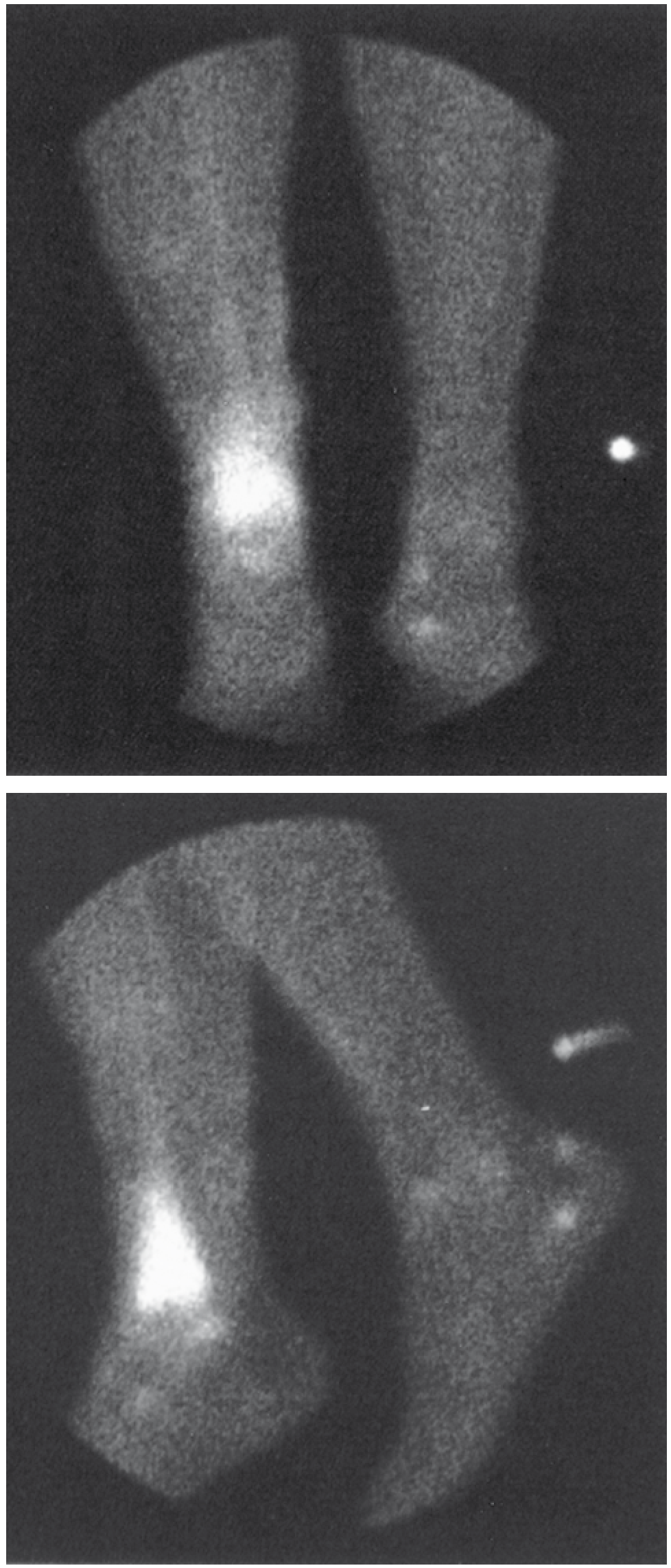
Three-Phase Bone Scan - The Most Important Concept for Exams
This is a single injection study that captures images at three different time points:
| Phase | Timing | What It Shows |
|---|---|---|
| Phase 1 - Blood Flow (Perfusion) | Immediately (few seconds) after injection | Arterial blood flow to the area |
| Phase 2 - Blood Pool (Equilibrium) | ~5 minutes after injection | Soft tissue hyperemia, capillary permeability |
| Phase 3 - Delayed (Bone phase) | 3-4 hours after injection | True bone uptake (osteoblastic activity) |
Three-Phase Scan Interpretation Table (High-yield!)
| Condition | Phase 1 (Flow) | Phase 2 (Blood Pool) | Phase 3 (Delayed) |
|---|---|---|---|
| Osteomyelitis | ↑ | ↑ | ↑ |
| Cellulitis | ↑ | ↑ | Normal (↔) |
| Osteoarthritis | Normal | Normal | ↑ |
- In osteomyelitis: all three phases are positive (hot in all phases)
- In cellulitis: only first two phases are positive (hot soft tissue, normal bone phase)
- This is the KEY way to differentiate osteomyelitis from cellulitis on a bone scan
Orthopaedic Uses of Bone Scan (MUST KNOW LIST)
1. Osteomyelitis
- Can detect osteomyelitis within 48 hours of clinical onset (before X-ray becomes positive)
- X-ray takes 10-21 days to show changes
- Use in patients with metallic implants where MRI is limited
- Sensitivity ~90% for detecting vertebral osteomyelitis
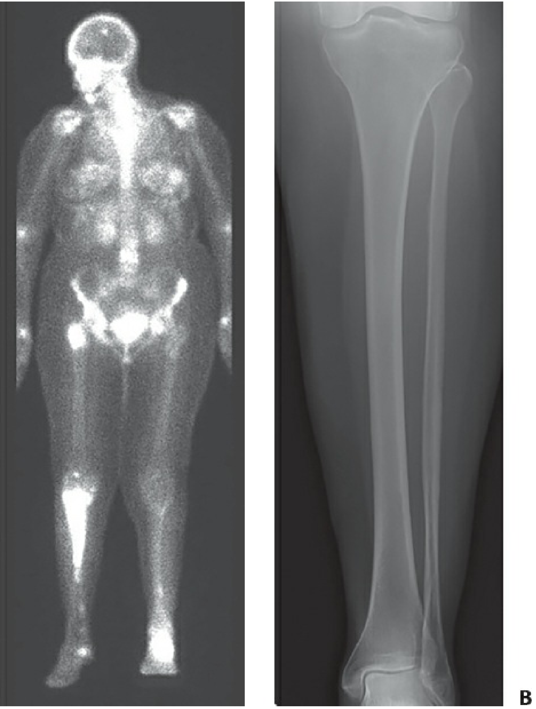
2. Stress Fractures
- Bone scan shows abnormality 1-2 weeks before it appears on plain X-ray
- Nearly 100% sensitive for stress injuries of bone
- Negative bone scan effectively rules out a stress fracture
- MRI is more specific, but bone scan is often used first due to availability
- Used for: pars interarticularis (spondylolysis), metatarsal stress fractures, tibial stress fractures
3. Bone Metastases
- Highly sensitive for osteoblastic (bone-forming) metastases (e.g., prostate, breast)
- Helps determine extent of metastatic disease throughout the entire skeleton at one shot (whole-body scan)
- Important limitation: Purely lytic lesions (renal cell, lung cancer) can be falsely negative because there is no osteoblastic activity to detect
- Multiple myeloma: Bone scan is unreliable - use skeletal survey with skull films instead
4. Avascular Necrosis (AVN)
- Early AVN (hypoperfusion stage): Bone scan shows a "cold" spot (decreased uptake) due to loss of blood supply
- Reparative phase: Shows increased uptake as revascularization occurs
- Bone scan can detect AVN earlier than X-ray
5. THA / TKA Component Loosening
- Used to evaluate painful joint replacements
- Best for femoral components
- Used in conjunction with gallium scan to distinguish aseptic loosening from infection
- Important: ⁹⁹ᵐTc + ¹¹¹In scans for detecting loosening have low specificity (78%) and sensitivity (64%) - not recommended for routine use
6. Reflex Sympathetic Dystrophy (Complex Regional Pain Syndrome)
- Three-phase bone scan is helpful
- Shows diffuse increased uptake in the affected limb in all phases
7. Spondylolysis
- SPECT (Single-Photon Emission CT) bone scan is useful
- Identifies active stress fracture at pars interarticularis even when X-ray is negative
8. Tumours
- Can detect primary bone tumours and assess polyostotic involvement
- Helps in staging and identifying skip lesions
The Three Radiotracer Agents - Summary (Exam High-Yield)
| Agent | Full Name | Key Feature | Use |
|---|---|---|---|
| ⁹⁹ᵐTc phosphate | Technetium-99m methylene diphosphonate | Most common; detects increased blood flow & osteoblastic activity; absorbed on hydroxyapatite | First-line for most indications |
| ⁶⁷Ga citrate | Gallium-67 | Localizes in inflammation and malignant tissue; slower clearance (24-72 hours for imaging); specificity 82% alone | Osteomyelitis (combined with Tc); better for axial skeleton infections |
| ¹¹¹In-labeled leukocytes | Indium-111 WBC scan | Labels patient's own WBCs; positive at earlier stages of osteomyelitis than Tc scan; positive in only ~60% of septic arthritis | Differentiating osteomyelitis from reactive bone formation; chronic infection |
Gallium scan special rules:
- Requires 24 hours delay for appendicular skeleton
- Requires 72 hours delay for axial skeleton
- Combined Tc + Gallium scan: sensitivity 70%, specificity up to 93% for osteomyelitis
- In inflammation: Gallium uptake exceeds Technetium uptake (or shows different distribution) - this distinguishes infection from reactive bone
Indium scan:
- Patient's blood drawn, WBCs separated and labeled with ¹¹¹In in the lab, then reinjected
- Images at 24-48 hours
- Reliable for acute osteomyelitis
- Unreliable for chronic osteomyelitis (which is lymphocyte-dominated, not neutrophil) - may give "cold" scan
- Also unreliable for aseptic vs. septic arthroplasty loosening
Cold Scan vs. Hot Scan
| Meaning | Seen In | |
|---|---|---|
| Hot scan (increased uptake) | More blood flow or osteoblastic activity than normal | Infection, tumour, fracture, degenerative disease |
| Cold scan (decreased uptake) | Reduced blood flow to area | Early AVN, early osteomyelitis (ischemic), pus/necrosis compressing vessels, some metastases |
Exam trap: Early osteomyelitis with vascular compromise (pus under pressure, necrosis, joint effusion, soft tissue swelling) can give a false-negative "cold" scan with ⁹⁹ᵐTc.
Advantages of Bone Scan
- Detects disease before X-ray becomes positive
- Surveys the entire skeleton in one study (whole-body)
- Useful when MRI is contraindicated (metallic implants)
- Relatively low cost vs. MRI
- High sensitivity (~90-100%) for many conditions
Limitations / Disadvantages
- Low specificity - many conditions look the same (infection, tumour, fracture, degenerative change all show increased uptake)
- Poor spatial resolution - cannot pinpoint exactly which bone in a region (e.g., cannot separate hindfoot from midfoot well)
- Radiation exposure
- False negatives with purely lytic lesions (myeloma, RCC)
- False positives in Charcot arthropathy (WBC scans)
- Delayed imaging needed for gallium (24-72 hours)
- Poor for differentiating infection from Charcot joint
SPECT and PET - Modern Additions
- SPECT (Single-Photon Emission CT): Combines scintigraphy with CT - better localization; used for spondylolysis, femoral head osteonecrosis, patellofemoral syndrome
- PET scan: Uses F-18 fluorodeoxyglucose (FDG); taken up by areas of high metabolic activity; best for small lesions especially in lung cancer; also useful for assessing treatment response
- PET/CT has gained favour for staging metastatic bone disease but indications are still evolving
Quick Exam Summary Table
| Condition | Bone Scan Finding | Exam Point |
|---|---|---|
| Osteomyelitis | All 3 phases ↑ | Detects within 48 hrs of onset |
| Cellulitis | Phase 1+2 ↑, Phase 3 normal | How to distinguish from osteomyelitis |
| Stress fracture | Hot spot | Positive before X-ray |
| Early AVN | Cold spot | First imaging sign |
| Late AVN (repair) | Hot spot | Revascularization |
| Bone metastases (prostate/breast) | Multiple hot spots | Whole body survey |
| Multiple myeloma | Often normal | Do skeletal survey instead |
| Lytic mets (RCC, lung) | May be cold/normal | False negative risk |
| CRPS/RSD | Diffuse uptake, all 3 phases | Three-phase scan helpful |
| THA loosening | Increased uptake around implant | Not routine due to poor specificity |
Sources: Campbell's Operative Orthopaedics 15th Ed (2026), p. 970-971; Miller's Review of Orthopaedics 9th Ed, p. 111; Rockwood and Green's Fractures in Adults 10th Ed (2025), p. 878
This is a shared conversation. Sign in to Orris to start your own chat.