
Ligaments of the hip joint
Reading File
Now let me search the medical library for hip joint ligaments content.
Finding Sources
Finding Sources
Good results. Let me read the detailed content from Gray's Anatomy for Students and the Imaging Anatomy atlas simultaneously.
Reading File
Reading File
Let me read more of the Gray's Anatomy content on the ligaments section and also fetch the key diagrams.
Reading File
Reading File
Loading Image
Excellent - I have a great image and detailed content. Let me also fetch the ligament of the head of femur image and the Thieme atlas content on the zona orbicularis and actions.
Reading File
Loading Image
Finding Sources
I now have excellent comprehensive content. Let me also read the Thieme Atlas section on hip joint ligaments for its clear tabular breakdown and the ligamentum teres section from the imaging atlas.
Reading File
Reading File
I now have thorough content from multiple authoritative sources. Let me compile the comprehensive answer.
Ligaments of the Hip Joint
The hip joint (a ball-and-socket synovial joint) is stabilized by a strong fibrous capsule reinforced by four main external ligaments and one intracapsular ligament. The capsule itself attaches proximally to the acetabular rim and distally to the intertrochanteric line anteriorly and femoral neck posteriorly.
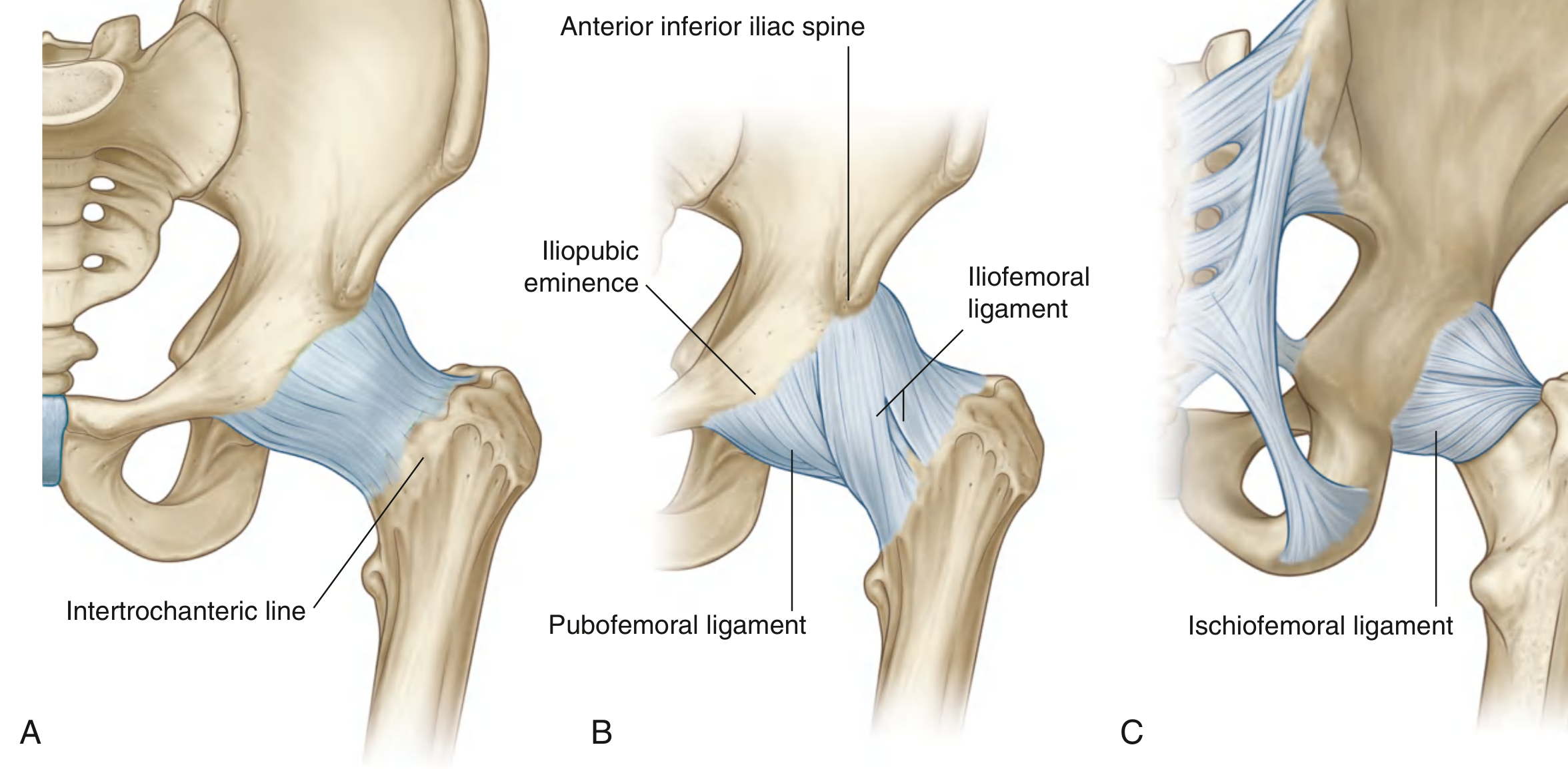
Fig. 6.33 - Fibrous membrane and ligaments of the hip joint. (A) Fibrous membrane. (B) Iliofemoral and pubofemoral ligaments - anterior view. (C) Ischiofemoral ligament - posterior view. (Gray's Anatomy for Students)
1. Iliofemoral Ligament (Ligament of Bigelow)
- Position: Anterior to the hip joint
- Shape: Inverted Y (triangular); the strongest ligament in the body
- Proximal attachment: Apex at the anterior inferior iliac spine (AIIS) and margin of the acetabulum
- Distal attachment: Along the entire intertrochanteric line; thicker bands attach above and below the line, giving it the classic Y shape
- Two bands:
- Superior (lateral-horizontal) band - taut in external rotation, lax in internal rotation; attaches to the upper intertrochanteric line and anterior base of greater trochanter
- Inferior (medial-vertical) band - taut in extension, lax in flexion; attaches to the distal-medial intertrochanteric line
- Function: Limits anterior hip translation during extension and external rotation; most important ligament for standing stability
- Clinical note: Also called the ligament of Bigelow; the strongest single ligament in the human body
2. Pubofemoral Ligament
- Position: Anteroinferior to the hip joint
- Shape: Triangular, relatively weak
- Proximal attachment: Base attached to the iliopubic eminence, obturator crest, superior pubic ramus, and obturator membrane
- Distal attachment: Blends with the fibrous membrane and the deep surface of the iliofemoral ligament; inserts medially near the lesser trochanter
- Function: Reinforces the inferior capsule; resists hyperextension and hyperabduction
- Tension: Taut with internal rotation and lax with external rotation
3. Ischiofemoral Ligament
- Position: Posterior to the hip joint; reinforces the posterior capsule
- Proximal attachment: Ischium, just posteroinferior to the acetabulum (ischial portion of the acetabular rim)
- Distal attachment: Greater trochanter, deep to the iliofemoral ligament
- Two bands:
- Superior band - spirals laterally and upward; inserts at the posterior base of the greater trochanter
- Inferior band - courses laterally and downward; inserts on the posteroinferior aspect of the femoral neck base
- Function: Limits posterior translation with internal rotation; prevents hyperextension
- Tension: Taut and appears longer with internal rotation; lax with external rotation
4. Zona Orbicularis
- Structure: A deeper layer of circular, transversely oriented fibers encircling the femoral neck - not a true separate ligament but a thickening of the deep capsule
- Attachments: No direct bony attachment; merges with and is reinforced by the three main capsular ligaments
- Function: Encircles the femoral neck like a collar; forms the narrowest portion of the articular cavity; resists inferior distraction of the femoral head
- Imaging: Best seen on MR arthrography as a natural tightening of the joint capsule around the mid femoral neck
5. Ligament of the Head of the Femur (Ligamentum Teres / Round Ligament)
- Position: Intracapsular but extrasynovial (synovium forms a sleeve around it)
- Proximal attachment: Fovea capitis on the femoral head
- Distal attachment: Acetabular fossa, transverse acetabular ligament, and margins of the acetabular notch
- Function: Plays a role in hip stabilization; resists subluxation forces; most taut when the hip is flexed, adducted, and externally rotated
- Key clinical point: Carries the artery of ligamentum teres (a branch of the obturator artery), which contributes to blood supply to the femoral head (important in children; less significant in adults)
- Tears: Prevalence at hip arthroscopy is 4-15%; third most common cause of hip pain in athletes; strongly associated with labral tears and cartilage lesions
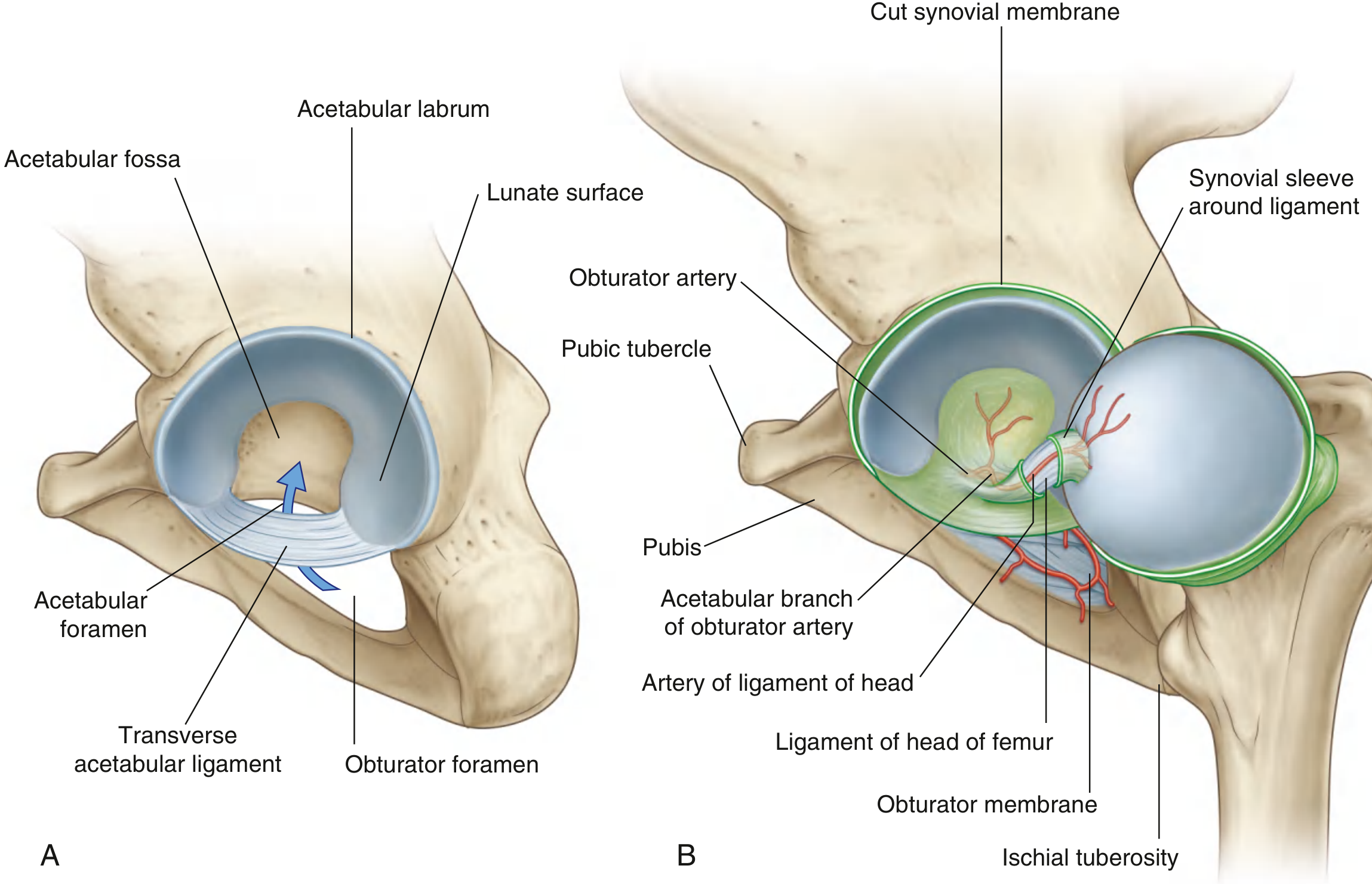
Fig. 6.31 - (A) Transverse acetabular ligament. (B) Ligament of the head of femur with the acetabular branch of the obturator artery. (Gray's Anatomy for Students)
6. Transverse Acetabular Ligament
- A band of fibrocartilage that bridges the acetabular notch inferiorly, converting it into a foramen
- Serves as a partial distal attachment for the ligamentum teres
- Deepens the acetabulum and contributes to joint stability
Functional Summary Table
| Ligament | Position | Prevents | Taut With |
|---|---|---|---|
| Iliofemoral (Bigelow) | Anterior | Extension, external rotation | Extension |
| Pubofemoral | Anteroinferior | Hyperextension, hyperabduction | Internal rotation |
| Ischiofemoral | Posterior | Hyperextension, internal rotation | Internal rotation |
| Zona orbicularis | Circumferential (deep) | Inferior distraction | - |
| Ligamentum teres | Intracapsular | Subluxation | Flexion + adduction + ER |
Key Functional Principle
All three capsular ligaments (iliofemoral, pubofemoral, ischiofemoral) are oriented in a spiral/helical fashion around the joint. In extension, they wind upon themselves, tightening and drawing the femoral head firmly into the acetabulum - stabilizing the standing posture with minimal muscle energy. In flexion, they unwind and become lax, permitting greater mobility. This is why the hip is most stable (and dislocations less common) in extension, and most vulnerable in flexion.
Sources:
- [Gray's Anatomy for Students](Gray's Anatomy for Students, pp. 662-663)
- [Imaging Anatomy: Bones, Joints, Vessels and Nerves, Vol. 3](Imaging Anatomy Vol. 3, pp. 737-740)
- [Thieme Atlas of Anatomy: General Anatomy and Musculoskeletal System](Thieme Atlas, p. 6723)
This is a shared conversation. Sign in to Orris to start your own chat.