
parts of stomach
Reading File
Now let me search the medical library for stomach anatomy content in parallel.
Finding Sources
Finding Sources
Reading File
Reading File
Reading File
Now let me fetch the key anatomy diagram and read the Bailey & Love section on anatomy and physiology:
Loading Image
Reading File
Reading File
I now have excellent, detailed content from multiple authoritative textbooks. Let me compile the full answer.
Parts of the Stomach
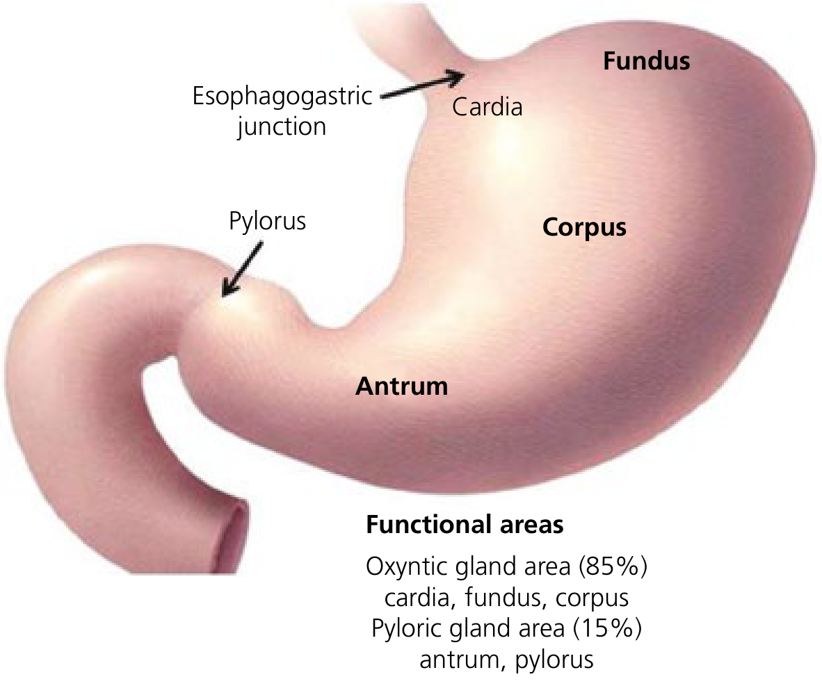
The stomach is a J-shaped muscular organ of the digestive tract, continuous with the esophagus above and the duodenum below. It holds approximately 30 mL at birth and 1.5-2 L in adulthood. Anatomically, it is divided into 4 main regions.
1. Cardia
- The small region immediately surrounding the esophagogastric junction
- Contains cardiac glands lined with columnar epithelium (distinct from the squamous epithelium of the esophagus)
- Cardiac glands contain mucous, endocrine, and undifferentiated cells, but notably lack parietal and chief cells
- Part of the oxyntic gland area (functionally)
2. Fundus
- The dome-shaped, most superior portion of the stomach, projecting upward above the esophagogastric junction
- Lies in contact with the left hemidiaphragm above and the spleen to the left
- Blood supply mainly from the vasa brevia (short gastric arteries) off the splenic artery
- Contains oxyntic glands with parietal cells and chief cells
- Functions as the reservoir for swallowed air (gas bubble visible on imaging)
3. Body (Corpus)
- The largest portion of the stomach, immediately below and continuous with the fundus
- Extends down to the incisura angularis - a fixed sharp indentation two-thirds of the way down the lesser curvature
- Contains the majority of parietal (oxyntic) cells - secrete HCl via the H+/K+-ATPase proton pump
- Contains chief cells in gastric crypts - secrete pepsinogen (converted to active pepsin in acidic conditions)
- Contains enterochromaffin-like (ECL) cells throughout, which produce histamine - a key driver of acid secretion
- D cells producing somatostatin are also present (negative regulatory role)
4. Antrum (Pyloric Antrum)
- Extends from its indistinct border with the body to the pyloric junction
- The border with the body lies roughly at the incisura angularis on the lesser curvature
- Contains predominantly G cells that secrete gastrin (stimulates acid production)
- Contains pyloric glands (mucous secreting, no parietal or chief cells)
- Contains the "antral pump" - smooth muscle that mixes food and controls gastric emptying
5. Pylorus
- A tubular structure joining the stomach to the duodenum, located ~2 cm to the right of midline at L1
- Contains the pyloric sphincter - the palpable circular muscle that controls gastric emptying
- Regulates the release of chyme from the stomach into the duodenum
Curvatures
| Structure | Description |
|---|---|
| Lesser curvature | Right upper border; site of gastroheaptic ligament (lesser omentum) |
| Greater curvature | Left lower border; longer; gives attachment to gastrocolic, gastrosplenic, and gastrophrenic ligaments |
| Incisura angularis | Sharp notch on the lesser curvature marking body-antrum junction |
Functional Zones (Glandular)
| Zone | Regions | % of Stomach | Gland Type |
|---|---|---|---|
| Oxyntic gland area | Cardia, Fundus, Corpus | ~75-85% | Oxyntic glands (parietal + chief cells) |
| Pyloric gland area | Antrum, Pylorus | ~15-25% | Pyloric glands (mucous + G cells) |
Key Cell Types by Region
| Cell | Location | Secretion | Function |
|---|---|---|---|
| Parietal (oxyntic) cells | Body/fundus glands | HCl + Intrinsic Factor | Digestion; B12 absorption |
| Chief (zymogenic) cells | Base of oxyntic glands | Pepsinogen I & II | Protein digestion (as pepsin) |
| G cells | Antral mucosa | Gastrin | Stimulates acid secretion |
| ECL cells | Body | Histamine | Amplifies acid secretion |
| D cells | Throughout | Somatostatin | Inhibits acid secretion |
| Surface mucous cells | Entire stomach surface | Mucus | Mucosal protection |
Blood Supply (Summary)
- Lesser curvature: Left gastric artery (celiac axis) + Right gastric artery (common hepatic)
- Greater curvature: Right gastroepiploic artery (gastroduodenal) + Left gastroepiploic artery (splenic)
- Fundus: Vasa brevia / short gastric arteries (splenic artery)
Sources: Sleisenger & Fordtran's Gastrointestinal and Liver Disease, Bailey & Love's Short Practice of Surgery 28th Ed., Yamada's Textbook of Gastroenterology 7th Ed.
This is a shared conversation. Sign in to Orris to start your own chat.