
Short notes on Structure of liver for 5 marks
liver structure anatomy lobule portal triad
liver lobule histology hepatocytes
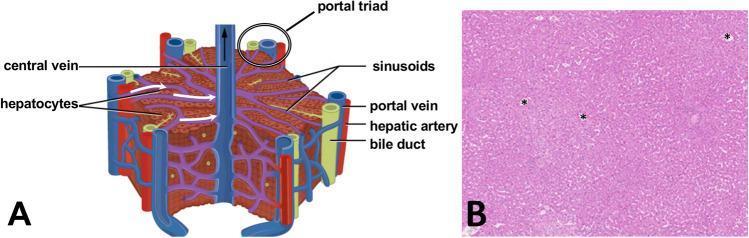
This educational image combines a schematic diagram (A) and a light microscopy photograph (B) illustrating human liver microanatomy. Part A is an anatomical diagram of a typical hepatic lobule, demonstrating its hexagonal structure. Key labeled components include the central vein at the lobule's core and the portal triad at the vertices, which consists of a hepatic artery, portal vein, and bile duct. It shows hepatocytes arranged in cords radiating toward the central vein, interspersed with sinusoids that facilitate fluid transport. Part B is a diagnostic histology image of human liver tissue stained with hematoxylin and eosin (H&E). The parenchyma is visible as a dense arrangement of pink-stained hepatic cords and clear, white-appearing sinusoids. Several central veins are marked with asterisks (*). The image provides a side-by-side comparison between the idealized anatomical model and actual clinical histology, suitable for medical students learning hepatology and tissue architecture.
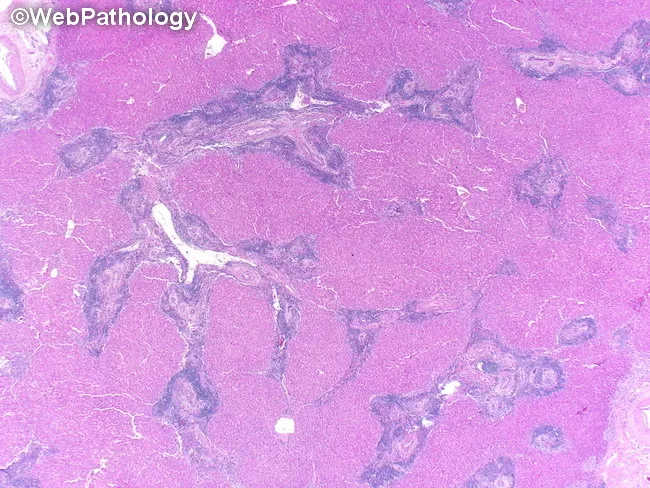
Brightfield light microscopy of a liver biopsy stained with hematoxylin and eosin (H&E). The specimen shows hepatic parenchyma with cords of polygonal hepatocytes separated by sinusoidal capillaries and occasional central veins. The overall lobular architecture appears preserved, with uniform cytoplasmic eosinophilia and round, centrally located nuclei. Portal tracts are not clearly delineated within this low magnification field, but no marked interface activity or interface hepatitis is visible. There is no appreciable steatosis, hepatocellular ballooning, necrosis, or inflammatory infiltrate evident at this magnification. Fibrous septa or cirrhotic nodules are not observed. The tissue appears well preserved with minimal artifact from sectioning. Overall, the histology is compatible with benign hepatic parenchyma in a non-diseased state at this plane of view. This image serves as a reference for normal hepatic architecture and cellular morphology, useful in educational comparisons with diseased liver specimens. Potential clinical applications include baseline histology evaluation, quality control in biopsy processing, and teaching scenarios illustrating hepatocyte cords, sinusoidal arrangement, central veins, and portal areas. Relevant keywords: liver biopsy, hepatic lobule, hepatocytes, sinusoids, central vein, portal tract, H&E, brightfield, normal liver.
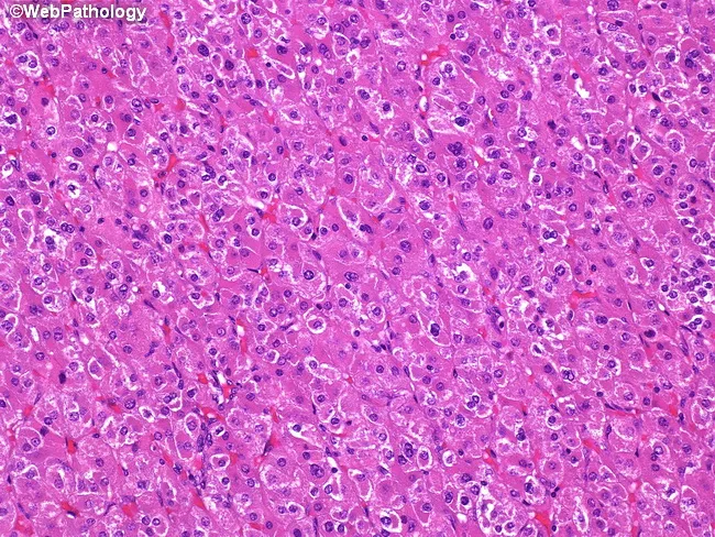
Imaging modality: Light microscopy of a Hematoxylin and Eosin (H&E) stained liver biopsy section examined under brightfield illumination. Primary site: liver parenchyma, hepatic lobule, hepatocytes arranged in cords separated by sinusoids. The cellular morphology shows polygonal hepatocytes with abundant eosinophilic granular cytoplasm and centrally placed round nuclei, occasional binucleation, and well-preserved cytoplasmic borders. Sinusoidal spaces appear patent with mild vascular congestion; there is no conspicuous steatosis, confluent necrosis, or prominent apoptosis in this field. Intercellular boundaries are intact; there is no significant inflammatory infiltrate or fibrotic septa evident in this limited view. Overall architecture is consistent with normal hepatic parenchyma, though a small sampling may miss portal tracts or fibrotic changes. No overt cytologic atypia or neoplastic features are obvious in this image. Diagnostic relevance: this image documents typical hepatocellular morphology suitable for educational reference and for distinguishing normal liver histology from inflammatory, degenerative, or neoplastic processes in hepatology and pathology practice. Potential clinical applications include baseline histology assessment in liver biopsies, evaluation of suspected fatty liver disease, viral hepatitis, or hepatocellular carcinoma if cytologic atypia present. Educational relevance extends to medical students, residents, and fellows; helps practice pattern recognition, differential diagnosis, and correlation with serology, imaging, and clinical course.
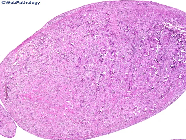
This is a light-field histopathology image of a liver biopsy section stained with Hematoxylin and Eosin (H&E). The image presents a low-magnification overview of hepatic parenchyma, showing cords of polygonal hepatocytes arranged in plates separated by sinusoidal spaces. The lobular architecture is visible, with a dense pink cytoplasmic matrix and basophilic nuclei; there is uniform cellular density without obvious necrosis, conspicuous inflammatory infiltrate, or confluent fibrotic bands at this field of view. The portal tracts are not clearly delineated in this orientation, and bile ducts or arterial branches are not readily identified. Overall liver architecture appears preserved, consistent with non-specific or normal hepatic tissue in this field. This image is suitable as an educational reference for basic liver histology, including identification of hepatocytes, sinusoids, and the hepatic lobule. Potential clinical uses include teaching hepatology and pathology, cross-reference for normal hepatic histology, and comparison against fatty change, steatosis, hepatitis, fibrosis, or cirrhosis in broader sampling. The image may be used to illustrate histological features of normal hepatic parenchyma, guide differential diagnosis when pathology is present, and enhance radiology-pathology correlation exercises in medical education. This description emphasizes reliable terminology for search indexing and clinical education.
Structure of the Liver
General Features
- Guyton and Hall Textbook of Medical Physiology
Structural Components
- Parenchyma - plates of hepatocytes
- Connective tissue stroma
- Sinusoidal capillaries (hepatic sinusoids)
- Perisinusoidal spaces (Space of Disse)
Functional Units of the Liver
| Unit | Shape | Description |
|---|---|---|
| Classic Lobule | Polygonal/hexagonal | Hepatocyte plates radiate from a central vein; portal triads at corners |
| Portal Lobule | Triangular | Emphasizes bile secretion; portal tract at centre |
| Liver Acinus | Diamond-shaped | Best correlates with blood perfusion, metabolic activity, and pathology; divided into Zones 1, 2, 3 |
Classic Lobule - Key Details
- Hepatocytes form irregular anastomosing plates radiating toward a central vein
- At each corner of the hexagon lies the portal triad, which contains:
- Branch of the portal vein
- Branch of the hepatic artery
- Bile duct
- Small lymphatic vessels
- The central vein drains into hepatic veins and then the inferior vena cava
Hepatic Sinusoids
- Irregular vascular channels running between hepatocyte plates
- Receive mixed blood: 75% from portal vein (venous) + 25% from hepatic artery (arterial)
- Total hepatic blood flow ~ 1,350 mL/min (27% of cardiac output)
- Lined by thin, discontinuous (fenestrated) endothelium
Space of Disse (Perisinusoidal Space)
- Narrow space between the sinusoidal endothelium and hepatocytes
- Site of exchange of materials between blood and hepatocytes
- Large pores in endothelium allow free diffusion of plasma proteins into this space
- Connects with lymphatic vessels in interlobular septa - excess fluid drains as lymph
Special Cell Types in Sinusoids
| Cell | Location | Function |
|---|---|---|
| Kupffer cells (sinusoidal macrophages) | Line sinusoidal wall | Phagocytose bacteria, worn-out RBCs; recycle iron |
| Hepatic stellate cells (Ito cells) | Perisinusoidal space | Store Vitamin A in lipid droplets; become myofibroblasts in pathology (fibrosis) |
Hepatocytes
- Constitute 80% of liver cells
- Large, polygonal cells with spherical nuclei (often binucleated)
- Acidophilic cytoplasm with abundant sER, rER, mitochondria, peroxisomes, and Golgi complexes
- Basal surface: faces Space of Disse (exchange)
- Apical surface: forms bile canaliculi with adjacent hepatocyte
Bile Drainage Pathway
- Canals of Hering harbor hepatic stem cells
- Biliary tree is lined by cholangiocytes (cuboidal/columnar epithelium)
Diagram: Liver Lobule and Histology
Short notes on Tongue for 5 marks
tongue anatomy papillae taste buds diagram
Structure of the Tongue
Definition & General Features
Surfaces and Parts
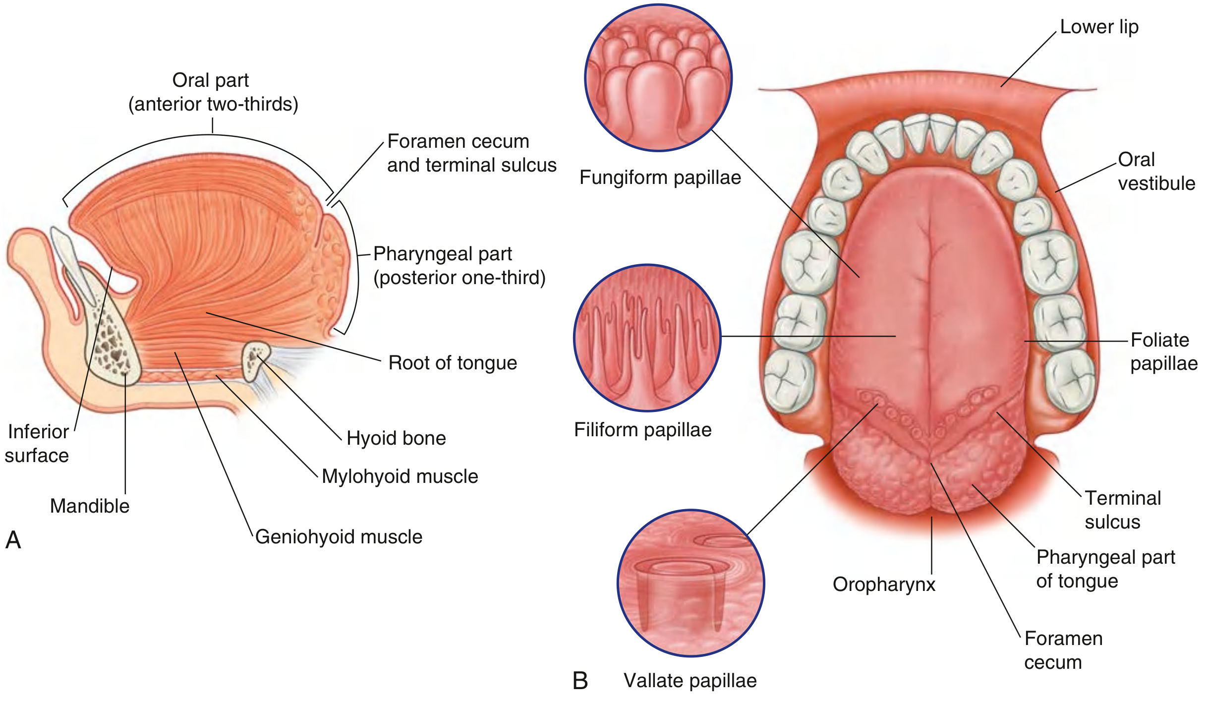
| Feature | Description |
|---|---|
| Dorsal (superior) surface | Anterior 2/3 bears papillae; posterior 1/3 has lingual tonsils |
| Inferior surface | No papillae; has the frenulum (median fold) and lingual veins |
| Oral part (anterior 2/3) | Lies in the oral cavity; horizontal plane |
| Pharyngeal part (posterior 1/3) | Forms anterior wall of oropharynx; vertical plane |
- The two parts are separated by a V-shaped groove - the sulcus terminalis
- At the apex of the V is the foramen cecum - embryological remnant marking the origin of the thyroid gland (thyroglossal duct invagination)
- The root of the tongue is attached to the mandible and hyoid bone
Muscles of the Tongue
Intrinsic Muscles (confined to the tongue, no external attachment)
| Muscle | Function |
|---|---|
| Superior longitudinal | Shortens tongue; curls apex upward |
| Inferior longitudinal | Shortens tongue; turns apex downward |
| Transverse | Narrows and elongates tongue |
| Vertical | Flattens and widens tongue |
Extrinsic Muscles (one attachment outside the tongue)
| Muscle | Origin | Action |
|---|---|---|
| Genioglossus | Mental spine of mandible | Protrudes tongue (main protruder) |
| Hyoglossus | Hyoid bone | Depresses tongue |
| Styloglossus | Styloid process | Retracts and elevates tongue |
| Palatoglossus | Palatine aponeurosis | Elevates posterior tongue |
All muscles innervated by CN XII (Hypoglossal nerve) except palatoglossus (innervated by CN X - Vagus via pharyngeal plexus).
Papillae of the Tongue (Dorsal surface - Anterior 2/3)
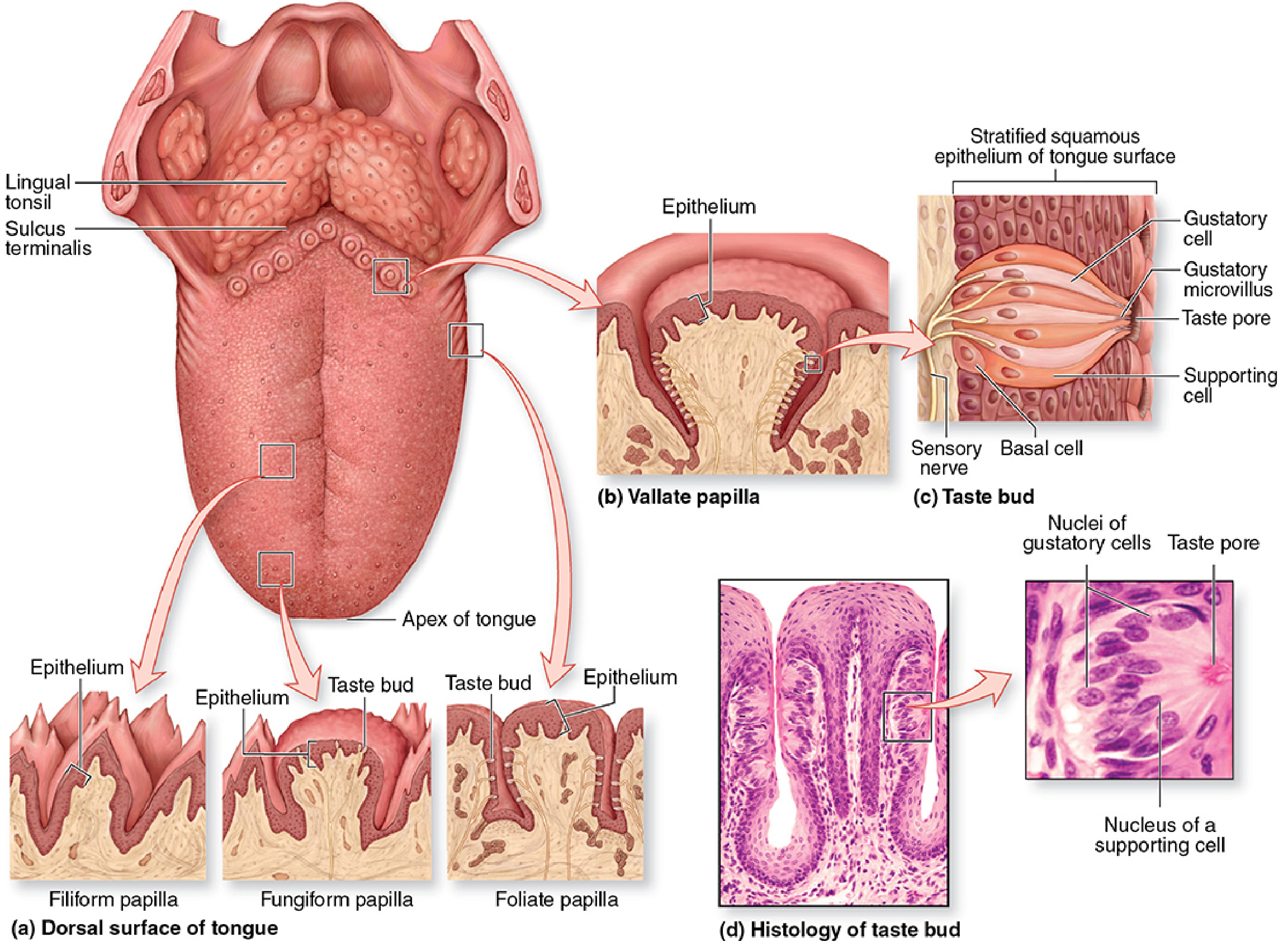
| Type | Shape | Location | Taste Buds | Key Feature |
|---|---|---|---|---|
| Filiform | Cone/thread-shaped, pointed | All over anterior dorsum | None | Most numerous; heavily keratinized; provide friction for food movement |
| Fungiform | Mushroom/bulbous-shaped | Scattered across dorsal surface, concentrated at margins | Present | Well-vascularized lamina propria core; lightly keratinized |
| Foliate | Leaf-like folds/ridges | Lateral sides of tongue | Present | Rudimentary in adults; best developed in children |
| Vallate (Circumvallate) | Large, blunt cylinders with surrounding trench | V-shaped row just anterior to sulcus terminalis | Most abundant (in trench walls) | Only 8-12 in number; largest papillae; serous (von Ebner's) glands open into trench |
All papillae except filiform bear taste buds and are called gustatory papillae.
Taste Buds
- Oval structures embedded in the epithelium of papillae
- Contain three cell types:
- Gustatory (taste) cells - bear microvilli projecting through the taste pore
- Supporting cells - unclear function
- Basal stem cells - replace worn gustatory cells
- Taste is detected when dissolved chemicals contact microvilli at the taste pore
Posterior Third (Pharyngeal Surface)
- No papillae
- Contains lingual tonsil - nodules of lymphoid tissue in the submucosa giving it an irregular, bumpy appearance
Nerve Supply Summary
| Function | Area | Nerve |
|---|---|---|
| General sensation (touch, pain, temp) | Anterior 2/3 | Lingual nerve (CN V3 - Mandibular) |
| Taste | Anterior 2/3 | Chorda tympani (CN VII - Facial) |
| General sensation + Taste | Posterior 1/3 | Glossopharyngeal nerve (CN IX) |
| General sensation | Root/epiglottis | Vagus nerve (CN X) |
| All muscles | Both parts | Hypoglossal nerve (CN XII) |
Essay on pitiatry hormone for 10 marks question
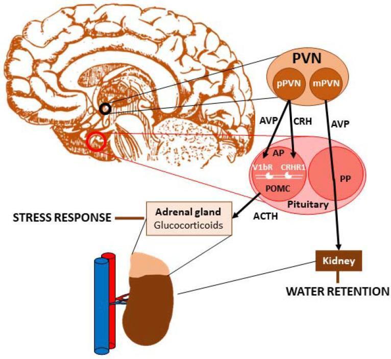
Pituitary Hormones - Essay (10 Marks)
Introduction
- Anterior lobe (Adenohypophysis) - ~80% of the gland
- Posterior lobe (Neurohypophysis) - ~20% of the gland
PART I: ANTERIOR PITUITARY (ADENOHYPOPHYSIS)
Cell Types of the Anterior Pituitary
| Cell Type | Hormone Produced | Staining |
|---|---|---|
| Somatotrophs | Growth Hormone (GH) | Acidophil |
| Mammosomatotrophs | GH + Prolactin | Acidophil |
| Lactotrophs | Prolactin (PRL) | Acidophil |
| Corticotrophs | ACTH | Basophil |
| Thyrotrophs | TSH | Basophil |
| Gonadotrophs | FSH + LH | Basophil |
Hormones of the Anterior Pituitary
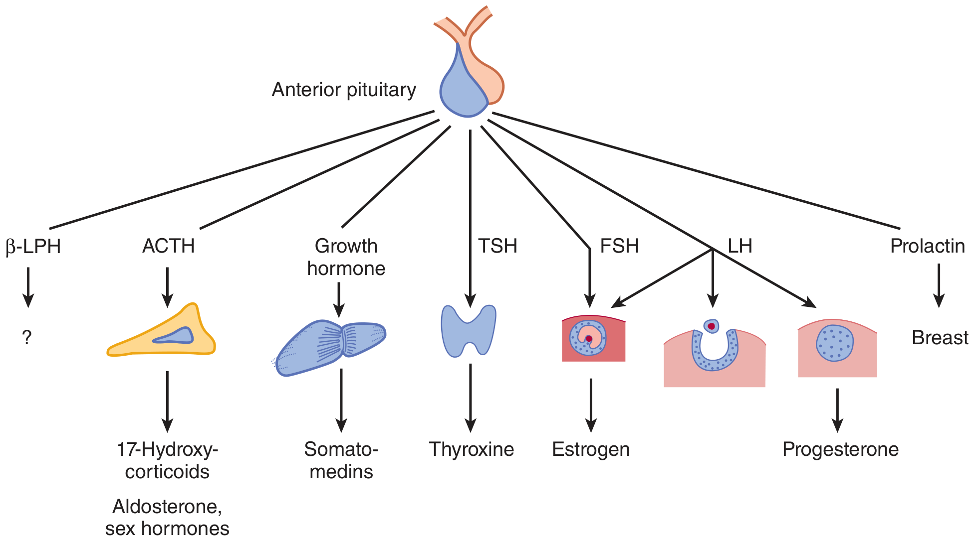
1. Growth Hormone (GH) / Somatotropin
- Chemical nature: Protein (191 amino acids)
- Actions:
- Stimulates linear bone growth via somatomedins (IGF-1, produced by liver)
- Promotes protein synthesis (anabolic effect)
- Anti-insulin / diabetogenic - raises blood glucose
- Promotes lipolysis (fat mobilization)
- Stimulates uptake of amino acids into cells
- Regulation:
- Stimulated by: GHRH (Growth Hormone-Releasing Hormone)
- Inhibited by: Somatostatin (GIH)
- Excess: Gigantism (before epiphyseal fusion), Acromegaly (after fusion)
- Deficiency: Dwarfism (pituitary dwarfism)
2. Thyroid-Stimulating Hormone (TSH) / Thyrotropin
- Chemical nature: Glycoprotein (alpha + beta subunits)
- Actions:
- Stimulates synthesis and release of T3 and T4 from thyroid gland
- Promotes thyroid gland growth (hypertrophy and hyperplasia)
- Regulation:
- Stimulated by: TRH (Thyrotropin-Releasing Hormone)
- Inhibited by: T3/T4 (negative feedback), somatostatin
3. Adrenocorticotropic Hormone (ACTH) / Corticotropin
- Chemical nature: Polypeptide (39 amino acids); derived from pro-opiomelanocortin (POMC)
- Actions:
- Stimulates synthesis and secretion of glucocorticoids (cortisol) and sex steroids from adrenal cortex
- Mild stimulation of mineralocorticoid (aldosterone) secretion
- Has melanocyte-stimulating activity (shares sequence with MSH)
- Regulation:
- Stimulated by: CRH (Corticotropin-Releasing Hormone), stress, low cortisol
- Inhibited by: cortisol (negative feedback)
- Excess: Cushing's disease
4. Follicle-Stimulating Hormone (FSH)
- Chemical nature: Glycoprotein
- Actions (Females): Stimulates growth of ovarian follicles and estrogen secretion
- Actions (Males): Stimulates spermatogenesis and Sertoli cells
- Regulation: Stimulated by GnRH; inhibited by inhibin (from gonads)
5. Luteinizing Hormone (LH)
- Chemical nature: Glycoprotein
- Actions (Females): Triggers ovulation, formation of corpus luteum, secretion of progesterone
- Actions (Males): Stimulates Leydig cells of testes → testosterone production (hence called ICSH - Interstitial Cell-Stimulating Hormone in males)
- Regulation: Stimulated by GnRH; inhibited by sex steroids
6. Prolactin (PRL)
- Chemical nature: Protein (198 amino acids)
- Actions:
- Stimulates breast development and lactation (milk production)
- Inhibits GnRH secretion → suppresses ovulation during breastfeeding
- Regulation: Unique - predominantly under inhibitory control
- Inhibited by: Dopamine (PIH - Prolactin Inhibiting Hormone) from hypothalamus
- Stimulated by: TRH, VIP, suckling reflex
- Excess (Hyperprolactinemia): Galactorrhea, amenorrhea, infertility
PART II: POSTERIOR PITUITARY (NEUROHYPOPHYSIS)
| Hormone | Synthesized in | Transported by |
|---|---|---|
| ADH (Vasopressin) | Primarily supraoptic nucleus | Neurophysin II |
| Oxytocin | Primarily paraventricular nucleus | Neurophysin I |
7. Antidiuretic Hormone (ADH) / Vasopressin
- Chemical nature: Nonapeptide (9 amino acids): Cys-Tyr-Phe-Gln-Asn-Cys-Pro-Arg-GlyNH₂
- Actions:
- Increases water reabsorption in collecting ducts of kidneys (via V2 receptors - inserts aquaporin-2 channels)
- At high doses: vasoconstriction (via V1 receptors) → raises blood pressure
- Antidiuresis - concentrates urine
- Stimuli for release:
- Increased plasma osmolality (most potent)
- Decreased blood volume/pressure
- Stress, nausea, nicotine
- Deficiency: Diabetes Insipidus (massive dilute urine output)
- Excess: SIADH (Syndrome of Inappropriate ADH secretion) → hyponatremia
8. Oxytocin
- Chemical nature: Nonapeptide (9 amino acids): Cys-Tyr-Ile-Gln-Asn-Cys-Pro-Leu-GlyNH₂ (differs from ADH in positions 3 and 8)
- Actions:
- Stimulates uterine contractions during labor (used clinically to induce labor)
- Stimulates milk ejection (let-down reflex) from breast - acts on myoepithelial cells
- Plays a role in social bonding and maternal behavior
- Stimuli for release:
- Stretching of the cervix during labor (Ferguson reflex)
- Suckling of the nipple
- Clinical use: Synthetic oxytocin (Pitocin) used to induce/augment labor and reduce postpartum hemorrhage
Hypothalamic Control Summary
| Hypothalamic Hormone | Effect on Anterior Pituitary |
|---|---|
| CRH | ↑ ACTH |
| TRH | ↑ TSH, ↑ Prolactin |
| GHRH | ↑ GH |
| Somatostatin (GIH) | ↓ GH, ↓ TSH |
| GnRH | ↑ FSH, ↑ LH |
| Dopamine (PIH) | ↓ Prolactin |
Summary Table: All Pituitary Hormones
| Lobe | Hormone | Chemical Nature | Target | Main Action |
|---|---|---|---|---|
| Anterior | GH | Protein | Liver, bone | Growth, anabolism |
| Anterior | TSH | Glycoprotein | Thyroid | T3/T4 secretion |
| Anterior | ACTH | Polypeptide | Adrenal cortex | Cortisol secretion |
| Anterior | FSH | Glycoprotein | Gonads | Follicle growth/spermatogenesis |
| Anterior | LH | Glycoprotein | Gonads | Ovulation/testosterone |
| Anterior | Prolactin | Protein | Breast | Lactation |
| Posterior | ADH | Nonapeptide | Kidneys | Water retention |
| Posterior | Oxytocin | Nonapeptide | Uterus/breast | Labor/milk ejection |