
Is slide ki har line ko samjao aur har difficult word ki definition aur roman Urdu ma bh samjao aur exam tip bh do aur related cheezen bh batao aur end ma easy trick bh batana yaad rakhny ki
cd /tmp/workspace/attachments/dea44dff-2171-46e9-960a-8f4d745049bc/ && python3 -c " from pptx import Presentation prs = Presentation('Spinal cord Fiinal.pptx') for i, slide in enumerate(prs.slides, 1): print(f'\n========== SLIDE {i} ==========') for shape in slide.shapes: if shape.has_text_frame: txt = shape.text_frame.text.strip() if txt: print(txt) "
spinal cord anatomy cross section gray white matter
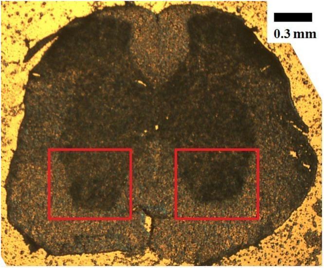
This microphotograph (5X objective) displays a transverse cross-section of a mouse spinal cord at the lumbar level (L1-L2), an important model for studying neurodegenerative conditions like ALS. The section demonstrates the classic internal architecture of the spinal cord, featuring a central butterfly-shaped region of darker gray matter surrounded by peripheral lighter-colored white matter. The image is oriented with the dorsal half at the top and the ventral half at the bottom. Two red boxes highlight the bilateral ventral horns within the gray matter, which are the specific anatomical locations where motor neuron cell bodies reside. The visual distinction between the lipid-rich white matter and the protein-rich gray matter is evident through the variation in tissue density and staining. A scale bar indicating 0.3 mm is provided in the top right corner for morphometric reference. This image serves as a foundational anatomical reference for histological analysis and spectroscopic probing of spinal cord pathology.
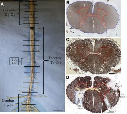
This composite educational image illustrates spinal cord gross anatomy and histopathological analysis of demyelination in Multiple Sclerosis (MS). Panel A shows a gross spinal cord dissection with labeling of cervical (C1-C8), thoracic (T1-T12), and lumbar (L1-L5) segments, indicating axial sectioning levels. Panels B and C present healthy control sections at the cervical and upper thoracic levels respectively. Panel B (SMI-31 stain) outlines the gray matter (GM) in red and cross-sectional area (CSA) in blue to calculate white matter (WM) volume. Panel C (MBP stain) demonstrates normal myelin distribution. Panel D displays an MS upper thoracic section stained for Myelin Basic Protein (MBP), highlighting localized pathology. Five demyelinated plaques are manually delineated: one large gray matter lesion (GM Lesion 1) and four distinct white matter lesions (WM Lesions 1-4). These microscopic sections (4x magnification) serve to teach quantitative assessment of demyelination and atrophy in neuroinflammatory disease. Key concepts include neuroanatomical mapping, immunohistochemical staining for axonal integrity (SMI-31) and myelin (MBP), and the identification of focal MS plaques.
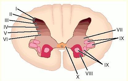
This medical anatomical diagram illustrates a cross-section of the human spinal cord at the cervical level (C7), specifically detailing the Rexed laminae within the gray matter. The image depicts the characteristic butterfly-shaped internal gray matter surrounded by peripheral white matter. The gray matter is partitioned into ten distinct functional layers labeled with Roman numerals I through X. Laminae I to VI are situated in the dorsal (posterior) horn, associated with sensory processing, and are shown with color gradients from dark to light. Laminae VII through IX are located in the ventral (anterior) horn and intermediate zone, representing motor and autonomic functions; lamina IX specifically identifies clusters of motor neurons. Lamina X is located centrally, surrounding the central canal. This diagram serves as a key educational resource for neuroanatomy and pain physiology, demonstrating the organized cytoarchitectonic distribution of neurons within the spinal cord.
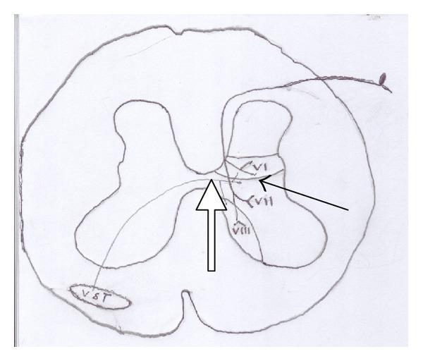
Anatomical diagram of a spinal cord cross-section illustrating the pathway of the ventral spinothalamic tract (VST). The illustration depicts the central H-shaped gray matter and surrounding white matter. Key structures labeled include Rexed laminae VI, VII, and VIII in the dorsal and intermediate horns of the gray matter. A black arrow indicates the origin of secondary sensory neurons within these deep laminae. A white arrow tracks the decussation (crossing over) of these nerve fibers through the anterior white commissure to the contralateral side. The fibers terminate in the ventral white column, labeled as the 'VST' tract. This diagram serves as a neuroanatomical representation of the ascending somatosensory pathway for crude touch and pressure, highlighting its origin in non-opioid-rich Rexed layers compared to the lateral spinothalamic tract.
spinal cord meninges dura mater arachnoid pia mater layers
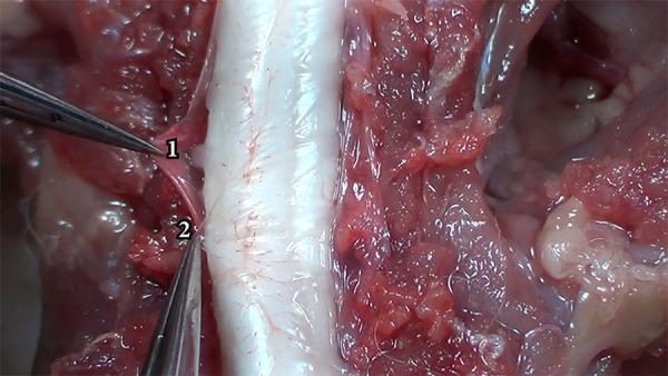
This clinical photograph captures a microsurgical dissection of the spinal meninges, demonstrating the anatomical differentiation between the dura mater and the arachnoid mater. In the center of the frame, the spinal cord appears as a prominent, whitish, cylindrical structure with visible fine vascularization on its surface. Surgical forceps are used to retract and distinguish two distinct layers: the dura mater (labeled '1') and the arachnoid mater (labeled '2'). The dura mater (1) is visualized as a thicker, more opaque, and slightly reddish fibrous membrane, reflecting its dense collagenous composition and relative resistance to manipulation. In contrast, the arachnoid mater (2) is depicted as a thinner, more translucent, and whiter membrane underlying the dura. The surrounding surgical field contains exposed paraspinal muscles and subcutaneous tissues, appearing reddish and fleshy with a glistening texture due to the presence of biological fluids. This image serves as an educational reference for neurosurgical anatomy, specifically the layered structure of the spinal meninges and the plane of dissection required to access the subarachnoid space.
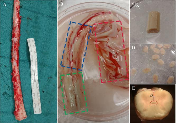
This composite of clinical photographs illustrates the surgical extraction and micro-dissection of a human spinal cord specimen for neural stem/progenitor cell (NSPC) research. Panel A shows the full length of an extracted adult human spinal cord alongside a medical ruler for scale. Panel B displays the spinal cord submerged in dissection buffer, with colored dashed boxes identifying the three meningeal layers: the thick, opaque dura mater (red), the translucent arachnoid mater (blue), and the thin pia mater (green) closely adhering to the cord surface. Panel C shows a spinal cord segment after complete removal of the meninges, while Panel D depicts multiple transverse sections (1–2 mm thick) prepared for dissection. Panel E provides a high-magnification cross-sectional view of a single spinal cord segment, featuring an anatomical overlay: a red dashed line indicates the dissection path from the ventral sulcus toward the central canal, which is highlighted by a small red ellipse. This series demonstrates a laboratory protocol for isolating specific neuroanatomical regions from post-mortem human tissue.
cauda equina conus medullaris spinal cord termination
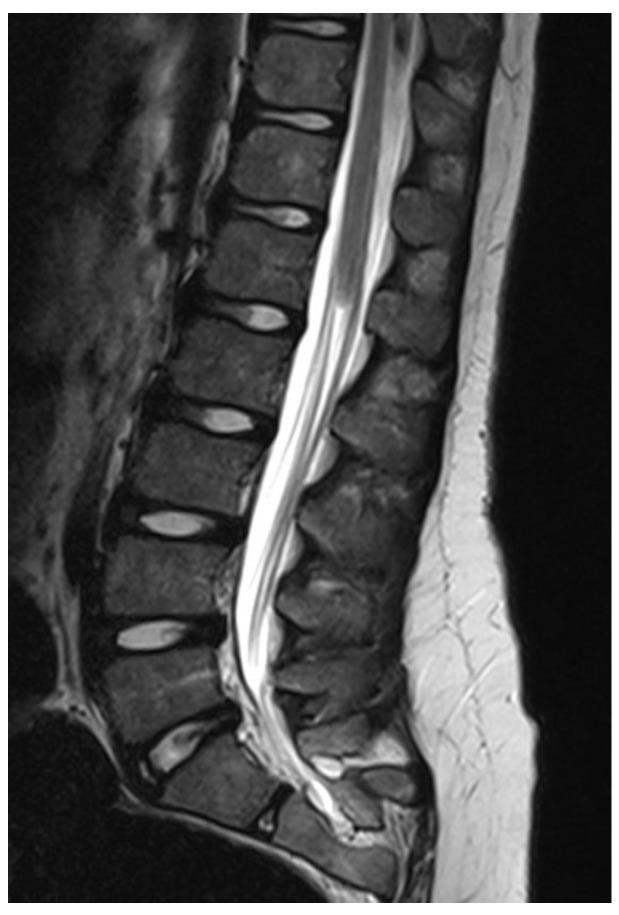
This diagnostic image is a T2-weighted sagittal MRI of the lumbosacral spine demonstrating a classic presentation of caudal regression syndrome. The imaging shows a blunt, high-riding termination of the spinal cord (conus medullaris) above the expected physiological level. There is a notable absence of the distal nerve roots of the cauda equina and the filum terminale within the lower thecal sac, which appears filled only with hyperintense cerebrospinal fluid (CSF). The lumbar vertebral bodies maintain relatively normal rectangular morphology and alignment; however, there is evidence of sacral dysgenesis or partial agenesis inferiorly. This finding is characteristic of a failure in the process of secondary neurulation, leading to complete agenesis of the caudal neural elements. The visual evidence supports a diagnosis associated with occult spinal dysraphism and significant clinical implications for lower limb and sphincter function.
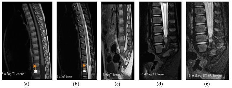
This diagnostic image set consists of sagittal MRI sequences (T1, T2, T1 Fat-Suppressed, and STIR) of the spine, illustrating features of Caudal Regression Syndrome (CRS) Type 1. The conus medullaris is high-set, appearing truncated and nodular, terminating prematurely at the level of the intrapedicular T11 vertebra. The distal spinal canal demonstrates a thickened cauda equina and a sagittal cleft in the thecal sac. The lower spine shows significant developmental anomalies, including a rudimentary, dysgenetic sacrum with a median sagittal cleft and absence of the coccyx. Additionally, rudimentary pelvic apophyses are visible. The STIR sequence (e) highlights increased signal intensity in the region of the truncated conus, while the T1-weighted images (a, c) best define the morphology of the osseous defects and the blunt termination of the cord. This visual documentation serves as a classic educational example of terminal spinal cord dysgenesis and sacral hypoplasia, essential for distinguishing between types of caudal regression in pediatric neuroradiology.
🧠 Spinal Cord - Slide by Slide Complete Guide
Roman Urdu Mein Samjhaya Gaya | Exam Tips + Yaad Karne Ki Tricks
📌 SLIDE 1 - Title Slide
📌 SLIDE 2 - Index
- Introduction
- Definitions
- Meninges
- Blood Supply
- Nerves
- Anatomy
📌 SLIDE 3 - Introduction
- Hamara nervous system (aasabi nizam) do hisson mein taqseem hai: CNS aur PNS
- CNS (Central Nervous System) = Markazi Aasabi Nizam - Brain aur Spinal Cord
- PNS (Peripheral Nervous System) = Irdgird ka Aasabi Nizam - baki saare nerve
- CNS mein sirf brain (dimaag) aur spinal cord (riidh ki haddi ka marghz) hota hai
- Spinal nerves = Riidh se niklne wali naasain
- Cranial nerves = Khopri se niklne wali 12 jori naasain
- Visceral nerves = Andar ki anghon (dil, meta wagera) ki naasain
- Plexuses = Nasoun ka jaal (network) - jaise brachial plexus, lumbar plexus
- Enteric system = Aant (intestine) ka apna alag aasabi nizam
- Spinal cord brain aur baqi jisam ke darmiyan rabita (connection) karta hai - jaise telephone exchange!
🎯 Exam Tip: CNS = Brain + Spinal Cord. PNS = baaki sab. Yeh basic distinction exam mein zaroor aata hai!
📌 SLIDE 4 - Anatomy of Spinal Cord
- Spinal cord ek lamba cylindrical (naali jaisa) hissa hai jo sir ke neeche hota hai
- Elongated = Lamba - Roman Urdu mein "Khechha hua"
- Cylindrical = Naali/Dabbe jaisi shakal
- Yeh Vertebral Canal mein band hai
- Vertebral Canal = Riidh ki haddiyon ke andar bani ek naali/tunnel
- Vertebral Column = Riidh ki haddiyaan (spine)
- Lambaai: 45 centimeter
- Wazan: 30 gram
🎯 Exam Tip: 45 cm aur 30 gm - yeh numbers MCQ mein aksar aate hain!
- SC (Spinal Cord) upar se Foramen Magnum ke qareeb shuru hoti hai
- Foramen Magnum = Khopri ke neeche ka bara suraakh (foramen = suraakh, magnum = bara)
- C1 = Gardan ki pehli haddi (Atlas)
- Medulla Oblongata = Brain ka sabse nichla hissa; yahan se spinal cord shuru hoti hai
- Adults mein: L1 haddi ke neeche khatam hoti hai
- Neonates mein: L3 ke upar khatam hoti hai
- Terminate = Khatam hona
- Neonates = Nawazeeda bachche (0-28 din ke bachche)
- L1, L3 = Kamar ki haddiyaan (Lumbar vertebrae)
🎯 Exam Tip: Adult = L1, Neonate = L3 - yeh confuse karne wala sawaal exam mein aata hai!
- Neeche jaake patlaa ho jaata hai aur Conus Medullaris banta hai
- Tapers = Patlaa hona (cone ki shakal mein)
- Conus Medullaris = Spinal cord ka sabse nichla naoka jaisa hissa (cone = naoka)
- Do jagah pe chaura (mota) hota hai
- Enlargements = Phelaawa / Motaapan
- Cervical = Gardan ka hissa
- Lumbar = Kamar ka hissa
💡 Related Cheez: Yeh enlargements isliye hote hain kyunki haath aur paon ki bari naasain yahan se nikalti hain!
📌 SLIDE 6 - External Anatomy
- Fissures = Gahrhi dararain (deep grooves)
- Sulci = Chhoti dararain (shallow grooves) - ek ko sulcus kehte hain
- Cauda Equina = Ghori ki poonch (Latin mein - ek group of nerve roots)
📌 SLIDE 7 - Fissures and Sulci
- Anterior Median Fissure = Saamne ke darmiyaan ki gahrhi daraar
- 2 Anterolateral Sulci = Saamne ke donon taraf ki dararain (2)
- Posteromedian Sulci = Peeche ke darmiyaan ki daraar
- 2 Posterolateral Sulci = Peeche ke donon tarafon ki dararain (2)
- 2 Postero-intermediate Sulci = Peeche ke darmiyan wali 2 chhoti dararain
- Anterior = Saamne wala
- Posterior = Peeche wala
- Median = Darmiyaan mein
- Lateral = Kinaray par
- Intermediate = Beech wala
🎯 Exam Tip: Anterior mein 1 bari fissure hai, Posterior mein chhoti sulci hain. Fissure > Sulcus (size mein).
📌 SLIDE 8 - Enlargements
- Gardan wali bari jagah: SC segment C5-T1 mein
- Haddi ka level: C4 se T2 tak
- C = Cervical (gardan), T = Thoracic (seena)
- Kamar wali bari jagah: SC segment L1 se S3 tak
- Haddi ka level: T10 se T12 tak
- L = Lumbar (kamar), S = Sacral (koolhe/pelvis)
🎯 Exam Tip: Cervical enlargement = Haath ki naasain (brachial plexus). Lumbar enlargement = Paon ki naasain (lumbosacral plexus). Yeh clinical MCQs mein aata hai.
📌 SLIDE 9 - Spinal Nerves
- 31 jori naasain hoti hain kul mila ke
| Section | Number |
|---|---|
| Cervical (C) | 8 |
| Thoracic (T) | 12 |
| Lumbar (L) | 5 |
| Sacral (S) | 5 |
| Coccygeal | 1 |
| Total | 31 |
- Har spinal nerve ke do roots (jaray) hote hain
- Dorsal Root = Peeche wala jaraa (root)
- Ventral Root = Saamne wala jaraa
- Afferent = Baahir se andar ki taraf aane wala (A = Arrives)
- Sensory = Ehsaas ka - jaise dard, garmi, thandi
- Peripheral receptors = Jism ke kinaray par mahsoos karne wale chhote organs (skin, muscles, joints)
- Dorsal root = Feeling/sensation andar laata hai
- Efferent = Andar se baahir ki taraf jaane wala (E = Exits)
- Motor fibers = Harkat karne wali naasain (muscles ko chalane ke liye)
- Skeletal muscles = Haddiyon se juri hue muscles (haath paon waali)
- Preganglionic = Ganglion se pehle ki naas
- Autonomic = Apne aap kaam karne wali naasain (dil ki dhadkan, saans wagera)
- Ventral root = Command baahir le jaata hai
- Dono root milke ek mixed spinal nerve banate hain
- Mixed = Sensory bhi motor bhi - dono
- Intervertebral Foramen (IVF) = Do haddiyoon ke darmiyan ka suraakh
- IVF se thoda pehle dorsal root par ek ganglion banta hai
- Ganglion = Nasoun ke cell bodies ka guccha (jab baahar ho spinal cord se)
🎯 Exam Tip: "SAME - DAVE" yaad karo:
- Sensory = Afferent = Dorsal = Efferent wala nahi
- Motor = Efferent = Ventral = Efferent
📌 SLIDE 11 - Cauda Equina
-
Spinal cord L1 par khatam hoti hai (Conus Medullaris)
-
Lekin L2 se neeche waali haddiyoun ke foramen tak naasain pahuchwi chahiye
-
Toh yeh naasain SC se nikal ke neechay tak late hain jaise ghori ki poonch ki tarha
-
Yeh guccha = Cauda Equina
-
Cauda = Poonch (Latin)
-
Equina = Ghori ki (Latin)
-
Filum Terminale = Pia mater ka ek patlaa dhaga jo cord ko anchor karta hai
-
Conus Medullaris = Spinal cord ka naoka aakhri hissa
🎯 Exam Tip: LP (Lumbar Puncture) L3-L4 ya L4-L5 par ki jaati hai KYUNKI wahan sirf Cauda Equina hai, spinal cord nahi - isliye cord ko nuqsaan nahi hoga!
💡 Clinical Importance: Cauda Equina Syndrome ek emergency hai - iss mein paoon mein kamzori + peeshab/pakhana ka control nahi rehta!
📌 SLIDE 12 - Meninges
- Pura CNS teen parton se dhaka hota hai
- Meninges = Woh parein jo brain aur spinal cord ko dhakti hain (Singular = Meninx)
- Baahir se andar ki taraf:
- Dura Mater = Sakht motii baahrli parat (Dura = Sakht)
- Arachnoid = Darmiyan wali parat (Arachnoid = makri ke jaal jaisi)
- Pia Mater = Andaruni narm parat (Pia = Narm)
- Yeh teen parten Foramen Magnum se le kar puri vertebral column tak jaati hain
- Teen parten alag alag jagah khatam hoti hain (yeh Slide 15-17 mein detail mein hai)
🎯 Exam Tip: DAP = Dura, Arachnoid, Pia - baahir se andar. Yeh order yaad rakhna zaroori hai!
📌 SLIDE 13 - Spaces in Spinal Meninges
- Pia mater = Sabse andar wali parat jo spinal cord se chipki hoti hai
- Innermost = Sabse andar wali
- Arachnoid aur Pia ke darmiyan Subarachnoid Space hai
- Subarachnoid Space = Makri ke jaal ke neeche ki jagah
- CSF (Cerebrospinal Fluid) = Dimaaghh aur riidh ka paani jo cushion karta hai
- Dura aur Arachnoid ke darmiyan Subdural Space hai
- Subdural = Dura ke neeche
- Dura ke baahir aur haddi ke andar = Epidural Space
- Epidural = Dura ke upar/baahir
- Venous Plexus = Nasoun (veins) ka jaal
- Yahan fat (charbi) bhi hoti hai
🎯 Exam Tip: Spaces yaad karne ka tarika - Baahir se andar: E-S-S = Epidural - Subdural - Subarachnoid. CSF sirf Subarachnoid mein hota hai!
💡 Clinical: Epidural anesthesia (dardon mein injection) Epidural Space mein di jaati hai. LP (spinal puncture) Subarachnoid Space mein ki jaati hai.
📌 SLIDE 15 - Dura Mater
- External = Baahrla / Baahir wala
- Fibrous sheet = Reksha (fibres) se bana sakht kaghaz jaisa kapra
- Spinal cord ko ghayra hua hai
- Upar ki taraf Foramen Magnum se guzar kar brain ki Dura se jud jaata hai
- Superiorly = Upar ki taraf
- Meningeal layer = Brain ki jhilli
- Neeche ki taraf S2 haddi par Filum Terminale par khatam hoti hai
- Inferiorly = Neeche ki taraf
- Dono tarafon se har spinal nerve ko IVF par ghayra hota hai
- Laterally = Kinaron ki taraf
- Epidural space mein yeh cheezain hoti hain:
- Extradural fat = Dura ke baahir charbi
- Venous plexus = Nasoun ka jaal
- Chhoti arteries, naasain aur lymph vessels
🎯 Exam Tip: Dura terminates at S2 - yeh number MCQ mein bahut aata hai!
📌 SLIDE 16 - Arachnoid Mater
- Note: Yahan "dura" likh diya lekin matlab Arachnoid hai
- Thin = Patlaa
- Transparent = Saaf/Shaffaf (aankh se aar paar nazar aaye)
- Yeh Dura ke saath chalti hai, Subdural Space se alag
- Arachnoid ke neeche ek wasi (wide) jagah hai = Subarachnoid Space
- Deep to = Neeche/Andar ki taraf
- Arachnoid ke chhote dhagay Pia Mater se milte hain
- Strands = Dhagay/Reshe
- Trabeculae = Chhote dharaay / pillars (like columns)
🎯 Exam Tip: Arachnoid = Makri ka jaal jaisi - transparent, patlii. CSF iske neeche (Subarachnoid space) mein hota hai.
📌 SLIDE 17 - Pia Mater
- Sabse andar, khooni nasoun wali parat jo cord se chipki hai
- Vascular = Khooni naasain wali (khoon ki supply wali)
- Tightly adhered = Mazbooti se chipki
- Pia ke 2 hisse hain jo cord ko jagah par rokh kar rakhte hain
- Stabilize = Jagah par rakhna / rokna
- Shuru: Foramen Magnum
- Khatam: L1 par
- Ligamentum = Ligament (bandhan / rassi)
- Denticulatum = Daanton jaisa (daanted kinara) - yeh naoki shakal mein Dura se jaati hai
-
Do hisse hain:
- Filum Terminale Internum = L1 se S2 tak
- Filum Terminale Externum (Coccygeal Ligament) = S2 se coccyx tak
-
Filum = Dhaga (Latin)
-
Terminale = Aakhri / Khatam karne wala
-
Internum = Andar wala
-
Externum = Baahir wala
-
Coccyx = Riidh ki sabse nichli chhoti haddi (Tail bone)
🎯 Exam Tip: Pia terminates at COCCYX (via Filum Externum). Dura terminates at S2. Arachnoid bhi S2 par. Yeh level differences MCQ mein aate hain!
📌 SLIDES 18-20 - Blood Supply
Slide 18 - Longitudinal aur Feeder Arteries
- Arterial supply = Khoon pohochana
- Longitudinal arteries = Lambay aur sidhe chalain wali blood vessels
- Upar se shuru ho kar neeche utarti hain
- Feeder arteries = Khoon dene wali patli naasain
- Vertebral arteries = Gardan ki haddiyoun se guzrne wali bari arterias
- Posterior intercostal = Pasliyon ke peeche se
- Lumbar arteries = Kamar ki arterias
- Segmental = Har hisse ke liye alag alag
- Radicular arteries = Root se aane wali arterias (radix = root)
- Yeh haddi ke suraakh (vertebral foramen) se andar aati hain
Slide 19 - Anterior Spinal Artery
- Feeder arteries se dono taraf arterias nikalti hain
- Baaein aur dayen vertebral artery se ek ek nikalti hai
- Dono mil kar ek bann jaati hai
- Fuse = Milna / Ek ho jaana
- Saamne wali gahrhi daraar se guzarti hai
- Chhoti shaakhain andar jaati hain aur SC ko khoon deti hain
Slide 20 - Posterior Spinal Artery & Artery of Adamkiewicz
- Do hain (pair) - peeche ke dono kinaron par chalti hain
- Anterior spinal artery = 1, Posterior = 2 - yeh yaad rakho!
- Main khoon neeche ke hisson ko Radicular arteries se milta hai
- Sabse bari radicular artery = Artery of Adamkiewicz
- Isko Arteria Radicularis Magna bhi kehte hain
- Magna = Bari (Latin)
- Yeh aksar T9-T12 ke darmiyan hoti hai, baen taraf se
- Yeh thoracic aur lumbar SC ko khoon deti hai
🎯 Exam Tip: Anterior Spinal = 1, Posterior Spinal = 2. Artery of Adamkiewicz = Sabse bari radicular artery, baaeen taraf. Yeh MCQ ka favorite sawaal hai!
💡 Clinical: Artery of Adamkiewicz bandh ho jaye to ANTERIOR SPINAL ARTERY SYNDROME ho jata hai - motor function jaata hai lekin sensation rehti hai!
📌 SLIDE 22 - Definitions: Gray Matter & White Matter
- SC ka andaar se H ya titli jaisi shakal ka gray hissa
- Cell bodies = Neuron ka woh hissa jahan nucleus hota hai
- Gray color isliye: myelin nahi hota
- Myelinated = Myelin sheath se dhaka hua
- Myelin = Ek safed charbi jaisi coating jo nas ke upar hoti hai
- Safed (white) isliye: Myelin ki wajah se
| Gray Matter | White Matter | |
|---|---|---|
| Kya hai | Cell bodies | Nerve fibers (axons) |
| Shakal | H / Butterfly | Baahri hissa |
| Color | Gira hua gray | Safed |
| Myelin | Nahi | Haan |
📌 SLIDE 23 - Definitions: Ascending & Descending Tracts
- Baahir se andar aane wali (sensory) information
- Ascending = Upar jaane wala (sensory = upar brain tak)
- Peripheral sensory receptors = Jism ke kinaron par mahsoos karne wale chhote organs
- Synapse = Do nasoun ka milne ka point
- Cerebral cortex = Dimaagh ki baahri parat
- Andar se baahir (brain se muscles tak) command le jaane wala
- Descending = Neeche aane wala (motor = neeche muscles tak)
- Voluntary movements = Apni marzi se karna (haath uthana)
- Postural movements = Body ka balance banaye rakhna
- Coordination = Milke kaam karna
🎯 Exam Tip: ASCENDING = SENSORY = AFFERENT. DESCENDING = MOTOR = EFFERENT. Yeh tino ek hi cheez ke naam hain!
🔑 SARI IMPORTANT DEFINITIONS EK JAGAH
| Lafz | Roman Urdu Mein |
|---|---|
| CNS | Markazi Aasabi Nizam |
| Foramen Magnum | Khopri ka bara suraakh |
| Conus Medullaris | SC ka naoka aakhri hissa |
| Cauda Equina | Ghori ki poonch jaisi naasain |
| Meninges | SC aur brain ko dhakne wali parein |
| Dura Mater | Sakht baahrli parat |
| Arachnoid | Makri ke jaal jaisi darmiyan parat |
| Pia Mater | Narm andaruni parat |
| Epidural | Dura ke baahir |
| Subarachnoid | Arachnoid ke neeche |
| CSF | Dimaaghh aur riidh ka raksha paani |
| Afferent | Baahir se andar aane wala |
| Efferent | Andar se baahir jaane wala |
| Gray Matter | H shakal wali cell bodies |
| White Matter | Myelin dhakke axons |
| Filum Terminale | Pia ka aakhri patlaa dhaga |
🧠 EASY TRICK - SAB YAAD KARNE KI
1. Meninges Order Yaad Karo - "DAP"
Dura → Arachnoid → Pia (baahir se andar) Ya: "Doctor Aur Patient" = Dura, Arachnoid, Pia
2. Spaces Yaad Karo - "E-S-S"
Epidural → Subdural → Subarachnoid CSF = Subarachnoid mein
3. Dorsal/Ventral Root Yaad Karo - "DAVE"
Dorsal = Afferent (sensory) = baahir se aaaye Ventral = Efferent (motor) = baahir export kare
4. Spinal Cord Numbers Yaad Karo - "8, 12, 5, 5, 1" = 31 total
C-8, T-12, L-5, S-5, Co-1 "8 teachers like school subjects, one coconut" 😄
5. SC Level Yaad Karo
Adult = L1 (A = 1 mein) Neonate = L3 (N baad mein = 3)
6. Arteries Yaad Karo
Anterior Spinal = 1 artery (1 = ek akela saamne) Posterior Spinal = 2 arteries (2 = jodi peeche) "1 saamne, 2 peeche"
7. Dura vs Pia Termination
Dura = S2 (D = 2nd) Pia = Coccyx (via Filum Externum) - Pia sabse zyada neeche jaati hai
8. Artery of Adamkiewicz
"Adam ki ek bari artery" = 1 bari radicular artery, baaeen taraf, T9-T12 ke paas
🎯 TOP 10 EXAM MCQ POINTS
- SC length = 45 cm, weight = 30 gm
- Adult mein SC khatam hoti hai = L1 lower border
- Neonate mein SC khatam hoti hai = L3 upper border
- Total spinal nerves = 31 pairs (C8, T12, L5, S5, Co1)
- Dorsal root = Sensory (Afferent), Ventral root = Motor (Efferent)
- CSF hota hai = Subarachnoid space mein
- Epidural space mein = Fat + Venous plexus
- Dura ends = S2, Pia ends = Coccyx
- Anterior spinal artery = 1 (fused), Posterior = 2 (pair)
- Artery of Adamkiewicz = Arteria Radicularis Magna = sabse bari radicular artery