
talk about stroke and presdisposition like cardioembolic events
"cardioembolic stroke" AND "predisposition" AND "atrial fibrillation"
"cardioembolic stroke" AND "atrial fibrillation"
"stroke" AND "cardioembolic" AND "risk factors"
PMID: 41834030
PMID: 38308431
cardioembolic stroke predisposing conditions 2024 2025 guidelines
Stroke and Cardioembolic Predisposition
Overview of Ischemic Stroke Subtypes
- Large-artery atherothrombosis (~33%) - thrombosis at ulcerated atherosclerotic plaques, especially at the ICA bifurcation
- Small-vessel (lacunar) disease (~20%) - lipohyalinosis of penetrating vessels, closely tied to hypertension and diabetes
- Cardioembolic (~20-25%) - embolism from a cardiac source
- Other determined cause - dissection, vasculitis, hypercoagulable states
- Cryptogenic / undetermined - >33% have no clear cause identified after full workup
- Harrison's Principles of Internal Medicine 22E, p. 3488; Rosen's Emergency Medicine, p. 2746
Cardioembolic Stroke: Core Mechanism
-
Sudden onset with maximal neurologic deficit at onset (no stuttering progression)
-
Large, cortical, wedge-shaped infarcts - often involving deep gray and white matter simultaneously
-
Multiple, bilateral, or simultaneous infarcts in different vascular territories
-
Hemorrhagic transformation - petechial hemorrhages from reperfusion into ischemic territory
-
Any vascular territory can be affected; the MCA, ICA terminus, and PCA are most frequently involved
-
Harrison's 22E, p. 3489; Bradley and Daroff's Neurology, p. 2194
Cardiac Sources of Embolism
High-Risk Sources
| Condition | Key Points |
|---|---|
| Atrial fibrillation (AF) | Most common cause of cerebral embolism overall. ~5% annual stroke risk untreated. Thrombus forms in the left atrial appendage. CHA₂DS₂-VASc score guides anticoagulation. |
| Recent MI with LV thrombus | Anterior/transmural MI: 1/3 to 1/2 develop LV thrombus. 85% of embolic events occur within 4 weeks. Anteroseptal MI carries the highest risk. |
| Dilated cardiomyopathy | Global ventricular impairment promotes stasis. Embolism in ~18% not on anticoagulants. |
| Mechanical prosthetic valves | Chronically elevated embolic risk; require long-term anticoagulation. |
| Infectious endocarditis | Septic emboli from mitral or aortic valve vegetations - risk of septic infarcts and mycotic aneurysms. |
| Rheumatic heart disease (mitral stenosis) | Emboli in 9-14% of patients; 60-75% cause stroke or TIA; risk dramatically elevated when AF coexists. |
Intermediate / Lower-Risk Sources
| Condition | Key Points |
|---|---|
| Patent foramen ovale (PFO) | Paradoxical embolism: venous thrombus crosses into systemic circulation. Leading cause of cryptogenic stroke in the young. |
| Atrial septal defect (ASD) | Similar paradoxical embolism mechanism. |
| Mitral annular calcification (MAC) | Approximately twofold increased stroke risk. |
| Non-bacterial thrombotic endocarditis | Marantic endocarditis - seen in malignancy, SLE (Libman-Sacks). |
| Atrial flutter | Classified as a cardiac source in TOAST criteria alongside AF. |
| Aortic arch atheroma | Plaques >4mm in the ascending aorta/arch are a recognized non-cardiac embolic source often grouped with cardioembolic workup. |
| Intracardiac tumors (atrial myxoma) | Rare but classic - myxoma fragments embolize; stroke may be the first presentation. |
| Left ventricular aneurysm | High LV thrombus prevalence but relatively lower systemic embolism rate. |
- Bradley and Daroff's Neurology, Box 65.2, p. 2194-2201; Goldman-Cecil Medicine, p. 3932; Fuster's The Heart, 15th Ed., p. 714-715
Predisposing Conditions to Cardioembolic Stroke
1. Atrial Fibrillation - The Dominant Risk Factor
| Factor | Points |
|---|---|
| Congestive heart failure | 1 |
| Hypertension | 1 |
| Age ≥75 years | 2 |
| Diabetes mellitus | 1 |
| Prior stroke / TIA / thromboembolism | 2 |
| Vascular disease (prior MI, PAD, aortic plaque) | 1 |
| Age 65-74 years | 1 |
| Sex category (female) | 1 |
- Harrison's 22E, p. 3489; Goldman-Cecil, p. 3932; [Clark et al., 2024 - PMID 38308431]
2. Left Atrial Myopathy
- Larger left atrial volume index
- Reduced LA emptying fraction
- Reduced LA reservoir strain
- [Clark et al., 2024 - Eur J Clin Invest - PMID 38308431]
3. Heart Failure and Reduced Ejection Fraction
4. Valvular Disease
- Mitral stenosis: even in sinus rhythm, the slow-moving blood in the enlarged left atrium predisposes to thrombus; risk is multiplied when AF supervenes
- Mitral valve prolapse: minor risk in isolation; risk elevated when combined with thickened leaflets, MR, or AF
- Prosthetic valves: mechanical valves carry higher thrombotic risk than bioprosthetics; require anticoagulation
5. Hypercoagulable and Prothrombotic States
-
Antiphospholipid antibody syndrome (lupus anticoagulant, anticardiolipin antibodies)
-
Protein S and C deficiencies
-
Factor V Leiden, prothrombin gene mutation
-
Sickle cell anemia, polycythemia vera
-
Cancer-associated hypercoagulability
-
Oral contraceptive use, pregnancy, and the postpartum state
-
Rosen's Emergency Medicine, p. 2754
6. Structural Cardiac Anomalies - PFO and Right-to-Left Shunts
7. Other Predisposing Conditions
- Infectious endocarditis: vegetations on native or prosthetic valves
- Non-bacterial thrombotic (marantic) endocarditis: malignancy, SLE
- Chagas cardiomyopathy: apical aneurysms with cerebral embolism
- Mitochondrial cardiomyopathies (MELAS, Kearns-Sayre): cerebral infarctions via embolism
- Atrial myxoma and other intracardiac tumors: rare but important - can present as stroke
- Sleep apnea: emerging evidence links OSA to cardioembolic risk via AF, nocturnal hemodynamic instability, and endothelial dysfunction ([Dharmakulaseelan & Boulos, Chest 2024 - PMID 38815623])
ESUS - Embolic Stroke of Undetermined Source
- Fuster's The Heart, p. 728-730; [Cozza et al. 2026 - PMID 41834030]
Diagnostic Workup for Suspected Cardioembolism
- Brain MRI (DWI) - pattern of multiple simultaneous infarcts in different vascular territories strongly suggests cardiac source
- 12-lead ECG and prolonged cardiac monitoring - detects AF (including paroxysmal); monitoring >24h detects occult AF in up to 25% of cryptogenic cases
- Transthoracic echocardiography (TTE) - first-line for LV thrombus, EF, valvular disease
- Transesophageal echocardiography (TEE) - superior for left atrial appendage thrombus, prosthetic valves, PFO with bubble study, aortic arch atheroma
- CT Angiography / MR Angiography - large vessel assessment
- Hypercoagulable panel - especially in young patients
- Blood cultures - if endocarditis suspected
Treatment and Secondary Prevention
| Condition | Prevention Strategy |
|---|---|
| AF (valvular or non-valvular) | Oral anticoagulation - NOACs preferred for non-valvular AF; warfarin for mechanical valves or rheumatic MS |
| AF + high-risk but anticoagulation contraindicated | Left atrial appendage closure (LAAC) - [Oliva et al. 2024, network meta-analysis - PMID 39119987] |
| LV thrombus after MI | Anticoagulation for 3-6 months |
| PFO with cryptogenic stroke (selected young patients) | Percutaneous PFO closure vs. antiplatelet/anticoagulation per guidelines |
| Valvular vegetations (endocarditis) | Antibiotics; surgery if hemodynamically significant |
| Hypercoagulable state | Anticoagulation |
| Mechanical prosthetic valve | Long-term warfarin (INR target 2.5-3.5) |
Key Takeaways
- Cardioembolism accounts for ~20-25% of ischemic strokes and carries the worst prognosis due to large infarct size and high recurrence risk
- Atrial fibrillation is the dominant predisposing condition; the CHA₂DS₂-VASc score guides anticoagulation
- Left atrial myopathy is an emerging concept explaining why many cryptogenic stroke patients develop AF later
- A thorough cardiac workup - including prolonged monitoring, TTE, and TEE when indicated - is essential after any embolic-pattern stroke
- Identifying the cardiac source guides anticoagulation vs. antiplatelet decisions, which are mechanistically opposite therapies
tell me comprehensively about stroke
"ischemic stroke" AND "thrombolysis OR thrombectomy"
"stroke" AND "secondary prevention" AND "guidelines"
PMID: 40245349
PMID: 39882605
AHA ASA 2024 stroke primary prevention guideline key recommendations
PMID: 40401775
Stroke: A Comprehensive Review
1. Definition and Epidemiology
-
Stroke is the 4th leading cause of death and the #1 cause of adult disability worldwide
-
~600,000 first-ever ischemic strokes occur annually in the United States
-
Prevalence in the US is approximately 4% overall, rising to ~14% in adults over 80
-
By 2050, prevalence is projected to increase ~66% from 2020 levels
-
Blacks and Hispanics have significantly higher stroke rates than non-Hispanic whites
-
~3-4% of all strokes occur in patients aged 15-45; average age of first stroke is trending younger
-
30-day mortality from ICH approaches 50%; ischemic stroke carries ~10-15% 30-day mortality
-
Rosen's Emergency Medicine, p. 2746; Bradley and Daroff's Neurology, p. 668
2. Classification
2.1 Ischemic Stroke (~85%)
| Subtype | Proportion | Mechanism |
|---|---|---|
| Large-artery atherothrombosis | ~33% | Thrombosis at atherosclerotic plaque, usually carotid bifurcation or intracranial vessels |
| Small-vessel (lacunar) | ~20% | Lipohyalinosis of penetrating arteries; hypertension and diabetes |
| Cardioembolic | ~20-25% | Embolus from cardiac source (see previous discussion) |
| Other determined cause | ~5% | Dissection, vasculitis, hypercoagulable state, drugs |
| Cryptogenic / undetermined | >33% | No cause identified despite full workup |
2.2 Hemorrhagic Stroke (~15%)
- Intracerebral hemorrhage (ICH): ~10% of all strokes
- Subarachnoid hemorrhage (SAH): ~5% of all strokes, usually from aneurysm rupture
2.3 Transient Ischemic Attack (TIA)
-
Defined as focal neurological deficit lasting <24 hours (most resolve in <1 hour), with no infarction on MRI
-
The ABCD² score (Age, Blood pressure, Clinical features, Duration, Diabetes) stratifies 2-day stroke risk after TIA
-
TIA carries ~10-15% risk of stroke in the first 90 days; the first 48 hours carry the highest risk
-
Fuster's The Heart 15E, p. 714; Goldman-Cecil Medicine, p. 3931
3. Pathophysiology
3.1 Ischemic Penumbra and Infarct Core
- Ischemic core: CBF <10 mL/100g/min - irreversibly infarcted within minutes ("dead tissue")
- Ischemic penumbra: CBF 10-20 mL/100g/min - electrically silent but metabolically viable; salvageable if reperfusion occurs within hours
3.2 Mechanisms of Neuronal Death
- Energy failure: ATP depletion from interrupted oxidative phosphorylation
- Ionic pump failure: Na⁺/K⁺-ATPase fails → cellular swelling → cytotoxic edema
- Excitotoxicity: Excessive glutamate release → NMDA receptor activation → calcium influx → protease and lipase activation → cell death
- Oxidative stress: Free radical generation, lipid peroxidation
- Inflammatory cascade: Leukocyte infiltration, cytokine release
- Apoptosis: Programmed cell death in peri-infarct zones
3.3 Large-Artery Atherothrombosis
3.4 Lacunar / Small-Vessel Disease
3.5 ICH Pathophysiology
- Exerts direct mechanical pressure and shear injury
- Disrupts the blood-brain barrier
- Triggers a perilesional inflammatory response
- Causes raised intracranial pressure (ICP)
3.6 SAH Pathophysiology
-
Sudden massive ICP elevation ("thunderclap" headache)
-
Transient global ischemia
-
Delayed cerebral ischemia (DCI) from vasospasm, peaking at days 4-14
-
Bradley and Daroff's, p. 2194-2200; Rosen's EM, p. 2748-2750
4. Risk Factors
Modifiable Risk Factors
| Risk Factor | Relative Risk for Stroke |
|---|---|
| Hypertension | ~4x (strongest modifiable RF) |
| Atrial fibrillation | ~5x |
| Smoking | ~2x |
| Diabetes mellitus | ~2x |
| Hyperlipidemia | Modest (stronger for atherosclerotic subtypes) |
| Carotid artery stenosis (>70%) | Significant |
| Heart failure / reduced EF | Elevated |
| Physical inactivity | Moderate |
| Obesity / sleep apnea | Emerging evidence |
Non-Modifiable Risk Factors
- Age (risk doubles each decade after 55)
- Male sex (higher incidence; females have higher lifetime risk due to longevity)
- Race/ethnicity (Black and Hispanic populations disproportionately affected)
- Family history / genetics (CADASIL, MELAS, PFO, factor disorders)
Special Populations
- Young adults: oral contraceptives, pregnancy/postpartum, antiphospholipid syndrome, protein S/C deficiency, sickle cell disease, fibromuscular dysplasia, drug use (cocaine, amphetamines), cervical artery dissection
- Older adults: atherosclerosis, AF, CAA, prosthetic valves
- Goldman-Cecil, p. 3944; Rosen's EM, p. 2754; 2024 AHA/ASA Primary Stroke Prevention Guideline
5. Clinical Presentation
5.1 Ischemic Stroke - By Vascular Territory
- Contralateral hemiparesis and hemisensory loss (arm > leg)
- Gaze deviation toward the lesion side (frontal eye field)
- Homonymous hemianopia
- Dominant hemisphere (left): Broca's aphasia (expressive), Wernicke's aphasia (receptive), or global aphasia
- Non-dominant hemisphere (right): Hemineglect, anosognosia, constructional apraxia
- Contralateral weakness and sensory loss - leg > arm
- Frontal lobe behavioral changes, urinary incontinence
- Apraxia of the left hand (disconnection syndrome)
- Contralateral homonymous hemianopia (macula often spared)
- Cortical blindness if bilateral
- Memory impairment (hippocampal involvement)
- Vertebrobasilar TIA/stroke can present with the "5 D's": Dizziness, Diplopia, Dysarthria, Dysphagia, Drop attacks
- Wallenberg (lateral medullary) syndrome: Ipsilateral face sensory loss, contralateral body sensory loss (crossed), ipsilateral Horner syndrome, ataxia, dysphagia - from PICA occlusion
- Weber syndrome: Ipsilateral CN III palsy + contralateral hemiplegia - from midbrain infarction
- Pure motor hemiparesis (internal capsule or pons)
- Pure sensory stroke (thalamus)
- Sensorimotor stroke
- Ataxic hemiparesis
- Dysarthria-clumsy hand syndrome
5.2 ICH Presentation
- Sudden onset focal neurological deficit during activity (vs. ischemic stroke which often occurs at rest or on awakening)
- Severe headache more common than in ischemic stroke
- Vomiting, elevated BP, rapid progression to decreased consciousness
- Location-specific deficits (putaminal: hemiplegia; thalamic: hemisensory + upgaze palsy; cerebellar: ataxia + vomiting ± obstructive hydrocephalus; pontine: coma, pin-point pupils, quadriplegia)
5.3 SAH Presentation
- Thunderclap headache - "worst headache of my life," sudden maximum-intensity onset
- Neck stiffness (meningismus) develops within hours
- Photophobia, nausea, vomiting
- Loss of consciousness (brief or prolonged)
- Focal deficits if associated intraparenchymal hemorrhage
- A sentinel headache ("warning leak") may precede the major rupture by days to weeks
6. Diagnosis
6.1 Initial Assessment
- LA Prehospital Stroke Screen (LAPSS)
- Cincinnati Prehospital Stroke Scale
- RACE scale (for detecting large vessel occlusion in the field)
- Standardized 15-item neurological assessment
- Score 0-42; scores ≥6 suggest LVO; used for triage decisions
- Score roughly estimates infarct volume and helps determine reperfusion eligibility
| Milestone | Target |
|---|---|
| Door to physician | 10 min |
| Door to CT completion | 25 min |
| Door to CT reading | 45 min |
| Door to tPA treatment | 60 min |
| Neurologic expertise access | 15 min |
6.2 Imaging
- Available 24/7, fast, rules out hemorrhage before thrombolysis
- Detects >95% of ICH and SAH (sensitivity for SAH is ~100% in first 3 days, falling with time)
- Early ischemic signs in up to 67% within 3 hours: hyperdense artery sign, loss of insular ribbon, gray-white blurring, sulcal effacement
- For acute ischemia, gross changes take 6-12 hours to appear
- Detects LVO for thrombectomy triage; can be done simultaneously with NCCT
- Detects intracranial aneurysms; first choice in acute SAH after NCCT
- Most sensitive for ischemic stroke - detects infarction within minutes of onset (hyperintense on DWI, hypointense on ADC)
- Superior for posterior fossa and brainstem strokes where CT is limited by artifact
- DWI-FLAIR mismatch used to estimate time of stroke onset in wake-up strokes
- GRE/SWI sequences detect microhemorrhages and chronic blood (SAH)
- Identifies penumbra vs. infarct core in extended time windows (>6 hours)
- Used for DAWN/DEFUSE 3-type patient selection for thrombectomy up to 24 hours
- Glucose (fingerstick immediately - hypoglycemia mimics stroke)
- CBC, electrolytes, BUN/Cr, coagulation (PT/PTT/INR), lipid profile
- ECG and telemetry (detect AF, STEMI)
- Troponin (neurogenic cardiac injury)
6.3 The SAH Workup
- Lumbar puncture: xanthochromia detectable 2 hours post-bleed, lasts weeks; elevated RBC count with xanthochromia confirms SAH
- 4-vessel catheter angiography with 3D reconstruction: gold standard for aneurysm localization when CTA negative or insufficient characterization
-
Grade 1: No blood detected
-
Grade 2: Thin diffuse blood, vertical layers <1mm
-
Grade 3: Localized clot or layers ≥1mm (highest vasospasm risk)
-
Grade 4: Diffuse or absent blood but intraparenchymal/intraventricular clot
-
Rosen's EM, p. 2979-2997; Bradley and Daroff's, p. 1820-1840
7. Acute Management
7.1 General Stabilization (All Stroke Types)
- Airway: Protect if brainstem involved or consciousness impaired; avoid routine intubation in alert patients
- Oxygen: Only if SpO₂ <95% - avoid hyperoxia
- IV access: Avoid dextrose solutions (hyperglycemia worsens ischemia); normal saline preferred
- Glucose: Treat hypoglycemia immediately; control hyperglycemia (target 140-180 mg/dL)
- Temperature: Treat fever aggressively; antipyretics for T>38°C; hypothermia not proven beneficial
- Swallowing assessment before any oral intake (aspiration risk)
- Positioning: Head-of-bed flat (or ≤30°) to maximize cerebral perfusion in ischemic stroke
7.2 Blood Pressure Management
- Permissive hypertension - withhold treatment unless SBP >220 or DBP >120 mmHg
- Rationale: elevated BP maintains collateral flow to ischemic penumbra
- BP must be <185/110 mmHg
- IV labetalol 10-20 mg over 1-2 min, or nicardipine infusion 5 mg/h (titrate to 15 mg/h), or clevidipine infusion
- Maintain BP <180/105 mmHg for first 24 hours (hemorrhagic transformation risk)
- Target SBP <140 mmHg when presenting SBP 150-220 mmHg (safe and recommended)
- More aggressive lowering (to <140 mmHg rapidly) may reduce hematoma expansion
7.3 IV Thrombolysis (tPA / alteplase)
- Odds of excellent outcome (mRS 0-1): OR 1.43 (95% CI 1.17-1.75)
- Odds of good outcome (mRS 0-2): OR 1.36 (95% CI 1.12-1.66)
- sICH increased (OR 4.25) but mortality not significantly different
- Age ≥18
- Clinical diagnosis of ischemic stroke with measurable deficit
- Symptom onset <3 hours (or 3-4.5 hours in selected patients)
- NCCT head showing no hemorrhage
- Any intracranial hemorrhage on CT
- Symptoms rapidly resolving (NIHSS score 0-1) or severe (relative)
- Prior stroke + diabetes combination
- Seizure at stroke onset
- Prior ICH or structural brain lesion
- Major surgery/trauma within 3 months
- INR >1.7, platelets <100,000
- Glucose <50 or >400 mg/dL
- BP >185/110 mmHg (not responsive to treatment)
- Active internal bleeding
7.4 Mechanical Thrombectomy (Endovascular Treatment)
- EVT vs. BMT: improved 90-day mRS (gOR 1.6, 95% CI 1.4-1.8)
- Independent ambulation (mRS 0-3): RR 1.9 (95% CI 1.5-2.5)
- Mortality reduction: RR 0.9 (95% CI 0.8-1.0)
- Higher sICH (RR 1.7) but mortality reduced
- Conclusion: EVT now benefits even patients with large infarct cores (ASPECTS 3-5 and even 0-2 in some analyses)
- LVO confirmed on CTA (ICA, M1, M2, basilar artery)
- ASPECTS score (CT-based ischemia scoring) guides decisions
- Pre-morbid function (mRS ≤2)
- Time from last seen well (clinical + imaging assessment in late window)
7.5 Antiplatelet and Anticoagulation in Acute Ischemic Stroke
- Aspirin 325 mg: Start within 24-48 hours of symptom onset (not within 24 hours of tPA)
- Dual antiplatelet therapy (DAPT - aspirin + clopidogrel): Now recommended for minor ischemic stroke or high-risk TIA (POINT and CHANCE trials) for 21 days, then single antiplatelet
- Anticoagulation: Not routinely initiated in acute ischemic stroke (hemorrhagic transformation risk); reserved for cardioembolism (start 4-14 days after, depending on infarct size)
7.6 Stroke Unit Care
8. Hemorrhagic Stroke Management
8.1 ICH Management
- Reverse coagulopathy immediately: warfarin → FFP + vitamin K; dabigatran → idarucizumab; anti-Xa agents → andexanet alfa; heparin → protamine
- BP target: SBP 140-160 mmHg (aggressive lowering to <140 safe in SBP 150-220 range)
- Seizure prophylaxis: treat clinical seizures with AEDs; routine prophylaxis not recommended (ESO 2025 guideline - PMID 40401775)
- ICP management: elevate HOB to 30°, osmotherapy (mannitol, hypertonic saline), intubation/sedation for GCS decline
- Neurosurgical consultation for all patients
- Cerebellar ICH >3 cm with neurological deterioration: emergent surgical evacuation (strong indication)
- Supratentorial ICH: Benefit of conventional craniotomy debated; minimally invasive approaches (endoscopic, stereotactic aspiration + tPA) show promise in recent trials (ENRICH, MISTIE III)
- Intraventricular extension with hydrocephalus: external ventricular drain (EVD) + intraventricular tPA
- Age, ICH volume, intraventricular extension, hemorrhage location (infratentorial worse), GCS on admission
- 30-day mortality ~50% overall; 80% for large pontine hemorrhages
8.2 SAH Management
- Aneurysm securing (coiling preferred over clipping in most anatomies - ISAT trial) within 24-72 hours to prevent rebleeding (highest rebleed risk in first 24 hours)
- Nimodipine (oral 60 mg every 4 hours for 21 days) - reduces vasospasm-related DCI
- Euvolemia; avoid hypovolemia (worsens DCI)
- External ventricular drain for hydrocephalus
- Vasospasm monitoring: TCD (transcranial Doppler) daily from day 3 to 14; CTA/DSA if vasospasm suspected
-
Grade I: Asymptomatic or minimal headache
-
Grade II: Moderate-severe headache, nuchal rigidity, no deficit
-
Grade III: Drowsiness, confusion, mild focal deficit
-
Grade IV: Stupor, moderate-severe hemiparesis
-
Grade V: Deep coma, decerebrate posturing
-
Rosen's EM, p. 3035-3060; Frameworks for Internal Medicine, p. 467-475; Bradley and Daroff's, p. 1820-1840; [ESO/EANS ICH Guideline 2025 - PMID 40401775]
9. Complications of Stroke
| Complication | Timeframe | Management |
|---|---|---|
| Brain edema / herniation | Days 2-5 (large hemispheric infarcts) | Hemicraniectomy for malignant MCA syndrome, osmotherapy |
| Hemorrhagic transformation | Hours to days | Monitor; anticoagulation delay |
| Seizures | Acute or late | AEDs for clinical seizures; no prophylaxis |
| Deep vein thrombosis / PE | Days to weeks | Compression stockings, early mobilization; LMWH when safe |
| Aspiration pneumonia | Early | Dysphagia screening, NPO until swallow assessed |
| Urinary tract infection | Early | Avoid indwelling catheters if possible |
| Cardiac complications | Acute | Neurogenic stunned myocardium, Takotsubo, arrhythmias |
| Depression | Subacute/chronic | Affects ~30-50% post-stroke; SSRIs beneficial |
| Spasticity | Weeks to months | Physiotherapy, botulinum toxin, baclofen |
| Post-stroke dementia | Months to years | Vascular cognitive impairment; aggressive risk factor control |
10. Secondary Prevention
Antiplatelet Therapy (non-cardioembolic ischemic stroke)
- Aspirin 50-325 mg daily OR
- Clopidogrel 75 mg daily (superior to aspirin in the CAPRIE trial) OR
- Aspirin + dipyridamole (extended-release)
- DAPT (aspirin + clopidogrel) for 21 days after minor stroke/high-risk TIA, then single antiplatelet
Anticoagulation (cardioembolic stroke)
- AF: NOACs (apixaban, rivaroxaban, dabigatran, edoxaban) preferred over warfarin for non-valvular AF
- Mechanical valves or rheumatic MS: warfarin (INR 2.5-3.5)
- LV thrombus: anticoagulation 3-6 months
- PFO + cryptogenic stroke: device closure (left atrial appendage anatomy-dependent)
Lipid Lowering
Blood Pressure Control
- Target BP <130/80 mmHg in most post-stroke patients
- Thiazide diuretics, ACE inhibitors, or ARBs are first-line choices with stroke-specific evidence
- Benefit begins 24-48 hours after acute stroke stabilization
Other Modifiable Risk Factors
-
Smoking cessation (most impactful single behavior change)
-
Glycemic control (HbA1c <7% in diabetes)
-
Weight management, dietary modification
-
Physical activity (≥30 min moderate intensity daily)
-
Carotid endarterectomy (CEA) or stenting for symptomatic stenosis ≥50% (CEA preferred for stenosis ≥70%)
-
Goldman-Cecil, p. 3944-3946; [ESO Lacunar Stroke Guideline 2024 - PMID 38380638]
11. Primary Prevention
- Screen all adults ≥18 for risk factors: hypertension, smoking, diabetes, dyslipidemia, physical inactivity, sleep disorders, and social determinants of health (a new addition)
- Routine ≥2 antihypertensive medications for most patients requiring pharmacologic BP treatment (only ~30% achieve BP control on monotherapy)
- Do not routinely screen asymptomatic patients for carotid artery stenosis
- Aspirin is not recommended for primary prevention in low-to-average risk individuals (net harm from bleeding outweighs benefit)
- The "Life's Essential 8" framework (AHA) encompasses: healthy diet, physical activity, no tobacco, healthy weight, blood pressure control, blood glucose control, lipid control, healthy sleep
12. Stroke Mimics
| Mimic | Key Differentiator |
|---|---|
| Hypoglycemia | Check fingerstick glucose immediately - may resolve with dextrose |
| Complex migraine with aura | Positive (spreading) symptoms, young patient, history |
| Todd's paralysis (post-ictal) | Witnessed seizure, resolves over minutes to hours |
| Brain tumor/abscess | Subacute, MRI enhancement, systemic signs |
| Subdural / epidural hematoma | Trauma history, gradual progression, CT |
| Bell's palsy | Peripheral CN VII (forehead spared in central = stroke) |
| Wernicke's encephalopathy | Triad: ophthalmoplegia, ataxia, confusion; alcohol use |
| Vestibular neuritis / Ménière's | Vertigo, hearing changes, HINTS exam |
| MS relapse | White matter lesions, younger patient, prior history |
| Hypertensive encephalopathy | Gradual onset, PRES pattern on MRI, responds to BP control |
- Rosen's EM, p. 2935-2937
13. Rehabilitation
- Begin within 24-48 hours (early mobilization improves outcomes)
- Multidisciplinary stroke team: neurology, physiotherapy, occupational therapy, speech-language therapy, nursing, neuropsychology, social work
- Speech therapy for aphasia and dysphagia
- Constraint-induced movement therapy (CIMT) for upper limb rehabilitation
- tDCS and TMS (transcranial magnetic stimulation) show promise in aphasia and motor recovery research
- Goal: maximize functional independence; prevent complications (DVT, aspiration, pressure ulcers)
Summary Reference Table
| Feature | Ischemic Stroke | ICH | SAH |
|---|---|---|---|
| Frequency | 85% | ~10% | ~5% |
| Onset | Often at rest, gradual | During activity, rapid | Thunderclap headache |
| Headache | Uncommon | Common | Severe, sudden-onset |
| Vomiting | Uncommon | Common | Common |
| CT finding | Normal early; hypodensity later | Hyperdense lesion (40-80 HU) | Blood in basal cisterns |
| Hemorrhagic? | No (unless transformation) | Yes | Yes (subarachnoid space) |
| BP management | Permissive hypertension | Aggressive lowering | Careful management |
| Specific treatment | tPA + EVT | Reverse anticoag; surgery | Coiling/clipping; nimodipine |
| 30-day mortality | ~10-15% | ~50% | ~30-40% |
consciousness and states give comprehensive info
"disorders of consciousness" AND "prognosis OR recovery"
"brain death" AND "determination" AND "criteria"
PMID: 39425733
PMID: 37821233
PMID: 38591650
disorders of consciousness spectrum vegetative minimally conscious state 2024 diagnostic criteria
Consciousness and States of Consciousness: A Comprehensive Review
1. What Is Consciousness?
1.1 Arousal (Wakefulness)
1.2 Awareness (Content of Consciousness)
A patient can have arousal (eyes open) without awareness (no meaningful interaction) - as in the vegetative state. You cannot, however, have awareness without some degree of arousal.
-
Alertness - brainstem/diencephalic arousal circuits
-
Attention - frontoparietal networks
-
Awareness - higher cortical integration and subjective experience
-
Neuroanatomy through Clinical Cases 3E, p. 655-659; Rosen's Emergency Medicine, p. 2370-2371
2. Neural Substrates of Consciousness
2.1 The Ascending Reticular Activating System (ARAS)
| Nucleus | Neurotransmitter | Projections |
|---|---|---|
| Locus coeruleus (rostral pons) | Norepinephrine | Entire forebrain (cortex + subcortex) |
| Raphe nuclei (midbrain/pons) | Serotonin | Cortex, limbic system, spinal cord |
| Pedunculopontine tegmental (PPT) / laterodorsal tegmental (LDT) nuclei | Acetylcholine | Thalamus, basal forebrain, hypothalamus |
| Pontomesencephalic reticular formation | Glutamate | Thalamus, hypothalamus |
| Substantia nigra / ventral tegmental area | Dopamine | Striatum, limbic, prefrontal cortex |
| Structure | Neurotransmitter | Role |
|---|---|---|
| Posterior hypothalamus (tuberomammillary nucleus) | Histamine | Wakefulness; antihistamines cause sedation |
| Lateral hypothalamus | Orexin (hypocretin) | Wakefulness stabilization; loss → narcolepsy |
| Intralaminar thalamic nuclei | Glutamate | Gating of cortical arousal |
| Basal forebrain (nucleus basalis of Meynert) | Acetylcholine | Cortical arousal and attention |
- Dorsal pathway: ARAS → thalamic relay nuclei → cortex (sleep-wake gating via thalamic reticular nucleus)
- Ventral pathway: ARAS → hypothalamus/basal forebrain → direct cortical activation
2.2 Where Lesions Cause Coma
- Brainstem lesion - specifically the upper brainstem reticular formation (midbrain/upper pons). Crucially, lower pontine and medullary lesions alone do NOT cause coma - they are below the critical ARAS zone
- Bilateral hemispheric dysfunction - extensive damage to both cortices simultaneously (as in diffuse anoxic injury, metabolic encephalopathy)
- They are large enough to cause transtentorial herniation compressing the upper brainstem
- They cause significant midline shift affecting the contralateral hemisphere
- Neuroanatomy through Clinical Cases 3E, p. 259-261; Kaplan and Sadock's Psychiatry, p. 3147-3152
3. The Spectrum of Consciousness States
3.1 Normal Wakefulness
3.2 Delirium (Acute Confusional State)
- Definition: Acute fluctuating disturbance in attention and awareness with cognitive dysfunction
- Core features: inattention, disorganized thinking, altered level of arousal, acute onset with fluctuation
- Two subtypes: hyperactive (agitated, combative) and hypoactive (withdrawn, somnolent - more commonly missed)
- Neural basis: diffuse cortical and subcortical dysfunction, often mediated by cholinergic deficiency and dopaminergic excess
- Most common cause of altered consciousness in hospitalized patients; especially in elderly
- Most common precipitant in ICU: sepsis, medications (opioids, benzos, anticholinergics), sleep deprivation, immobility
3.3 Obtundation
- Mild-to-moderate depression of alertness
- Patient is drowsy but arousable with verbal stimulation
- Responds to questions but slowly; may drift back to sleep
- Awareness is impaired but present
3.4 Stupor
- Deeper impairment - patient requires continuous vigorous stimulation (loud voice + physical stimulus) to maintain any responsiveness
- Arousal is present only with intense stimulation; lapses immediately when stimulation ceases
- Awareness markedly reduced; responds minimally or with only simple stereotyped movements
3.5 Coma
- Definition: State of unresponsiveness in which the patient cannot be aroused to purposeful behavior, eyes remain closed, no intelligible verbal responses
- GCS ≤8 conventionally used as a threshold
- No sleep-wake cycling detectable clinically
- Causes ~65% metabolic/toxic in etiology; structural causes account for ~35%
| Feature | Structural (focal) | Metabolic/Toxic (diffuse) |
|---|---|---|
| Onset | Often abrupt | Often gradual |
| Pupillary responses | Unequal, unreactive | Equal, reactive (usually) |
| Eye movements | Abnormal, asymmetric | Normal conjugate movements |
| Motor findings | Asymmetric (hemiplegia) | Symmetric |
| CT scan | Lesion present | Often normal |
3.6 Vegetative State (VS) / Unresponsive Wakefulness Syndrome (UWS)
- Eyes open spontaneously (sleep-wake cycling preserved) - distinguishes VS from coma
- No evidence of awareness of self or environment
- No purposeful movement; no consistent responses to commands
- No language comprehension or production
- Preservation of brainstem reflexes (pupillary, corneal, gag)
- Intact autonomic function (breathing, heart rate, BP maintenance with autonomic regulation)
- Preserved vegetative functions: spontaneous breathing, digestion, thermoregulation
- Persistent VS: >1 month after brain injury
- Permanent VS (now discouraged term): previously used at >3 months (non-traumatic) or >12 months (traumatic) - but recovery has occurred beyond these timepoints
3.7 Minimally Conscious State (MCS)
- Following simple commands
- Gestural or verbal yes/no responses (regardless of accuracy)
- Intelligible verbalizations
- Purposeful behavior (including: laughing/crying to emotional stimuli, reaching for objects, fixation/pursuit of moving stimuli, touching or holding objects)
- MCS-: Only low-level behaviors (visual pursuit, localization of pain, non-reflex movements)
- MCS+: Command following, intelligible verbalization, or intentional communication
3.8 Locked-In Syndrome (LIS)
- Cause: Ventral pontine lesion (basilar artery occlusion, central pontine myelinolysis) destroying the corticospinal and corticobulbar tracts but sparing the ARAS and cerebral hemispheres
- Preserved: vertical gaze (CN III/IV, midbrain), blinking, consciousness
- Lost: all voluntary motor function except vertical gaze and blinking
- Critical to distinguish from coma: the patient hears, thinks, feels pain, and can communicate
- EEG is normal; standard cognitive testing can be performed via eye movements
3.9 Akinetic Mutism
- A state of apparent wakefulness (eyes open, sleep-wake cycling) with severe reduction in movement and speech, but without true paralysis
- Patient appears awake but shows minimal spontaneous movement; can often follow objects with their eyes
- When stimulated, may briefly speak or move, then relapse
- Caused by bilateral damage to the anterior cingulate cortex or mesial frontal lobes (motivation/initiation circuitry) or bilateral thalamic lesions
- Differentiated from VS: more clearly aware, but motivational drive is profoundly impaired
3.10 Brain Death (Death by Neurologic Criteria - DNC)
- Definition: Irreversible cessation of all functions of the entire brain, including the brainstem
- Legally and medically equivalent to cardiorespiratory death in most countries
- Codified by the Harvard criteria (1968); updated by AAN 2010, and most recently by the 2023 AAN/AAP/SCCM/CNS Consensus Guideline ([Greer et al., Neurology 2023 - PMID 37821233])
- Established irreversible cause of brain injury (imaging or clinical context)
- Exclude confounders: hypothermia (core temp <36°C), metabolic derangements, drug intoxication (sedatives, NMBAs, opioids, alcohol), severe metabolic abnormalities
- Adequate oxygenation and perfusion restored
| Function Tested | Examination | Expected Finding in DNC |
|---|---|---|
| Cortical responsiveness | Pain stimulation, commands | No purposeful response; no motor response to pain above foramen magnum |
| Pupillary reflex (CN II/III) | Bright light to each eye | Fixed, non-reactive (mid-dilated 4-9mm) |
| Corneal reflex (CN V/VII) | Cotton wisp to cornea | Absent |
| Oculocephalic reflex (CN III/VI/VIII) | Doll's eye maneuver | Absent (eyes stay fixed in neutral position) |
| Oculovestibular reflex | Cold water calorics (30-50 mL ice water each ear) | Absent tonic deviation |
| Facial response to pain (CN V/VII) | Supraorbital pressure | Absent grimacing |
| Pharyngeal reflex (CN IX/X) | Gag, cough to suction | Absent |
| Breathing (brainstem respiratory center) | Apnea test | No respiratory effort at PaCO₂ ≥60 mmHg (or ≥20 mmHg rise above baseline) |
- Pre-oxygenate with 100% FiO₂ for 10 min
- Obtain baseline ABG
- Disconnect ventilator; provide passive O₂ via CPAP or insufflation catheter
- Observe for 8-10 minutes; check ABG
- Positive test (supports DNC): no respiratory effort + PaCO₂ ≥60 mmHg
- Cerebral angiography: gold standard - no intracranial blood flow
- EEG: electrocerebral inactivity (isoelectric)
- Technetium-99m brain perfusion scintigraphy: no cerebral perfusion ("hollow skull" sign)
- Transcranial Doppler: reverberating or to-and-fro flow pattern
- CT angiography: emerging as rapid alternative
4. Causes of Altered Consciousness
4.1 Metabolic / Systemic Causes (Diffuse Encephalopathy)
| Category | Examples |
|---|---|
| Glucose disorders | Hypoglycemia (most common reversible cause), DKA, HHS |
| Electrolyte disorders | Hyponatremia, hypercalcemia, hyperammonemia, hypomagnesemia |
| Organ failure | Hepatic encephalopathy, uremic encephalopathy, CO₂ narcosis |
| Endocrine | Myxedema coma, Addisonian crisis, pituitary apoplexy |
| Respiratory | Hypoxia, hypercarbia |
| Toxicologic | Opioids, benzodiazepines, alcohol, sedatives, antiepileptics, CO poisoning, methanol, ethylene glycol, salicylates |
| Infectious | Sepsis-associated encephalopathy, meningitis, encephalitis, cerebral malaria |
| Nutritional | Wernicke's encephalopathy (thiamine deficiency - triad: ophthalmoplegia, ataxia, confusion) |
| Temperature dysregulation | Hypothermia, heat stroke |
| Cardiac / circulatory | Global hypoperfusion (cardiac arrest, shock), hypertensive encephalopathy |
| Autoimmune | Anti-NMDAR encephalitis, limbic encephalitis, ADEM |
| Seizure | Non-convulsive status epilepticus (NCSE), post-ictal state |
4.2 Structural Causes
| Category | Examples |
|---|---|
| Vascular | Ischemic stroke (basilar artery occlusion = coma), ICH, SAH, bilateral thalamic infarction |
| Traumatic | Diffuse axonal injury (DAI), subdural hematoma, epidural hematoma, cerebral contusion |
| Infectious | Brain abscess, subdural empyema |
| Neoplastic | Primary brain tumor, leptomeningeal metastases |
| Herniation | Any mass lesion causing transtentorial or foramen magnum herniation |
- Rosen's Emergency Medicine, p. 2380-2382, Table 12.1
5. Herniation Syndromes
5.1 Uncal (Lateral Transtentorial) Herniation
- Early: Ipsilateral pupil dilation (CN III compression - parasympathetic fibers run on the outside of CN III and are compressed first → mydriasis before ophthalmoplegia)
- Progressive: CN III palsy (ipsilateral eye down and out, ptosis, fixed dilated pupil)
- Later: Contralateral hemiparesis (ipsilateral cerebral peduncle compression) - then Kernohan's notch (contralateral peduncle compressed against opposite tentorial edge = ipsilateral weakness - "false localizing sign")
- Advanced: Bilateral posturing, coma, bilateral fixed dilated pupils
- Terminal: Cushing's triad (hypertension, bradycardia, irregular respiration)
5.2 Central (Rostrocaudal) Transtentorial Herniation
- Diencephalic stage: Small reactive pupils, Cheyne-Stokes respirations, decorticate posturing
- Midbrain-upper pontine stage: Mid-size fixed pupils, hyperventilation, decerebrate posturing
- Lower pontine stage: Pin-point pupils, irregular breathing
- Medullary stage: Ataxic/gasping respirations, fixed dilated pupils, flaccidity, death
5.3 Tonsillar (Foramen Magnum) Herniation
5.4 Subfalcine Herniation
5.5 Upward (Transtentorial Upward) Herniation
6. Clinical Assessment of the Unconscious Patient
6.1 Rapid Initial Assessment
- Dextrose (after glucose check - if hypoglycemia confirmed)
- Oxygen
- Naloxone (if opioid toxidrome suspected: pinpoint pupils, bradypnea)
- Thiamine 500 mg IV (before dextrose in suspected alcoholism/malnutrition - prevents Wernicke's)
6.2 History
- Onset: Abrupt (stroke, seizure, cardiac) vs. gradual (metabolic, mass, infection)
- Preceding symptoms: headache, fever, trauma, prior episodes
- Medical history: seizures, diabetes, liver/kidney disease, psychiatric history
- Medications, substance use, potential toxin exposures
- Circumstances of discovery (suicide note, drug paraphernalia)
6.3 Vital Signs
- Temperature: Fever → infection, CNS infection, heat stroke; hypothermia → exposure, myxedema, drug overdose
- BP: Hypertension → hypertensive encephalopathy, Cushing's reflex, or pre-existing; Hypotension → sepsis, cardiac failure, adrenal crisis
- Respiration: Pattern provides localizing clues (see below)
- Pulse: Bradycardia + hypertension = Cushing's reflex (late ICP sign)
6.4 Glasgow Coma Scale (GCS)
| Component | Response | Score |
|---|---|---|
| Eye Opening (E) | Spontaneous | 4 |
| To voice | 3 | |
| To pain | 2 | |
| None | 1 | |
| Verbal (V) | Oriented | 5 |
| Confused | 4 | |
| Words only | 3 | |
| Sounds only | 2 | |
| None | 1 | |
| Motor (M) | Obeys commands | 6 |
| Localizes pain | 5 | |
| Withdraws | 4 | |
| Decorticate (abnormal flexion) | 3 | |
| Decerebrate (abnormal extension) | 2 | |
| None | 1 |
- Adds brainstem reflexes and respiratory pattern
- Does not include verbal component (useful in intubated patients)
- Scores eye responses (0-4), motor (0-4), brainstem reflexes (0-4), respiration (0-4)
- Maximum = 16; FOUR score 0 = brain death possible
6.5 Pupillary Examination
| Pupil Finding | Significance |
|---|---|
| Equal, reactive (3-4mm) | Normal; metabolic causes likely |
| Pinpoint, reactive | Pontine lesion; opioid toxicity; metabolic |
| Mid-size (4-5mm), fixed | Midbrain lesion (dorsal midbrain/herniation) |
| Unilaterally dilated, fixed (mydriasis) | CN III compression (uncal herniation) - emergency |
| Bilaterally dilated, fixed | Severe midbrain damage; atropine toxicity; brain death |
| Horner syndrome (ptosis, miosis, anhidrosis) | Lateral medullary/pontine lesion; carotid dissection |
| Sluggish, reactive | Sedative drugs; early herniation; metabolic |
6.6 Eye Movements
- Conjugate deviation toward the lesion = frontal lobe irritation or seizure focus
- Conjugate deviation away from the lesion = frontal lobe destructive lesion (eyes look away from hemiplegia)
- Downward deviation = thalamic/pretectal lesion (Parinaud syndrome)
- Skew deviation = posterior fossa lesion
- Contraindicated if C-spine injury not cleared
- Normal (in coma): eyes move opposite to head rotation (intact brainstem MLF)
- Absent: eyes move with the head (brainstem dysfunction)
- 30-50 mL ice water instilled into each ear (ensure intact tympanic membrane)
- Normal comatose response: tonic deviation toward cold water (no nystagmus)
- Absent: no deviation at all (brainstem dysfunction or brain death)
- Normal awake response: nystagmus with fast phase away from cold water
6.7 Motor Responses
- Arms flex at elbows, wrists, fingers; adducted at shoulder
- Legs extend and internally rotate
- Implies damage above the midbrain (level of red nucleus)
- Relatively better prognosis than decerebrate
- Arms extend, pronate, and internally rotate
- Wrists and fingers flex; teeth may clench; neck extends
- Implies midbrain/upper pontine damage
- Worse prognosis than decorticate
6.8 Respiratory Patterns
| Pattern | Localization |
|---|---|
| Cheyne-Stokes respiration | Bilateral hemispheric/diencephalic dysfunction; cardiac failure |
| Central neurogenic hyperventilation | Midbrain/upper pons (sustained rapid deep breathing) |
| Apneustic breathing (prolonged inspiratory pause) | Lower pons (pontine infarct) |
| Cluster breathing (irregular groups) | Lower pons/upper medulla |
| Ataxic (Biot's) breathing | Medullary dysfunction; preterminal sign |
| Apnea | Brain death; medullary failure |
7. Investigations
| Investigation | Purpose |
|---|---|
| Fingerstick glucose | First test - exclude hypoglycemia immediately |
| Noncontrast CT head | ICH, SDH, EDH, mass, hydrocephalus, early infarct |
| CTA head and neck | Basilar occlusion, aneurysm, vascular malformation |
| MRI with DWI | Ischemic stroke, encephalitis, demyelination, PRES |
| EEG | NCSE (non-convulsive status epilepticus), encephalopathy pattern |
| ABG | Hypoxia, hypercarbia, metabolic acidosis |
| CMP + LFTs + ammonia | Metabolic encephalopathy |
| CBC + coagulation | DIC, thrombocytopenia, anticoagulant toxicity |
| Tox screen + ethanol | Poisoning, drug intoxication |
| Thyroid function | Myxedema coma |
| Cortisol | Adrenal crisis |
| Blood cultures | Septic encephalopathy |
| LP (after CT) | Meningitis, encephalitis, SAH (xanthochromia) |
| Autoimmune panel | Anti-NMDAR, LGI1, CASPR2, GAD65 antibodies |
8. Specific Consciousness Disorders in More Detail
8.1 Anoxic-Ischemic Encephalopathy (AIE)
- Pupillary responses at 72h: bilateral absent = poor prognosis (specificity ~100%)
- SSEP: bilateral absent cortical N20 response at 24-72h = very poor prognosis
- EEG: malignant patterns (burst suppression with identical bursts, isoelectric) at 24-48h
- MRI DWI: extensive cortical/thalamic diffusion restriction
- NSE (neuron-specific enolase): serial values >60 μg/L at 48-72h associated with poor outcome
- Must account for TTM (targeted temperature management) which delays metabolism of sedatives and alters prognostic timelines
- Maintaining temperature at 32-36°C for 24 hours after cardiac arrest
- Reduces metabolic demand and excitotoxicity in the post-arrest brain
- No benefit from 33°C vs 36°C (TTM2 trial), but avoidance of fever remains strongly recommended
8.2 Traumatic Brain Injury (TBI) and Disorders of Consciousness
- Mild TBI (mTBI/concussion): GCS 13-15; brief LOC <30 min; PTA <24h
- Moderate TBI: GCS 9-12; LOC up to 24h; PTA 1-7 days
- Severe TBI: GCS ≤8; LOC >24h or focal neurological deficit
8.3 Prognosis of Prolonged DOC
- ~35% of patients regain consciousness per year (remarkably stable annual rate)
- MCS patients recover more frequently than UWS/VS patients
- Etiology matters: Hypoxic injury = worst prognosis; Traumatic and vascular = similar, better than hypoxic
- Age: Younger UWS patients recover more frequently; age does not significantly affect MCS recovery
- Sex: Male patients have moderately better recovery odds
- No clear time threshold after which recovery is impossible - neurologists should exercise great caution making negative predictions
8.4 Neuromodulation for DOC
- Deep Brain Stimulation (DBS) of the central thalamus showed the greatest improvements across DOC
- Repetitive TMS (rTMS) demonstrated clinical potential with much lower invasiveness
- tDCS and spinal cord stimulation also investigated
- Outcomes were mixed and confounded by natural recovery; larger RCTs are still needed
- Amantadine (dopaminergic/glutamatergic): best evidence in TBI-related DOC (Giacino NEJM 2012 trial) - accelerates recovery trajectory in TBI-related VS/MCS
- Zolpidem (GABA-A agonist): paradoxical activation in a small subset of UWS patients (likely through anterior forebrain mesocircuit modulation)
- Baclofen (intrathecal): improves motor aspects; some benefit in MCS
- Levodopa, bromocriptine: limited evidence
9. Summary Comparison of Consciousness States
| State | Eyes Open | Sleep-Wake Cycle | Awareness | Communication | Motor | Brainstem |
|---|---|---|---|---|---|---|
| Alert | Yes | Yes | Full | Full | Full | Intact |
| Delirium | Yes | Disturbed | Fluctuating | Impaired | Variable | Intact |
| Obtundation | May close | Partially preserved | Reduced | Reduced | Normal | Intact |
| Stupor | Closes without stimulation | Impaired | Markedly reduced | Absent | Reflex | Intact |
| Coma | No | Absent | Absent | Absent | Reflex/posturing | Variable |
| VS/UWS | Yes | Yes | Absent | Absent | Reflex | Intact |
| MCS | Yes | Yes | Partial | Inconsistent | Purposeful at times | Intact |
| EMCS | Yes | Yes | Present | Functional | Present | Intact |
| Locked-In | Yes | Yes | Full | Eye movements only | Absent (except vertical gaze) | Partially intact |
| Akinetic Mutism | Yes | Yes | Partial | Minimal | Minimal | Intact |
| Brain Death | No | Absent | Absent | Absent | None | Absent |
10. Key Clinical Pearls
-
Always check glucose first - hypoglycemia is the most common reversible cause of coma and mimics virtually any neurological syndrome
-
Pupil asymmetry in a comatose patient is a herniation emergency until proven otherwise - CN III compression from uncal herniation is the leading structural cause
-
NCSE is frequently missed - up to 8% of comatose patients in the ICU have non-convulsive status epilepticus; EEG is mandatory in unexplained persistent coma
-
Absence of brainstem reflexes ≠ brain death without strict protocol adherence - confounders (hypothermia, drugs, metabolic derangement) must be formally excluded
-
Vegetative state ≠ permanent - the 2024 mega-analysis shows ~35% annual recovery rate; the term "permanent VS" should be abandoned in favor of ongoing reassessment
-
Locked-in syndrome is conscious - these patients are often misdiagnosed as comatose or VS; eye movement testing and EEG are essential when there is any doubt
-
The FOUR score outperforms GCS in intubated patients and can detect brain death criteria that GCS cannot assess
-
Thiamine before glucose in any alcoholic or malnourished patient - administering dextrose without thiamine can precipitate acute Wernicke's encephalopathy
cerebrate and decerebrate posturing
decorticate decerebrate posturing neuroanatomy mechanism clinical significance 2024
Decorticate and Decerebrate Posturing
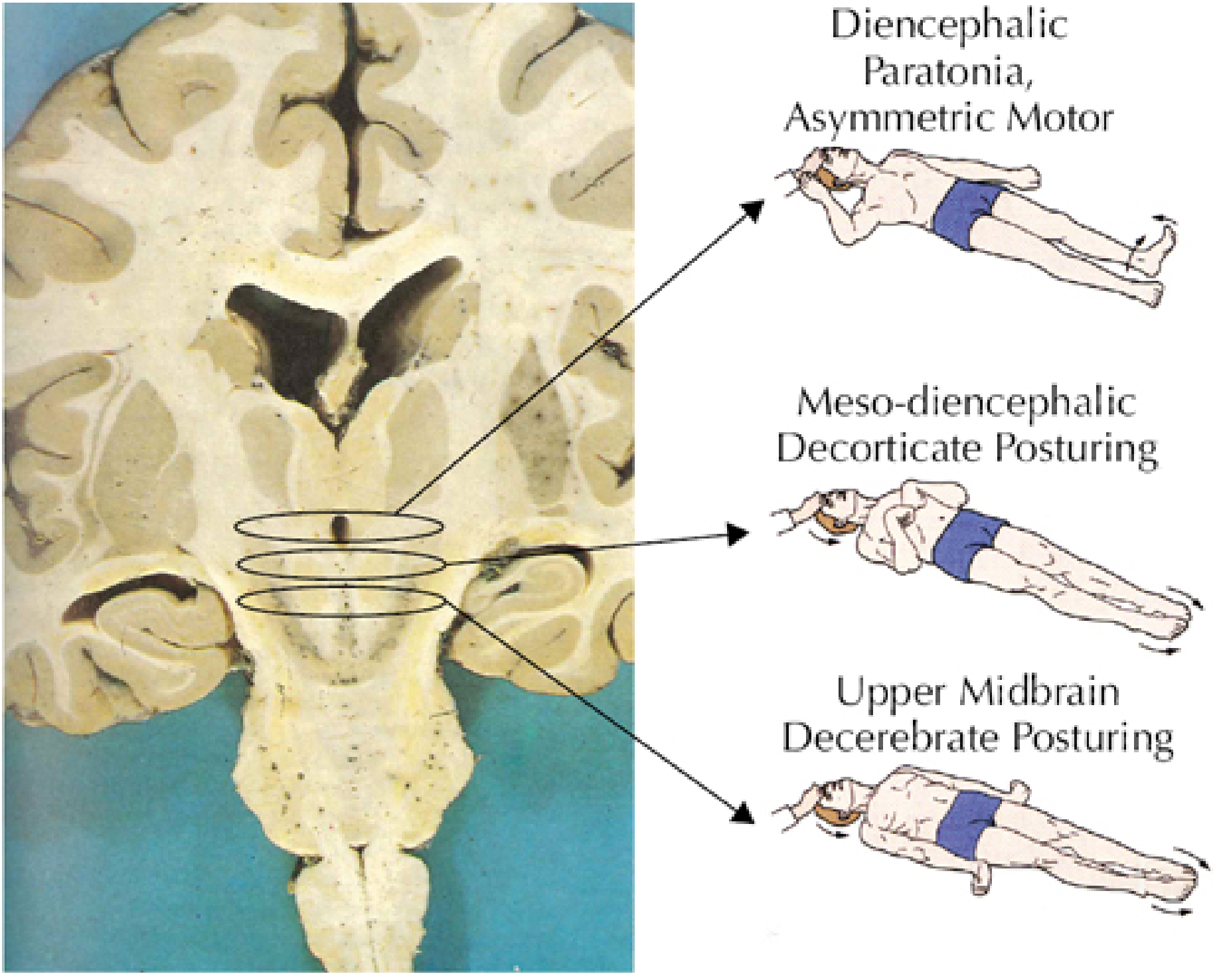
1. Background: Why These Postures Occur
- Not purposeful - they do not attempt to escape the painful stimulus in a goal-directed way
- Stereotyped - the same pattern repeats regardless of where the stimulus is applied
- Reflex-mediated - driven by brainstem vestibular, reticular, and tectospinal pathways
| Pathway | Origin | Effect |
|---|---|---|
| Rubrospinal tract | Red nucleus (midbrain) | Promotes flexion of upper limbs |
| Vestibulospinal tract | Vestibular nuclei (lower pons/medulla) | Promotes extension of all limbs |
| Pontine reticulospinal tract | Pontine reticular formation | Promotes extension (antigravity) |
| Medullary reticulospinal tract | Medullary reticular formation | Promotes flexion |
- Plum and Posner's Stupor and Coma, p. 161-163; Adams and Victor's Neurology 12E, p. 2592-2598
2. Decorticate Posturing (Abnormal Flexion)
Terminology
Physical Description
| Body Part | Position |
|---|---|
| Arms | Flexed at the elbows and wrists; fingers clenched into a fist |
| Shoulders | Adducted (arms held close to the body) |
| Forearms | Held against the chest |
| Legs | Extended, internally rotated; plantar flexed (feet pointed down) |
| Neck/trunk | Variable; less dramatic than decerebrate |
Neuroanatomical Basis
- The rubrospinal tract (originating in the red nucleus) normally promotes upper limb flexion
- When the cortex and corticospinal tract above the red nucleus are destroyed, the red nucleus remains intact and its flexion drive is unopposed in the upper limbs
- The vestibulospinal tract, originating below the red nucleus and still intact, promotes lower limb extension
- Result: upper limb flexion + lower limb extension
- Extensive bilateral cerebral hemisphere damage (white matter, internal capsule)
- Bilateral thalamic lesions
- High diencephalic compression (e.g., expanding supratentorial mass compressing the diencephalon - early herniation)
- Meso-diencephalic junction injury
Plum and Posner note that the fully developed response consists of relatively slow flexion of the arm, wrist, and fingers with adduction in the upper extremity, and extension with internal rotation and plantar flexion of the lower extremity.
Clinical Significance
-
Indicates less caudal (higher, more rostral) and often less severe brainstem involvement than decerebrate
-
Still indicates serious bilateral brain injury
-
After head trauma, only 37% of comatose patients with decorticate posturing recovered (Jennett and Teasdale series)
-
May be seen in metabolic encephalopathy (hepatic coma, severe hypoglycemia, anoxia) - important caveat
-
GCS motor score = 3 (abnormal flexion)
-
Plum and Posner, p. 2048-2050; Bradley and Daroff's Neurology, p. 2320
3. Decerebrate Posturing (Abnormal Extension)
Terminology
Physical Description
| Body Part | Position |
|---|---|
| Arms | Extended, adducted at shoulders |
| Elbows | Extended (stiffly straight) |
| Forearms | Fully pronated |
| Wrists | Flexed |
| Fingers | May be flexed |
| Legs | Extended, internally rotated; plantar flexed |
| Neck | May be hyperextended |
| Jaw | May be clenched |
| Trunk | Opisthotonos (arching backward) in severe cases |
Neuroanatomical Basis
- The red nucleus and its rubrospinal flexion drive are now destroyed or disconnected
- The vestibulospinal tract (from vestibular nuclei) and pontine reticulospinal tract are released from all supraspinal inhibition
- These tracts powerfully promote extensor (antigravity) tone throughout all limbs
- Result: extension of all four extremities with full pronation
- Bilateral midbrain or upper pontine lesions (most classic)
- Transtentorial herniation with midbrain compression
- Cerebellar or posterior fossa mass compressing the midbrain
- Metabolic causes (anoxia, severe hypoglycemia, hepatic coma) - these can also produce this pattern
- Bilateral supratentorial lesions with sufficient mass effect
- Rotating the head causes the arm on the side toward which the nose points to hyperextend, while the opposite arm flexes
- Extending the head → arm extension + leg relaxation
- Flexing the head → opposite
Clinical Significance
-
Indicates more caudal and more severe brainstem dysfunction than decorticate
-
Associated with disturbances of ocular motility (unlike decorticate, where ocular movements are typically normal)
-
Carries a significantly worse prognosis
-
GCS motor score = 2 (abnormal extension)
-
Patients who progress from decorticate to decerebrate posturing are showing rostrocaudal deterioration - a worsening herniation pattern requiring emergency intervention
-
Plum and Posner, p. 2056-2058; Adams and Victor's Neurology, p. 2592-2597
4. Comparison Table
| Feature | Decorticate (Flexor) | Decerebrate (Extensor) |
|---|---|---|
| Upper limbs | Flexion at elbow, wrist; adducted | Extension, adduction, full pronation |
| Lower limbs | Extension, internal rotation, plantar flexion | Extension, internal rotation, plantar flexion |
| Neck/trunk | Normal or mild | Possible opisthotonos, jaw clenching |
| GCS motor score | M3 (abnormal flexion) | M2 (abnormal extension) |
| Lesion level | Above red nucleus (diencephalon, high midbrain) | At/below red nucleus (midbrain, upper pons) |
| Tracts released | Vestibulospinal (legs) + rubrospinal flexion preserved (arms) | Vestibulospinal + pontine reticulospinal (all limbs) |
| Ocular movements | Usually normal | Usually abnormal (CN III palsy, disconjugate) |
| Severity | Serious - but relatively less severe | Very serious - worse prognosis |
| Reversibility | More likely reversible | Less likely reversible |
| Recovery rate (TBI) | ~37% | Much lower |
5. Progression: Rostrocaudal Deterioration
Normal motor response
↓
Purposeful (localizing) pain response ← Diencephalic stage
↓
Decorticate posturing ← Meso-diencephalic junction
↓
Decerebrate posturing ← Upper midbrain/pontine stage
↓
Flaccidity (no response) ← Lower pons/medullary stage
↓
Apnea → Brain death
6. Important Caveats and Common Pitfalls
6.1 Metabolic Causes Can Mimic Both
6.2 Not Always Bilateral
- Unilateral decorticate or decerebrate posturing is less ominous than bilateral - lesions causing unilateral posturing may be anywhere in the motor system from cortex to brainstem
- Unilateral extensor posturing is common immediately after stroke, followed later by a flexor response
- Diagonal posturing (flexion of one arm, extension of opposite arm and leg) usually indicates a supratentorial mass
6.3 Not Always "Rigidity"
6.4 Confusion with Seizures
- Posturing is tonic and relatively slow to develop - not the rapid clonic jerking of seizures
- Posturing is stimulus-driven - it occurs or worsens with painful stimulation
- No postictal period follows posturing
- EEG does not show ictal activity
6.5 Triple Flexion vs. Decorticate
- Triple flexion is a spinal reflex (preserved even with spinal cord transection)
- In triple flexion, the foot withdraws INTO the stimulus (dorsal foot pain → foot goes UP into the stimulus) - non-purposeful
- True decorticate posturing involves the upper limbs in flexion, which triple flexion does not
- Triple flexion can occur even in brain death - it is not a sign of brainstem function
6.6 "Forceful extensor arms + weak flexor legs"
7. Clinical Response to Posturing
- Treat as neurological emergency - especially if new or if progressing from decorticate to decerebrate
- Emergent NCCT head to identify mass lesion, hemorrhage, or herniation
- Airway protection - patients with posturing have GCS ≤5; intubation is often required
- ICP management: elevate head of bed 30°, maintain EtCO₂ 35 mmHg, avoid hypoxia/hyperthermia
- Osmotherapy (mannitol 0.5-1 g/kg IV or hypertonic saline 3%) if herniation suspected
- Neurosurgery consultation urgently for any reversible structural cause
- Treat metabolic causes: check glucose, sodium, ammonia, toxin screen
Summary
Decorticate = arms flex, legs extend = lesion above the red nucleus (diencephalon/high midbrain) = rubrospinal tract intact, vestibulospinal released = relatively less caudal, somewhat better prognosis = GCS M3
Decerebrate = all limbs extend, arms pronate = lesion at/below the red nucleus (midbrain/upper pons) = both rubrospinal and cortical inhibition lost, vestibulospinal + pontine reticulospinal fully released = more caudal, worse prognosis = GCS M2
Progression from decorticate → decerebrate = rostrocaudal deterioration = herniation is advancing = treat immediately
EEG graphs and waves comprehensive info
EEG waves types clinical significance epilepsy sleep patterns 2024 comprehensive
EEG: Comprehensive Guide to Waves, Patterns, and Clinical Interpretation
1. What Is an EEG?
- Adams and Victor's Principles of Neurology 12E, p. 762-768
2. Technical Setup
2.1 The 10-20 International Electrode System
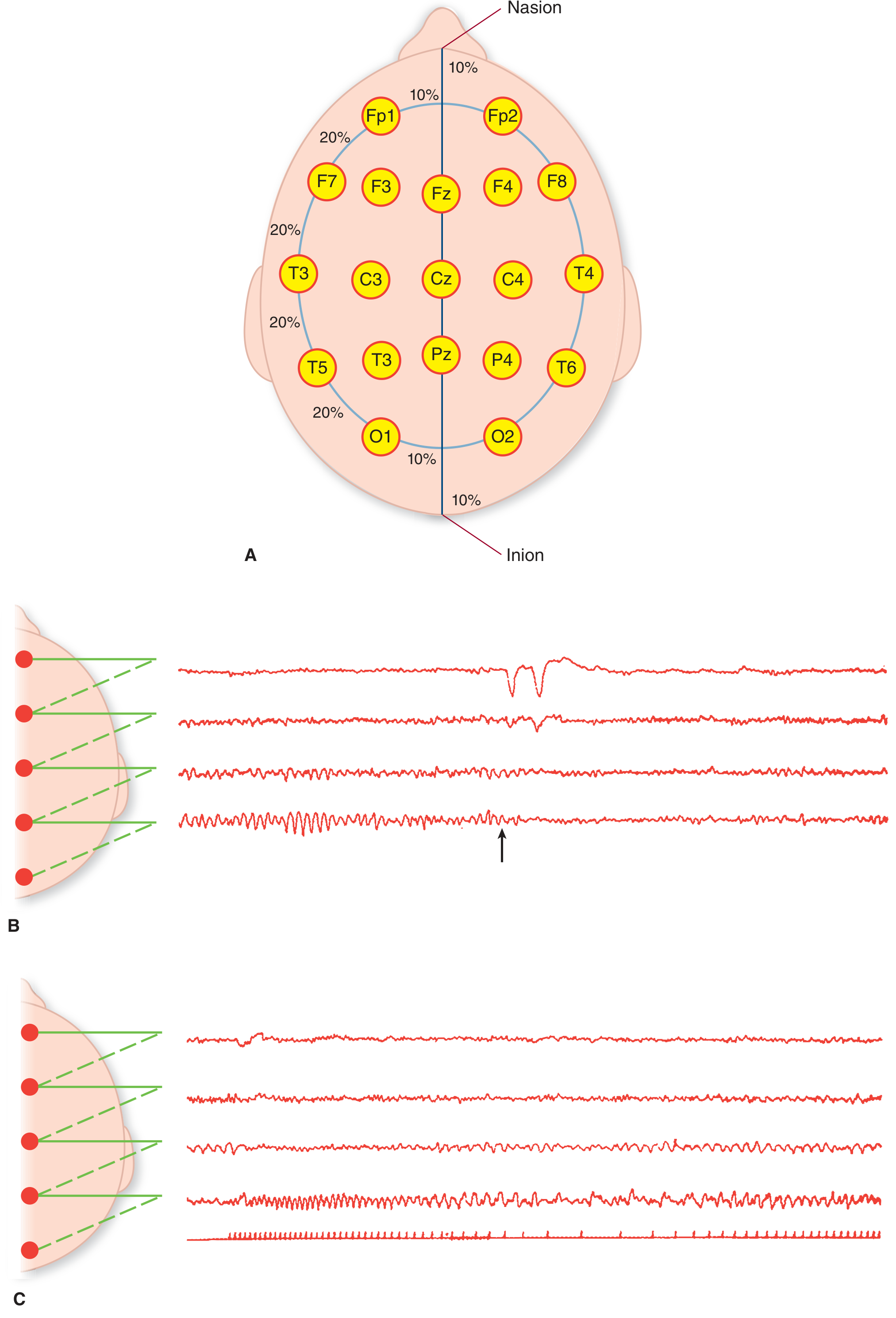
- Fp - Frontopolar
- F - Frontal
- C - Central
- P - Parietal
- O - Occipital
- T - Temporal
- z suffix = midline (Fz, Cz, Pz)
- Odd numbers = left hemisphere; even numbers = right hemisphere
2.2 Montages
- Bipolar (longitudinal/transverse): each channel = difference between two adjacent electrodes; allows phase reversal localization
- Referential (common average or linked-ears): each channel = difference between an electrode and a neutral reference
- Phase reversal: when a spike appears in opposite directions in two adjacent channels, the source is at the shared electrode - allows precise focal localization
2.3 Amplifier Settings
- Display speed: standard 3 cm/second (30 mm/sec)
- Frequency range recorded: 0.5 to 70 Hz
- Sensitivity: typically 7-10 μV/mm
- Low-frequency filter: 1 Hz; high-frequency filter: 70 Hz (or 35 Hz for standard recordings)
2.4 Activation Procedures
| Procedure | Duration | Provokes |
|---|---|---|
| Hyperventilation | 3 minutes (20 breaths/min) | Absence seizures (3 Hz spike-wave), focal slowing |
| Photic stimulation | Strobe light at 1-30 Hz | Photoparoxysmal response, photic driving |
| Sleep deprivation | Recorded after sleep deprivation | Temporal lobe spikes, absence discharges |
| Sleep recording | Natural or induced | Sleep architecture disorders, temporal lobe epilepsy |
| Eye opening/closing | Simple command | Alpha reactivity testing |
3. Normal EEG Wave Types
3.1 Beta Waves (β)
| Parameter | Value |
|---|---|
| Frequency | >13 Hz (typically 14-30 Hz) |
| Amplitude | Low, 10-20 μV |
| Normal distribution | Predominant frontally |
| State | Active wakefulness, mental activity, anxiety |
| Synchrony | Desynchronized (low amplitude, irregular) |
3.2 Alpha Waves (α)
| Parameter | Value |
|---|---|
| Frequency | 8-13 Hz |
| Amplitude | 20-100 μV (dominant rhythm in adults) |
| Normal distribution | Posterior - occipital and parietal regions |
| State | Relaxed wakefulness with eyes closed |
| Key feature | Alpha attenuation (blocking) with eye opening or mental effort |
- Appears in both hemispheres symmetrically
- Waxes and wanes in amplitude spontaneously
- Is invariant in frequency for a given individual (though slows slightly with normal aging)
- Appears around age 6 and reaches adult frequency by age 10-12 years
3.3 Theta Waves (θ)
| Parameter | Value |
|---|---|
| Frequency | 4-7 Hz (4-8 Hz by some classifications) |
| Amplitude | Variable, moderate |
| Normal distribution | Temporal regions (small amount normal, especially in elderly) |
| State | Drowsiness, early sleep (Stage N1), emotional states |
3.4 Delta Waves (δ)
| Parameter | Value |
|---|---|
| Frequency | <4 Hz (0.5-3 Hz) |
| Amplitude | High, 50-350 μV |
| Normal distribution | Not present in normal waking adults |
| State | Deep slow-wave sleep (Stage N3), infants |
- Focal delta over one region: underlying structural lesion (tumor, infarct, abscess, subdural hematoma)
- Diffuse delta: severe metabolic encephalopathy, anoxia, deep sedation, coma
3.5 Gamma Waves (γ)
| Frequency | 30-80+ Hz |
|---|---|
| Not visible on routine EEG; detected with specialized high-frequency filters or intracranial recordings. Associated with cognitive processing, attention, and sensory binding. Research relevance; limited routine clinical use. |
4. Summary: Frequency Bands
Slow ←————————————————————————→ Fast
Delta Theta Alpha Beta Gamma
<4 Hz 4-8 Hz 8-13 Hz 13-30 Hz >30 Hz
Coma / Drowsy / Relaxed Active Cognition
Deep Stage N1 awake thinking (intracranial)
sleep sleep (eyes / anxiety
(Stage closed)
N3)
5. Normal Sleep Architecture on EEG
Sleep EEG Waveforms
| EEG Feature | Characteristics | Stage |
|---|---|---|
| Alpha activity | 8-13 Hz, occipital; attenuates with eye opening | Relaxed wakefulness |
| Slow eye movements (SEM) | Conjugate rolling movements, initial deflection ≥0.5 sec | Stage N1 onset |
| Theta activity | 4-8 Hz, central and temporal leads; sawtooth variant in REM | Stage N1 / REM |
| Vertex sharp waves | Sharply contoured negative bursts in central (Cz) leads; stand out from background | Stage N1/N2 transition |
| Sleep spindles | 11-16 Hz bursts, 0.5-1.5 sec duration; central leads; spindle shape; represent thalamocortical oscillations | Stage N2 |
| K-complexes | Biphasic: negative sharp wave immediately followed by positive component; total duration ≥0.5 sec; maximal frontally; can be evoked by external stimuli | Stage N2 |
| Slow waves (delta) | High amplitude ≥75 μV, frequency ≤2 Hz | Stage N3 (slow-wave sleep) |
| Sawtooth waves | Notched/serrated theta activity (4-7 Hz); seen in central leads | REM sleep |
| Rapid eye movements (REM) | Conjugate saccades, initial deflection <0.5 sec; associated with dreaming | REM sleep |
Sleep Stage EEG Summary
| Stage | EEG Features | % of Night |
|---|---|---|
| Wake (W) | Alpha (eyes closed); beta (eyes open) | - |
| N1 (light NREM) | Theta; vertex sharp waves; SEM; alpha attenuates | 5% |
| N2 (NREM) | Sleep spindles + K-complexes on theta background | 45-55% |
| N3 (deep NREM) | High-amplitude slow delta waves (≥75 μV, ≤2 Hz) | 15-25% |
| REM | Low-voltage mixed frequency; sawtooth theta; REMs; muscle atonia | 20-25% |
6. Abnormal EEG Patterns
6.1 Pathological Slow Waves
- Arrhythmic, irregular delta over one region
- Indicates structural lesion directly beneath (cortical or subcortical white matter)
- Causes: brain tumor, infarct, abscess, intracerebral hemorrhage, subdural hematoma, encephalitis
- Rhythmic delta bursts (usually frontally - FIRDA; or occipitally - OIRDA)
- FIRDA: suggests deep midline or diencephalic dysfunction; hydrocephalus, metabolic encephalopathy
- OIRDA: more often seen in children; associated with idiopathic generalized epilepsies
- Bilateral high-voltage slow waves, frontally predominant
- Seen in deep coma from any cause (metabolic, structural)
- The more severe the encephalopathy, the more dominant and slower the delta
6.2 Epileptiform Discharges
- Sharp transient, duration 20-70 ms, pointed peak
- Strong association with epilepsy
- Ictal: during a seizure
- Interictal: epileptiform discharge between seizures (indicates epileptogenic zone)
- Similar to spike but duration 70-200 ms
- Also associated with epilepsy; slightly less specific than a spike
- A spike followed immediately by a slow wave
- Characteristic of generalized epilepsies
- 3 Hz (classic absence): Bilaterally synchronous, symmetric, sudden onset and offset; provoked by hyperventilation; clinically correlates with absence seizures in childhood absence epilepsy
- Slow spike-wave (< 2.5 Hz): Lennox-Gastaut syndrome
- Fast spike-wave (> 3 Hz): Juvenile myoclonic epilepsy and other IGEs
- Multiple spikes followed by a slow wave
- Characteristic of myoclonic seizures (juvenile myoclonic epilepsy)
- Chaotic, very high amplitude discharges from all regions without synchrony
- Pathognomonic of infantile spasms (West syndrome)
- Chaotic, large-amplitude spikes and slow waves from all areas in a disorganized manner
6.3 Disease-Specific EEG Patterns
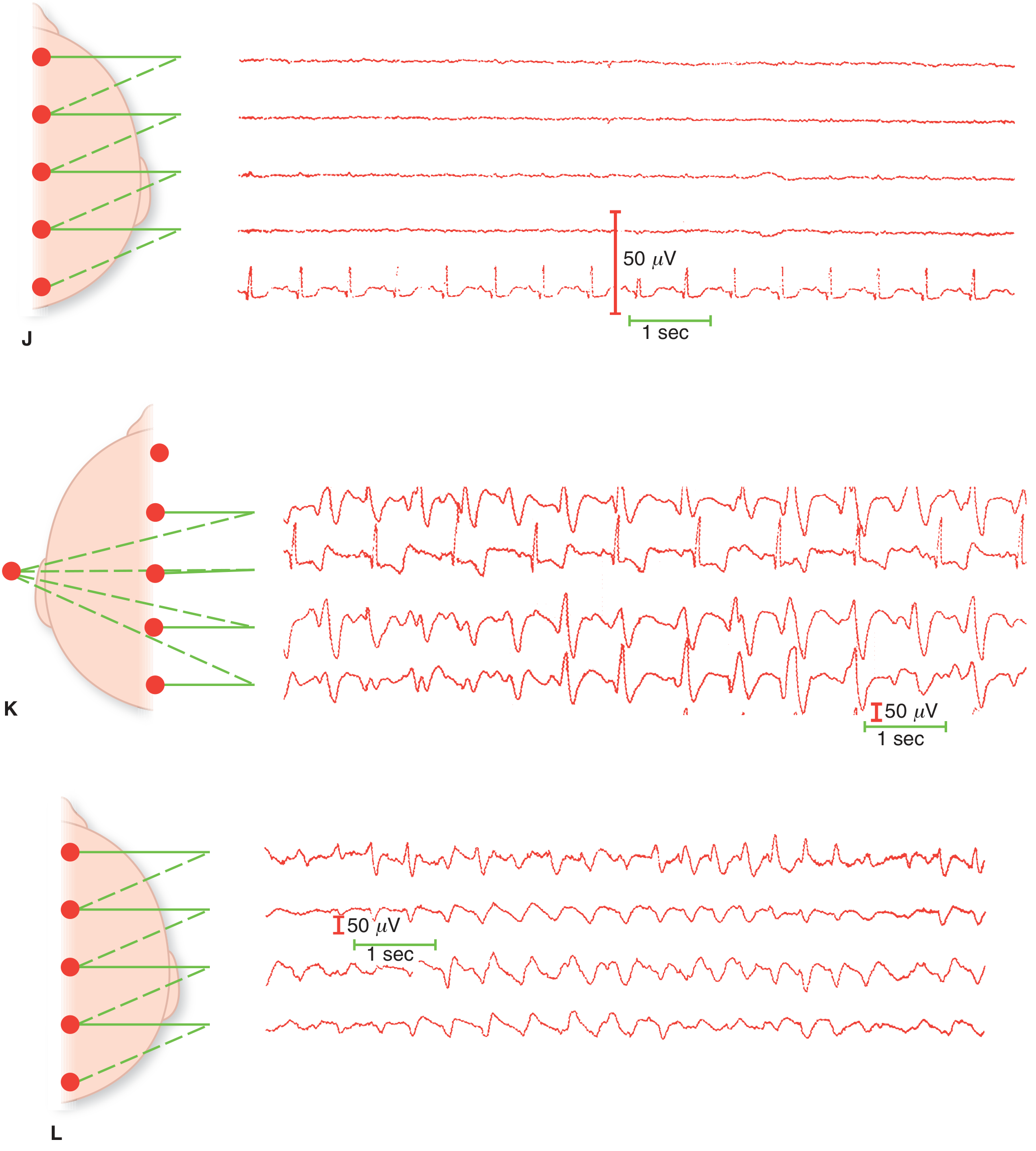
- No cerebral rhythms visible
- Only ECG artifact (cardiac electrical activity) visible on highest amplification
- Only EEG artifacts present at high gain
- Must exclude confounders: hypothermia, drug intoxication
- High-amplitude, bilaterally synchronous, pseudoperiodic sharp wave complexes at ~1/second
- Background activity grossly disorganized
- Pathognomonic of Creutzfeldt-Jakob disease in the appropriate clinical context (rapidly progressive dementia, myoclonus)
- Also seen in: SSPE (1-3/second), late SSPE evolves to burst-suppression
- Large, slow (~2/second), bilaterally synchronous waves with 3 components (positive-negative-positive or negative-positive-negative)
- Characteristically seen in hepatic encephalopathy; correlates roughly with degree of encephalopathy
- Also seen in: renal failure, septic encephalopathy, pulmonary failure, acute hydrocephalus
- Tend to have anterior predominance and a posterior-to-anterior lag
7. Graded EEG Changes in Coma
| Level of Consciousness | Dominant EEG Activity |
|---|---|
| Alert, active thought | Low-voltage beta (desynchronized) |
| Relaxed wakefulness | Posterior alpha (8-12 Hz) |
| Drowsiness | Theta (4-7 Hz) |
| Light sleep (N1/N2) | Theta, vertex waves, spindles, K-complexes |
| Deep sleep (N3) | High-amplitude delta |
| Mild encephalopathy/stupor | Diffuse theta, loss of alpha |
| Moderate encephalopathy/coma | Diffuse delta, loss of normal background |
| Severe coma (e.g., anoxia) | Burst suppression |
| Deep coma → Brain death | Electrocerebral silence (isoelectric) |
Burst Suppression
- Alternating periods of high-voltage, sharp/irregular delta activity ("bursts") followed by flat isoelectric periods ("suppression")
- Indicates profound global cortical dysfunction
- Causes: deep anesthesia, severe anoxia, hypothermia, barbiturate or propofol overdose (intentional for refractory status epilepticus)
- Bursts represent residual cortical activity; the suppression period reflects electrical silence
- Progression: burst-suppression → electrocerebral silence = approaching brain death in anoxia
Alpha Coma
- An apparent alpha-frequency rhythm (8-12 Hz) distributed diffusely rather than the normal posterior distribution
- Unlike normal alpha: more regular, less variable, not inhibited by eye opening
- May be driven by photic stimulation (unlike true coma where photic response is absent)
- Usually follows global anoxia (transitional pattern); less often with acute pontine lesions
- Carries poor prognosis in post-cardiac arrest patients
8. EEG in Specific Conditions
8.1 Epilepsy
- EEG sensitivity: ~50% of patients with epilepsy show epileptiform activity on a single routine recording
- Yield increases to ~90% with repeated recordings + sleep deprivation + activation procedures
- ~1% of neurologically normal adults and 3.5% of normal children have epileptiform-looking discharges without epilepsy (specificity limitation)
- A normal EEG during a convulsion strongly suggests psychogenic non-epileptic seizure (PNES)
| Epilepsy Syndrome | EEG Finding |
|---|---|
| Childhood absence epilepsy | 3 Hz generalized spike-and-wave; abrupt onset/offset; provoked by hyperventilation |
| Juvenile myoclonic epilepsy (JME) | 3.5-6 Hz polyspike-and-wave; enhanced by sleep deprivation; normal background |
| Lennox-Gastaut syndrome | Slow (<2.5 Hz) spike-and-wave; slow background; multifocal spikes |
| West syndrome (infantile spasms) | Hypsarrhythmia |
| Temporal lobe epilepsy (TLE) | Anterior temporal spikes or sharp waves; often unilateral |
| Benign epilepsy of childhood with centrotemporal spikes (BECTS/BCECTS) | High-amplitude centrotemporal spikes with characteristic horizontal dipole; activated by sleep |
| Tonic-clonic seizure (ictal) | Initial fast rhythmic discharge evolving to spike-wave, then post-ictal slowing |
| Focal (partial) seizure | Focal rhythmic discharge in the seizure onset zone |
8.2 Non-Convulsive Status Epilepticus (NCSE)
- Continuous or near-continuous seizure activity without clinical convulsions
- Patient appears merely confused, drowsy, or comatose
- 8% of comatose ICU patients have NCSE
- EEG is essential for diagnosis
- Classic pattern: continuous spike-wave or rhythmic sharp waves
- Treatment: IV benzodiazepine (if patient improves, confirms diagnosis); then IV AEDs
- Plum and Posner, p. 2242-2246
8.3 Herpes Simplex Encephalitis
- Periodic lateralized epileptiform discharges (PLEDs) - now called LPDs (lateralized periodic discharges)
- High-voltage sharp waves and slow-wave complexes at 1-3 per second over temporal regions (often unilateral)
- Highly characteristic when bilateral or pseudoperiodic temporal discharges
- Appears typically in the first 2 weeks; highly supportive of HSE diagnosis
8.4 Creutzfeldt-Jakob Disease (CJD)
- Generalized periodic sharp wave complexes (PSWCs) at ~1 per second
- Bilaterally synchronous, symmetric
- Background activity severely disorganized
- Appears in mid-to-late stages of illness
- Combined with clinical dementia and + 14-3-3 protein in CSF = diagnostic
- (See Panel K in image above)
8.5 Subacute Sclerosing Panencephalitis (SSPE)
- Repetitive, high-amplitude stereotyped burst complexes at regular intervals (4-15 second intervals)
- Bursts consist of 2-3 second runs of high-amplitude slow and sharp waves
- Each burst correlates with a myoclonic jerk
- Highly characteristic of SSPE
8.6 Stroke and Focal Lesions
- Large hemispheric stroke: focal polymorphic delta over affected region
- MCA territory infarct → large area of slow-wave activity; normalizes over 3-6 months in ~50%
- Lacunar stroke: often normal EEG despite prominent clinical deficits (deep lesion below the cortical surface)
- Brainstem lesion below the midbrain (pons, medulla): often normal or near-normal EEG despite severe clinical changes
8.7 Metabolic Encephalopathy
| Disorder | EEG Finding |
|---|---|
| Hepatic encephalopathy | Triphasic waves (2-3/sec, frontally dominant); progressive slowing |
| Uremic encephalopathy | Triphasic-like waves; diffuse slowing; may have burst suppression |
| Hypoglycemia | Progressive slowing → delta → burst suppression |
| Hyponatremia | Diffuse slowing; may have seizures |
| Septic encephalopathy | Diffuse slowing; may have triphasic waves |
| Hypothyroidism (myxedema) | Normal wave configuration but reduced amplitude and frequency |
| Alzheimer's disease | Initially normal; progresses to mild diffuse theta slowing |
9. Drug Effects on EEG
| Drug/Substance | EEG Effect |
|---|---|
| Benzodiazepines | Increased beta activity (fast activity) |
| Barbiturates | Increased beta (low dose); burst suppression (high dose); withdrawal → paroxysmal activity and spikes |
| Opioids | Decreased alpha; increased theta and delta voltage; overdose → diffuse slow waves |
| Alcohol (intoxication) | Decreased alpha; increased theta |
| Alcohol (withdrawal) | Decreased alpha; increased fast activity; paroxysmal discharges (seizure risk) |
| Propofol | Beta at low dose; burst suppression at high dose (intentional for refractory status epilepticus) |
| Lithium | Diffuse slowing or paroxysmal activity |
| Marijuana/Cocaine | Increased frontal alpha; overall slow alpha |
| Nicotine | Increased alpha; withdrawal = marked decrease in alpha |
| Caffeine withdrawal | Increased theta amplitude |
| Inhalants | Diffuse delta and theta slowing |
| Clozapine | Nonspecific changes; high-dose → seizure risk |
10. EEG in Brain Death
- No cerebral electrical activity (isoelectric)
- Technically defined as <2 μV over all parts of the head at highest amplification
- ECG artifact (cardiac) may be seen - this is NOT cerebral activity
- Must be confirmed at highest amplification to ensure it is not a technical failure
- At least 8 scalp electrodes + ear electrodes
- Interelectrode impedance <10,000 Ω (>100 Ω)
- Minimum amplification 2 μV/mm for 30 minutes
- No reactive change to sensory stimulation
- Patient normothermic; no CNS-depressant drugs; no severe metabolic derangement
- Adams and Victor's Neurology 12E, p. 855-856
11. Gamma/High-Frequency Oscillations
| Frequency | 30-100+ Hz |
|---|---|
| Gamma | 30-80 Hz: cognitive binding, attention, sensory processing |
| High-frequency oscillations (HFOs) | Ripples (80-250 Hz) and fast ripples (250-500 Hz) - only visible on intracranial EEG |
12. Quantitative EEG (QEEG) and Processed EEG
- Spectral analysis (FFT): power in each frequency band displayed as a spectrum
- Compressed spectral array (CSA) and density spectral array (DSA): 3D power-over-time displays used in anesthesia and ICU monitoring
- Brain mapping: topographic color maps of power distribution across the scalp
- Bispectral Index (BIS): a proprietary processed EEG number (0-100) used to monitor anesthetic depth; derived from EEG power, frequency, and phase relationships
- SEDLINE / Narcotrend: other commercial anesthesia depth monitors based on processed EEG
- Intraoperative neurological monitoring (carotid endarterectomy, cardiac bypass)
- ICU continuous monitoring (detecting silent seizures, monitoring sedation depth)
- Brain death support
- Anesthetic management (titrating depth of anesthesia)
13. Key Clinical Uses of EEG - Summary
| Clinical Scenario | EEG Role |
|---|---|
| Epilepsy diagnosis and classification | Primary tool; seizure type, focus localization |
| Non-convulsive status epilepticus | Essential - only way to diagnose NCSE |
| Coma evaluation | Grades severity; detects burst suppression, alpha coma, NCSE in coma |
| Brain death | Ancillary test (electrocerebral silence) |
| Metabolic encephalopathy | Triphasic waves (hepatic); diffuse slowing |
| CJD / prion disease | Periodic sharp wave complexes |
| HSV encephalitis | Periodic temporal discharges |
| SSPE | Periodic burst complexes with myoclonus |
| Sleep disorders (polysomnography) | Sleep staging, PLMD, RBD, narcolepsy |
| Intraoperative monitoring | Cerebral ischemia detection, anesthetic depth |
| Post-cardiac arrest prognostication | Burst suppression, malignant patterns |
| Neonatal seizures | Many neonatal seizures have no clinical correlate; EEG essential |
any abnormal chromosomal number other then normal is called as heteroploidy
heteroploidy definition genetics aneuploidy polyploidy classification chromosomal number
https://www.sciencedirect.com/topics/biochemistry-genetics-a…
Heteroploidy, Aneuploidy, and Euploidy: A Precise Breakdown
Is the Statement Correct?
The Correct Framework
HETEROPLOIDY
(any chromosome number ≠ normal diploid 2x)
|
——————————————————————————
| |
EUPLOIDY ANEUPLOIDY
(exact multiple of (NOT an exact multiple
basic number x) of basic number x)
| |
Haploid, Triploid, Monosomy, Trisomy,
Tetraploid, etc. Nullisomy, Tetrasomy, etc.
1. Heteroploidy - The Umbrella Term
- Coined and used primarily in plant genetics and crop science
- Less commonly used in clinical/medical genetics (where "aneuploidy" is preferred)
- Encompasses all deviations from the standard diploid number - both whole-genome duplications and single-chromosome gains/losses
2. Euploidy (a type of heteroploidy)
| Type | Formula | Chromosome count (if x=7) |
|---|---|---|
| Monoploid / Haploid | x or n | 7 |
| Diploid (NORMAL) | 2x | 14 |
| Triploid | 3x | 21 |
| Tetraploid | 4x | 28 |
| Hexaploid | 6x | 42 |
| Octaploid | 8x | 56 |
- Autopolyploidy: multiple identical genomes from the same species (e.g., 4x from one parent species)
- Allopolyploidy: genomes from two or more different species (e.g., wheat = allohexaploid, 6x from 3 species)
- Amphidiploid: an allopolyploid with two copies of each contributing genome; behaves like a diploid at meiosis
3. Aneuploidy (the other type of heteroploidy)
| Type | Formula | What it means | Human Example |
|---|---|---|---|
| Nullisomic | 2n - 2 | Missing an entire chromosome pair | Usually lethal in humans |
| Monosomic | 2n - 1 | Missing one chromosome from a pair | Turner syndrome (45,X) |
| Double monosomic | 2n - 1 - 1 | Two chromosomes missing (different pairs) | - |
| Trisomic | 2n + 1 | One extra chromosome | Down (trisomy 21), Edwards (trisomy 18), Patau (trisomy 13) |
| Double trisomic | 2n + 1 + 1 | Two extra chromosomes (different pairs) | - |
| Tetrasomic | 2n + 2 | Extra pair of one chromosome | Rare |
| Polysomic | 2n + extra multiple | Multiple extra copies of one chromosome | XXY = Klinefelter; XYY |
4. Why "Heteroploidy" Is Technically More Precise Than Just "Aneuploidy"
- Heteroploidy = aneuploidy + euploidy (polyploidy)
- Heteroploidy is the genus; aneuploidy and euploidy are the species
- Simply saying "any abnormal number = heteroploidy" is true, but it omits the crucial distinction between whether complete sets (euploidy) or individual chromosomes (aneuploidy) are affected
5. Clinical Relevance in Human Medicine
| Term Used Clinically | Meaning |
|---|---|
| Aneuploidy | Gain or loss of individual chromosomes (Down, Turner, Klinefelter, Edwards, Patau) |
| Polyploidy | Complete extra genome sets (triploidy 69,XXX/XXY/XYY → fetal hydrops; tetraploidy → lethal) |
| Euploidy | Normal diploid complement (46 chromosomes in humans) |
6. Summary Table
| Term | Definition | Chromosome formula | Notes |
|---|---|---|---|
| Euploidy (normal) | Exact multiple of x; includes normal diploid | 2n = 2x | Normal state |
| Heteroploidy | Any number ≠ normal diploid | Any ≠ 2x | Umbrella term |
| Euploidy (abnormal) | Exact multiple of x, but not 2x | 3x, 4x, 6x... | Polyploidy; normal in plants |
| Aneuploidy | Not an exact multiple of x; individual chr. gained/lost | 2n±1, 2n±2... | Clinically most relevant in humans |
Conclusion
Heteroploidy = Euploidy (polyploidy - whole genome multiples) + Aneuploidy (individual chromosome gains/losses)
talk about genomic imprinting of chromosomes and it's significance in appiled genetics
"genomic imprinting" AND "disease" AND "epigenetics"
genomic imprinting mechanism significance applied genetics clinical 2024 2025
PMID: 40613785
PMID: 40695263
Genomic Imprinting: Mechanisms and Significance in Applied Genetics
1. Definition and Concept
Key distinction: genomic imprinting is not mutation - the DNA sequence is intact. The silencing is achieved by epigenetic marks that are heritable through cell division but potentially reversible.
- Thompson & Thompson Genetics 9E, p. 160; Robbins Pathologic Basis of Disease, p. 2100; Emery's Elements of Medical Genetics, p. 78
2. Genomic Map of Imprinted Genes
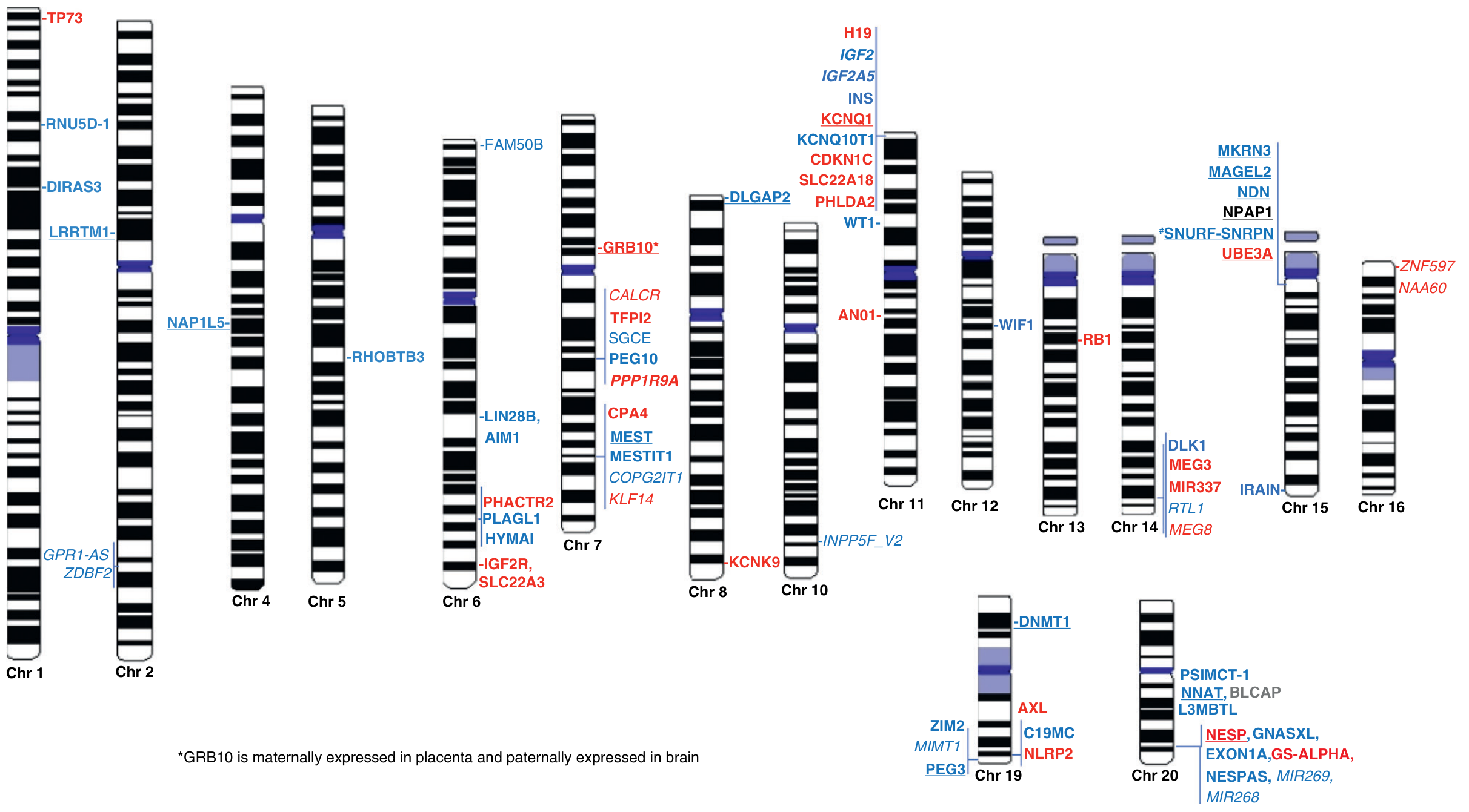
3. Molecular Mechanisms of Imprinting
3.1 DNA Methylation - The Primary Mark
- The allele that is methylated = silenced
- The allele that is unmethylated = expressed
- Methylation is established during gametogenesis (in the germline - either ovum or sperm) before fertilization
- Once established, the methylation pattern is stably maintained through mitosis in all somatic cells via the enzyme DNMT1 (maintenance methyltransferase)
- Erasure: All prior imprints are wiped in primordial germ cells (PGCs) via active demethylation
- Re-establishment: Sex-specific methylation marks are placed during gametogenesis
- In oogenesis (female germline): maternal imprints are established
- In spermatogenesis (male germline): paternal imprints are established
- Maintenance: After fertilization, the imprints are maintained in somatic cells through all subsequent cell divisions
3.2 Histone Modifications
- Active allele: histone H3 acetylation (H3Ac) and H3K4 methylation
- Silent allele: H3K9 methylation and H3K27 trimethylation (repressive marks)
- These form a self-reinforcing epigenetic code that stabilizes the imprinted state
3.3 Non-Coding RNAs (ncRNAs)
- H19 (maternally expressed lncRNA): regulates IGF2 in the 11p15 domain
- KCNQ1OT1: paternally expressed antisense ncRNA that silences multiple genes across the IC2 domain in 11p15
- SNURF-SNRPN antisense transcript: silences maternally expressed genes in 15q11-q13
- These ncRNAs recruit chromatin-modifying complexes (e.g., Polycomb Repressive Complex 2) to silence entire domains in cis
3.4 CTCF Insulator Model (IC1/IGF2-H19 locus)
- The unmethylated maternal allele of IC1 is bound by the CTCF insulator protein
- CTCF creates a chromatin loop that insulates IGF2 from its enhancers → IGF2 silenced on maternal allele; H19 expressed instead
- On the paternal allele, IC1 is methylated → CTCF cannot bind → IGF2 is expressed from paternal allele; H19 is silenced
- Pandey & Raman, Cytogenet Genome Res 2026 - PMID 40695263
4. Imprinted Gene Domains: Key Loci
4.1 Chromosome 15q11-q13 (PWS/AS Region)
| Gene | Expression | Function |
|---|---|---|
| SNRPN / SNURF | Paternal only | RNA splicing |
| MKRN3 | Paternal only | Puberty timing |
| NDN (Necdin) | Paternal only | Neuronal survival |
| MAGEL2 | Paternal only | Circadian rhythm, neuronal function |
| UBE3A | Maternal only (in neurons) | E3 ubiquitin ligase; protein degradation |
4.2 Chromosome 11p15 (BWS/RSS Region)
| Domain | Gene | Expression | Function |
|---|---|---|---|
| IC1 | IGF2 | Paternal | Growth factor; promotes cell proliferation |
| IC1 | H19 | Maternal | lncRNA; tumor suppressor |
| IC2 | CDKN1C (p57KIP2) | Maternal | Cell cycle inhibitor; growth suppressor |
| IC2 | KCNQ1 | Maternal | Potassium channel |
| IC2 | KCNQ1OT1 | Paternal | lncRNA; silences IC2 genes in cis |
4.3 Chromosome 14q32 (Temple Syndrome / Kagami-Ogata)
| Gene | Expression |
|---|---|
| DLK1 | Paternal only |
| MEG3 | Maternal only (lncRNA) |
| RTL1 | Paternal only |
4.4 Chromosome 20q13 (GNAS locus)
| Gene/Transcript | Expression | Disease |
|---|---|---|
| GNAS (Gsα exon 1A) | Maternal (in certain tissues) | Pseudohypoparathyroidism type Ia |
| NESP55 | Maternal only | - |
| XLαs | Paternal only | - |
| NESPAS | Paternal only (antisense) | - |
5. Disorders of Genomic Imprinting
5.1 Prader-Willi Syndrome (PWS) - Loss of Paternal 15q11-q13
| Mechanism | Frequency | Detail |
|---|---|---|
| Deletion of paternal chr 15q11-q13 | ~70% | ~5-6 Mb interstitial deletion; always on paternal chromosome |
| Maternal uniparental disomy (UPD15mat) | ~25-30% | Two maternal copies of chr 15; functionally same as deletion of paternal genes |
| Imprinting center (IC) defect / epimutation | ~1% | Paternal ICR methylated like maternal → paternal genes silenced |
5.2 Angelman Syndrome (AS) - Loss of Maternal 15q11-q13
| Mechanism | Frequency | Detail |
|---|---|---|
| Deletion of maternal chr 15q11-q13 | ~70% | Same 5-6 Mb deletion, but on MATERNAL chromosome |
| Paternal UPD (UPD15pat) | ~2-5% | Two paternal copies of chr 15 |
| IC defect (epimutation) | ~3% | Maternal ICR unmethylated like paternal → maternal genes silenced |
| UBE3A pathogenic variant | ~10-15% | Direct mutation in the maternally expressed UBE3A gene |
- Robbins Pathologic Basis of Disease, p. 2113-2116; Emery's Medical Genetics, p. 89
5.3 Beckwith-Wiedemann Syndrome (BWS) - Gain of Paternal 11p15
- Loss of methylation at IC1 on maternal allele (~5%): CTCF cannot bind → both alleles now express IGF2 → growth excess
- Gain of methylation at IC1 on maternal allele (~5%): maternal allele now acts like paternal
- Paternal UPD of 11p15 (~20%): two copies of paternal-type genes (excess IGF2, absent CDKN1C)
- Loss of imprinting / hypomethylation at IC2 (~50%): KCNQ1OT1 not silenced → silences CDKN1C → loss of growth brake
5.4 Russell-Silver Syndrome (RSS) - Loss of Paternal 11p15
- Loss of methylation at IC1 on paternal allele (~40%): CTCF now binds paternal allele too → IGF2 silenced on both → growth restriction
- Maternal UPD of chromosome 7 (UPD7mat) (~10%)
- Chromosome 11p15 imprinting defects at IC1 (~40%)
5.5 Temple Syndrome (14q32 - Paternal loss)
- Loss of paternal DLK1 expression
- Features: IUGR, short stature, hypotonia, small hands/feet, premature puberty
- Mechanisms: maternal UPD14, del(14)(q32)pat, IC epimutation
5.6 Kagami-Ogata Syndrome (14q32 - Maternal loss)
- Loss of maternal MEG3 expression; excess paternal DLK1 and RTL1
- Features: coat-hanger rib cage, large placenta, polyhydramnios, intellectual disability
- Mechanisms: paternal UPD14, del(14)(q32)mat
5.7 Pseudohypoparathyroidism Type Ia (PHP1a) - GNAS locus 20q13
- Caused by maternal inheritance of GNAS mutations
- The Gsα exon 1A isoform is maternally expressed in certain tissues (kidney, thyroid, pituitary)
- Maternal mutations → Albright hereditary osteodystrophy (AHO) phenotype + hormone resistance (PTH, TSH, GHRH)
- The same mutation inherited paternally causes only AHO without hormone resistance (PHP1c/PPHP) - classic imprinting effect
6. Uniparental Disomy (UPD) - A Mechanism Linked to Imprinting
- Maternal UPD of an imprinted region → absence of paternally expressed genes → disease
- Paternal UPD of an imprinted region → absence of maternally expressed genes → disease
- Trisomy rescue (the commonest mechanism): a trisomic embryo loses one chromosome; if the lost copy came from the normal parent, UPD results
- Monosomy rescue: a monosomic cell gains a chromosome by duplication
- Post-fertilization error in mitosis
7. Significance in Applied Genetics
7.1 Genetic Counseling and Recurrence Risk
- A deletion in 15q11-q13 inherited from the father → PWS; same deletion from mother → AS
- The same karyotype in a parent may produce completely different diseases in offspring depending on which parent transmits it
- For IC defect (epimutation) forms: low recurrence risk if de novo; up to 50% risk if the parent carries an IC mutation
- UPD forms have very low recurrence risk (~1%) since they arise from sporadic chromosomal errors
7.2 Prenatal and Preimplantation Diagnosis
- Methylation-specific PCR (MS-PCR) and methylation-specific MLPA (MS-MLPA): detect abnormal methylation at DMRs → identify PWS, AS, BWS, RSS, Temple, Kagami-Ogata from a single test
- SNP array: detects UPD by identifying regions of long contiguous stretches of homozygosity (LCSH)
- FISH and chromosomal microarray (CMA): detect deletions and duplications in imprinted domains
- Preimplantation genetic testing (PGT-M): for families with known imprinting center mutations, PGT can select unaffected embryos
7.3 Cancer - Loss of Imprinting (LOI)
- IGF2 LOI (biallelic expression of IGF2 on chromosome 11p15): one of the most common epigenetic alterations in human cancers
- Wilms tumor (nephroblastoma) - the first cancer where LOI was described
- Colorectal cancer, hepatocellular carcinoma, lung, ovarian, and breast cancers
- CDKN1C silencing: loss of this maternal cell cycle brake promotes proliferation
- H19 hypermethylation: silences this tumor suppressor lncRNA
- LOI is often an early event in carcinogenesis and may correlate with disease severity
- Hypermethylation of MLH1 promoter (a tumor suppressor) drives microsatellite-unstable sporadic colorectal cancer - a direct application of imprinting-related epigenetic silencing
Imprinted genes are functionally haploid - only a single genetic or epigenetic hit is needed to eliminate their expression, making them unusually sensitive to both mutation and epimutation. - Jirtle RL, Essays Biochem 2025 (PMID 40613785)
7.4 Assisted Reproductive Technologies (ART) and Imprinting Disorders
- The window of IVF/ICSI and embryo culture coincides with the critical period of post-fertilization imprint maintenance
- BWS risk is increased ~5-9x in IVF pregnancies (particularly IC2 hypomethylation)
- RSS, AS, and Temple syndrome have also been reported at increased frequency after ART
- Superovulation, embryo culture media composition, cryopreservation, and the hormonal environment all potentially disturb imprint maintenance
- Testing for imprinting disorders should be considered in any ART-conceived child presenting with growth anomalies
7.5 Pharmacogenomics and Therapeutic Targets
- Angelman syndrome: UBE3A is imprinted only in neurons (the paternal copy is silenced by SNHG14 antisense RNA). Efforts to un-silence the paternal UBE3A using antisense oligonucleotides (ASOs) that block the repressive paternal antisense transcript represent an active therapeutic strategy
- PWS: growth hormone treatment is effective for short stature and body composition; research into SNRPN/SNORD restoration
- Epigenetic drug targets: DNMT inhibitors (azacitidine, decitabine) and HDAC inhibitors can reactivate silenced imprinted alleles - used in cancer; being explored in imprinting disorders
- 5-Azacytidine and zebularine (DNMT inhibitors) used in cancer therapy work partly by reversing aberrant LOI events
7.6 Evolutionary Significance - The Kinship/Conflict Theory
- In polygamous species, paternal genes in the fetus "want" to maximize resource extraction from the mother (at the expense of future offspring by other males)
- Maternal genes "want" to conserve resources for future pregnancies
- Paternally expressed genes tend to promote growth (e.g., IGF2, PEG1/MEST, PEG3)
- Maternally expressed genes tend to restrain growth (e.g., CDKN1C/p57, H19, IGF2R)
- This neatly explains BWS (paternal excess) vs. RSS (maternal excess) and PWS/AS
7.7 Behavioral and Neurological Consequences
- Many imprinted genes are expressed in the brain and regulate behavior, metabolism, and social function
- MAGEL2 (paternal) is involved in hypothalamic function; mutations cause Schaaf-Yang syndrome (autism, neonatal hypotonia)
- MKRN3 (paternal) controls the onset of puberty; loss-of-function mutations cause central precocious puberty
- UBE3A (maternal, in neurons): critical for synaptic plasticity; loss → Angelman syndrome; duplication → autism spectrum disorder
- DIRAS3/ARHI (paternal): imprinted tumor suppressor; implicated in ovarian cancer dormancy
7.8 Transgenerational Epigenetic Inheritance
- Maternal nutrition, endocrine disruptors, chemical toxicants, and ionizing radiation during embryogenesis can alter imprint stability
- The Agouti viable yellow (Avy) mouse model (Jirtle) demonstrates that nutritional supplementation (folic acid, betaine, genistein) in pregnant mice alters offspring epigenome and phenotype through metastable epialleles
- This has implications for developmental origins of health and disease (DOHaD) and the fetal programming hypothesis
- ICRs associated with chronic diseases (cancer, diabetes, obesity, autism, schizophrenia, bipolar disorder) can now be profiled using the human imprintome array from peripheral blood samples
8. Summary Table: Key Imprinting Disorders
| Disorder | Chromosome | Expressed Allele Lost | Mechanism | Key Features |
|---|---|---|---|---|
| Prader-Willi | 15q11-q13 | Paternal (SNRPN, NDN, MKRN3) | Del(pat), UPD(mat), IC defect | Hypotonia, obesity, hyperphagia, hypogonadism |
| Angelman | 15q11-q13 | Maternal (UBE3A in neurons) | Del(mat), UPD(pat), IC defect, UBE3A variant | Seizures, ataxia, absent speech, laughter |
| Beckwith-Wiedemann | 11p15 | IC1: IGF2 OFF (maternal); IC2: CDKN1C (maternal) | IC1 methylation gain, IC2 methylation loss, UPD(pat) | Overgrowth, macroglossia, tumor risk |
| Russell-Silver | 11p15, chr 7 | Paternal IGF2 | IC1 methylation loss, UPD7(mat) | IUGR, postnatal growth failure, asymmetry |
| Temple | 14q32 | Paternal (DLK1) | UPD14(mat), del(pat), epimutation | IUGR, short stature, precocious puberty |
| Kagami-Ogata | 14q32 | Maternal (MEG3) | UPD14(pat), del(mat) | Coat-hanger ribs, polyhydramnios, ID |
| PHP type Ia | 20q13 | Maternal GNAS | GNAS mutation (mat) | AHO + PTH/TSH resistance |
9. Key Principles Summary
- Imprinting is an epigenetic, not genetic mechanism - the DNA sequence is normal but expression is altered by methylation and histone marks
- The same chromosomal deletion produces completely different syndromes depending on parental origin (PWS vs AS)
- Imprinted genes are functionally haploid - one allele is always silent; a single hit eliminates the gene's function
- Four mechanisms can disrupt imprinting: deletion, UPD, IC epimutation, or direct gene mutation
- Loss of imprinting is an early and common event in human cancers (especially IGF2)
- ART increases risk of imprinting disorders by disrupting the post-fertilization imprint maintenance window
- Imprinting evolved under parental conflict - paternal genes promote growth; maternal genes restrain it
- The human imprintome links imprinted gene ICRs to chronic disease susceptibility including cancer, metabolic disease, and behavioral disorders
give me information on klienfelter syndrome, triple X syndrome, jacobs sundrome, tuner syndrome
Klinefelter Turner triple X syndrome XYY Jacobs syndrome clinical features management 2024 2025
"sex chromosome aneuploidy" AND "Klinefelter OR Turner OR trisomy X OR XYY" AND "management"
"Turner syndrome" AND "management" AND "guidelines"
Sex Chromosome Aneuploidies: A Comprehensive Review
Quick Comparison Table
| Feature | Klinefelter (47,XXY) | Jacobs (47,XYY) | Triple X (47,XXX) | Turner (45,X) |
|---|---|---|---|---|
| Sex | Male | Male | Female | Female |
| Prevalence | 1 in 650 male births | 1 in 1000 male births | 1 in 1000 female births | 1 in 2500-4000 female births |
| Height | Tall, long legs | Tall | Taller than average | Short stature |
| Cognition | Verbal IQ reduced; educational difficulties | Verbal IQ reduced; language delay | Normal to low-normal | Normal; performance IQ < verbal IQ |
| Fertility | Azoospermia, infertile | Normal | Reduced in some; premature ovarian failure | Gonadal dysgenesis; infertile |
| Behavior | Poor social adjustment; no major disorders | Subset with behavioral problems, ADHD, ASD | Anxiety, low self-esteem, reduced social skills | Impaired social adjustment |
| Key variant | 48,XXXY; 49,XXXXY | - | 48,XXXX; 49,XXXXX | 45,X/46,XX mosaic; 46,Xi(Xq) |
1. Klinefelter Syndrome (47,XXY)
Genetics and Epidemiology
- Karyotype: 47,XXY - most common; variants include 48,XXXY, 49,XXXXY, 48,XXYY
- Prevalence: ~1 in 650 male births - the most common sex chromosome aneuploidy in males and the most common cause of male hypogonadism/infertility
- Origin: ~50% paternal nondisjunction (meiosis I), ~50% maternal; rarely mitotic
- Discovered by Harry Klinefelter in 1942
- Nuclear sex chromatin: Barr body POSITIVE (female pattern) despite male anatomy - one Barr body per extra X
Clinical Features
- Slightly tall for age, long legs
- Mild developmental delays, language/speech difficulties
- Often undiagnosed until puberty or adulthood
- Small testes (hallmark) - typically <4 mL volume (normal >15 mL)
- Azoospermia - the leading cause (~10-15% of all azoospermic men have KS)
- Hypergonadotropic hypogonadism: elevated LH and FSH; low testosterone
- Gynecomastia (~50%) - increased breast cancer risk (~20x normal males, similar to average females)
- Eunuchoid proportions: arm span > height; upper:lower segment ratio reduced
- Sparse facial, axillary, and pubic hair
- Reduced libido, erectile dysfunction
- Metabolic syndrome (increased risk): central obesity, type 2 diabetes, dyslipidemia
- Osteoporosis (testosterone deficiency)
- Autoimmune diseases: SLE, Sjögren syndrome, rheumatoid arthritis (increased risk)
- Venous thromboembolism (2-4x increased risk)
- Cardiovascular disease risk increased
- Mitral valve prolapse in some
- Intelligence generally in the normal range but verbal IQ slightly reduced; learning disabilities
- 48,XXXY: intellectual disability, more severe hypogonadism, radio-ulnar synostosis
- 49,XXXXY: severe intellectual disability, distinctive facies, congenital heart defects, skeletal abnormalities
Diagnosis
- Karyotype (gold standard)
- Hormonal profile: ↑ FSH, ↑ LH, ↓ testosterone, ↑ estradiol
- Semen analysis: azoospermia or severe oligospermia
- Buccal smear (Barr body): positive (historically used, now replaced by karyotype)
Management
- Testosterone replacement therapy (TRT): started at puberty (11-12 years) - restores secondary sex characteristics, prevents osteoporosis, improves energy, mood, and body composition; lifelong
- Fertility: testicular sperm extraction (TESE) + ICSI can achieve paternity in ~50% of men with KS (rare residual spermatogenesis)
- Gynecomastia: mastectomy if significant/psychologically distressing
- Educational support: speech therapy, learning support from early childhood
- Psychological support: counseling for body image and relationships
2. Jacobs Syndrome (47,XYY)
Genetics and Epidemiology
- Karyotype: 47,XYY
- Prevalence: ~1 in 1000 male births
- Origin: Almost exclusively paternal nondisjunction during meiosis II - both Y chromosomes end up in one sperm (XY sperm fertilizes X egg → XYY); OR post-zygotic mitotic nondisjunction
- Named after Patricia Jacobs, who described it in 1965
- The extra Y is lost during normal meiosis in the affected male (since only one Y is transmitted at a time) - so fertility is usually preserved
Clinical Features
- Tall stature (often >6 feet) - most consistent finding; due to extra copies of growth-regulating genes on the Y chromosome
- Otherwise typical male appearance
- Macrocephaly (large head circumference) in some
- Severe acne in adolescence
- Large teeth (macrognathia)
- Tremors, motor clumsiness
- Delayed speech and language (most consistent developmental finding)
- Reading difficulties, dyslexia
- ADHD - significantly increased prevalence (~50%)
- Autism Spectrum Disorder (ASD) - increased prevalence (~15-20%)
- Low average to average intelligence; verbal IQ slightly reduced
- Normal fertility (spermatogenesis intact - extra Y not transmitted)
- Historical controversy: early studies from institutionalized populations in the 1960s-70s suggested aggressive/antisocial tendencies - this was largely discredited as sampling bias
- Current evidence: increased risk of impulsivity and poor frustration tolerance related to lower IQ and ADHD, NOT inherently aggressive
- [D'Acunto et al., Brain Sciences 2026 - PMID 41750232]: neuropsychiatric phenotype includes ADHD, ASD, anxiety; treatment challenges in adolescents include low medication adherence
- Asthma (increased risk)
- Seizure disorders (increased risk vs. general population)
- Hypotonia in infancy
Diagnosis
- Karyotype - most are diagnosed incidentally (amniocentesis) or not at all
- ~90% of affected males are never diagnosed in their lifetime
Management
- No hormonal treatment needed (testosterone normal; fertility intact)
- Developmental support: early speech/language therapy, educational intervention
- ADHD management: behavioral therapy; stimulant medications if indicated
- ASD support: social skills training, occupational therapy
- Genetic counseling: very low recurrence risk; offspring not at increased risk
3. Triple X Syndrome (47,XXX) - Trisomy X
Genetics and Epidemiology
- Karyotype: 47,XXX; variants: 48,XXXX (tetrasomy X); 49,XXXXX (pentasomy X)
- Prevalence: ~1 in 1000 female births - one of the most common female chromosomal disorders, yet heavily underdiagnosed (~90% never diagnosed)
- Origin: Usually maternal nondisjunction during meiosis I (~60%), giving two X chromosomes to the egg; less commonly meiosis II or paternal
- X-inactivation: two of the three X chromosomes are inactivated (two Barr bodies per cell), which explains the relatively mild phenotype
Clinical Features
- Tall stature - most consistent physical feature (often >75th percentile)
- Hypotonia in infancy (low muscle tone)
- Hypertelorism (wide-set eyes), epicanthal folds
- Clinodactyly (curved 5th finger)
- Delayed motor milestones
- Tremors (fine motor tremor, sometimes mistaken for essential tremor)
- Normal to low-normal intelligence
- Language and speech delays - most consistent developmental finding
- Verbal and performance IQ both mildly reduced (~10-15 points below siblings)
- Learning disabilities (reading, math)
- Executive function difficulties
- Generally fertile, though some have premature ovarian insufficiency (POI) or early menopause
- Menstrual irregularities in some
- Normal pregnancy outcomes are possible
- Anxiety and depression - significantly elevated prevalence
- Low self-esteem
- Reduced social skills, social withdrawal
- No major behavioral disorders in most
- 48,XXXX: moderate intellectual disability, more dysmorphic features
- 49,XXXXX: severe intellectual disability, major dysmorphic features, congenital heart defects
Diagnosis
- Karyotype - majority never diagnosed
- Buccal smear: two Barr bodies (vs. one in normal 46,XX females)
Management
- No sex hormone replacement usually required
- Early intervention: speech/language therapy from infancy
- Educational support: individualized education plans (IEPs)
- Psychological support: counseling for anxiety, social skills training
- Monitor for premature ovarian failure (annual FSH from late teens)
- Genetic counseling: risk to offspring is not significantly increased (extra X usually not transmitted due to normal meiosis with preferential inactivation)
4. Turner Syndrome (45,X / 45,X0)
Genetics and Epidemiology
- Karyotype: 45,X (classic) - the only viable human monosomy
- Prevalence: ~1 in 2500-4000 female births; however, 98-99% of 45,X conceptuses are spontaneously aborted - it is actually the most common chromosomal abnormality in spontaneous abortions
- Nuclear sex: Barr body NEGATIVE (male pattern) despite female anatomy - only one X present
- Mosaicism is common: ~50% of Turner syndrome patients are mosaic:
- 45,X/46,XX (milder phenotype)
- 45,X/46,XY (risk of gonadoblastoma → gonadectomy required)
- 45,X/46,Xi(Xq) - isochromosome Xq (most common structural variant)
- Origin: ~80% due to paternal nondisjunction (loss of paternal sex chromosome); paternal X is lost more often than maternal X
Clinical Features
- Lymphedema of hands and feet at birth (characteristic; due to jugular lymphatic obstruction during fetal life)
- Cystic hygroma remnants = loose skin folds at the back of the neck
- Low birth weight, low birth length
- Feeding difficulties, hypotonia
- Short stature - universal; average adult height ~143-145 cm without treatment; due to loss of one copy of SHOX gene (short stature homeobox gene, on Xp22.3)
- Gonadal dysgenesis (streak gonads) - ovaries replaced by fibrous streaks; no primordial follicles
- Primary amenorrhea and infertility - universal in classic 45,X; some mosaics may have spontaneous puberty and fertility
- Webbed neck (pterygium colli) - classic feature
- Low posterior hairline
- Shield chest (broad chest with widely-spaced nipples)
- Cubitus valgus (increased carrying angle)
- Short 4th metacarpal (and 4th metatarsal)
- High-arched palate, low-set ears
- Epicanthal folds, ptosis
- Multiple pigmented nevi
- Nail dysplasia (hypoplastic, upturned nails)
- Bicuspid aortic valve (~30%) - most common
- Coarctation of the aorta (~5-10%)
- Aortic dilatation and aortic dissection risk (major concern in adulthood - annual surveillance with MRI/CT recommended)
- Septal defects (ASD, VSD)
- Elongated transverse aortic arch
- Horseshoe kidney - most characteristic
- Duplex collecting system, malrotation
- Urinary tract infections are common
- Hypothyroidism (Hashimoto thyroiditis; ~30-50%) - most common autoimmune disease in Turner
- Type 2 diabetes / insulin resistance (increased risk)
- Hypertension (common, even without cardiac defect)
- Osteoporosis (estrogen deficiency)
- Sensorineural hearing loss (high-frequency; common; Eustachian tube dysfunction)
- Recurrent otitis media in childhood
- Intelligence generally normal (unlike many other aneuploidies)
- Characteristic nonverbal learning disability: performance IQ < verbal IQ
- Difficulties with math, spatial reasoning, visual-motor skills, attention
Diagnosis
- Karyotype (gold standard) - sufficient cells needed (~50 cells if mosaicism suspected)
- Prenatal: increased nuchal translucency on first-trimester ultrasound (cystic hygroma); abnormal NIPT
- Neonatal: lymphedema, cystic hygroma, coarctation of aorta
- Childhood: short stature (most common presentation)
- Adolescence: delayed puberty, primary amenorrhea
- Hormones: ↑ FSH and ↑ LH (hypergonadotropic hypogonadism), ↓ estradiol
Management
| Domain | Intervention |
|---|---|
| Short stature | Recombinant GH (somatropin) - started early (2-5 years); increases adult height by ~5-10 cm |
| Puberty induction | Estrogen replacement - low-dose starting ~11-12 years; gradually increase over 2-3 years; then cyclic estrogen/progesterone |
| Adult HRT | Estrogen + progesterone (HRT) until ~50 years (natural menopause age) - protects bones, cardiovascular system, sexual function |
| Fertility | Egg donation + IVF (most common route); in some mosaics, own eggs possible; oocyte cryopreservation explored |
| Cardiac surveillance | Echocardiography at diagnosis and every 5 years; cardiac MRI every 5-10 years for aortic root; annual BP monitoring |
| Thyroid | Annual TSH from diagnosis |
| Renal | Ultrasound at diagnosis; UTI prevention |
| Hearing | Audiometry every 3-5 years |
| Bone health | DXA scan, calcium, Vitamin D; bisphosphonates if needed |
| Psychological | Cognitive behavioral therapy; social skills training; educational support |
5. Mechanism: How Sex Chromosome Aneuploidies Arise
Meiotic Nondisjunction (most common)
MEIOSIS I NONDISJUNCTION:
Normal: X X Abnormal: X X | (none)
↓ ↓ ↓ ↓
X X X X (nullisomic)
+ Y → XXY (Klinefelter)
→ XX + Y → Turner if Y lost
MEIOSIS II NONDISJUNCTION (paternal, for XYY):
Normal: Y | Y Abnormal: Y Y | (none)
↓ ↓ ↓
Sperm: Y Y Sperm: YY
X egg + YY sperm → XYY (Jacobs syndrome)
Why Are Sex Chromosome Aneuploidies Better Tolerated Than Autosomal?
- X-inactivation (Lyon hypothesis): all extra X chromosomes are inactivated (Barr bodies), minimizing excess X-gene expression. This explains why 47,XXX and 47,XXY are viable and relatively mild.
- Y chromosome gene content: the Y chromosome has very few genes (~200), mostly involved in spermatogenesis and male sex determination. An extra Y (XYY) causes minimal gene dosage imbalance.
- SHOX and PAR genes (pseudoautosomal region) escape X-inactivation - this explains why loss of one X (Turner) still causes short stature despite the remaining X being active.
6. Key Mnemonics
- KleinFelter = Feminine Barr body (Barr body positive, extra X) in a male = "Feels Feminine"
- Turner = "Turns" the Barr body off (Barr body negative in a female) = only one X
- Triple X = 3 X chromosomes, 2 Barr bodies (always n-1 Barr bodies)
- XYY = the only SCA with a Y-origin error (meiosis II paternal nondisjunction)
- All four → tall except Turner (who is short due to SHOX haploinsufficiency)
7. Forensic Significance (Barr Body / Nuclear Sexing)
| Karyotype | Barr Bodies | Nuclear Sex |
|---|---|---|
| 46,XX (normal female) | 1 | Female |
| 46,XY (normal male) | 0 | Male |
| 47,XXY (Klinefelter) | 1 | Female (in male body) |
| 47,XYY (Jacobs) | 0 | Male |
| 47,XXX (Triple X) | 2 | Female |
| 45,X (Turner) | 0 | Male (in female body) |
- Essentials of Forensic Medicine & Toxicology 36E, p. 75
- Thompson & Thompson Genetics 9E, Table 6.8
- Robbins & Cotran Pathologic Basis of Disease
- Langman's Medical Embryology
- [D'Acunto et al., Brain Sci 2026 - PMID 41750232] | [Society for Endocrinology Guideline, Clin Endocrinol 2024 - PMID 39031660]
give me information on klinefelter syndrome, Triple X syndrome, Jacobs syndrome, Turner syndrome
What Was Already Covered
1. Klinefelter Syndrome (47,XXY)
- Most common male SCA - 1 in 650 male births
- Features: tall stature, small testes, azoospermia, gynecomastia, hypergonadotropic hypogonadism, infertility
- Barr body positive (female nuclear sex) in a male
- Management: testosterone replacement therapy from puberty, TESE+ICSI for fertility, educational support
2. Jacobs Syndrome (47,XYY)
- 1 in 1000 male births; caused by paternal meiosis II nondisjunction
- Features: tall stature, ADHD, ASD, speech delay, normal fertility
- The historical "aggressive male" claim was discredited as sampling bias
- Management: ADHD/ASD support, speech therapy - no hormonal treatment needed
3. Triple X Syndrome (47,XXX)
- 1 in 1000 female births; ~90% never diagnosed
- Features: tall stature, speech/language delay, mild cognitive difficulties, anxiety, generally fertile
- Two Barr bodies per cell
- Management: speech therapy, educational support, monitor for premature ovarian failure
4. Turner Syndrome (45,X)
- 1 in 2500-4000 female births; the only viable monosomy; 98% of 45,X fetuses spontaneously abort
- Features: short stature (SHOX haploinsufficiency), webbed neck, shield chest, gonadal dysgenesis, primary amenorrhea, bicuspid aortic valve, coarctation of aorta, horseshoe kidney
- Barr body negative (male nuclear sex) in a female
- Management: recombinant GH for height, estrogen for puberty induction, lifelong HRT, cardiac surveillance, egg donation for fertility
A married couple seeks genetic counseling because the wife was diagnosed with breast cancer at a very young age. Her husband’s family history is not suggestive of cancer predisposition, but several of the wife’s relatives developed malignancies relatively early in life. The affected relatives include a sister with colon cancer at age 42, a brother with colon cancer at age 46, a mother with breast cancer at age 56, a maternal aunt with leukemia at age 45, a maternal uncle with soft-tissue sarcoma at age 49, and a nephew, through the brother with colon cancer, who developed leukemia at age 8. a. A hereditary cancer predisposition is unlikely because the affected relatives had different types of cancer. b. The pattern may suggest an inherited familial cancer predisposition, possibly involving autosomal dominant or multifactorial inheritance. c. All of the above. d. None of the above. e. The pattern is best explained by mitochondrial inheritance, because several affected relatives are from the maternal side.
Li-Fraumeni syndrome TP53 breast cancer sarcoma leukemia colon cancer family history autosomal dominant
A woman with genetic counseling after being diagnosed with breast cancer at a young age. Her husband has no known family history of malignancy, but several members of her family have developed cancer relatively early in life. Reported cases include a sister with colorectal cancer at age 42, a brother with colorectal cancer at age 46, a mother with breast cancer at age 56, a maternal aunt with leukemia at age 45, a maternal uncle with soft-tissue sarcoma at age 49, and a nephew through the affected brother with leukemia at age 8. Based on this pattern, which conclusion is most appropriate? a. A hereditary cancer predisposition is unlikely because the relatives had different cancer types. b. The family history is compatible with a possible inherited cancer predisposition, such as autosomal dominant inheritance or multifactorial familial susceptibility. c. The pattern is best explained by germline activation of an oncogene, followed by somatic mutations that suppress oncogene activity. d. The cancers are most likely due only to chemical or environmental exposure, with no hereditary component. e. The pattern strongly supports mitochondrial inheritance because several affected individuals are on the maternal side. f. The family history proves X-linked recessive inheritance of cancer susceptibility. g. A hereditary cancer syndrome can be excluded because both males and females are affected. h. The occurrence of cancer in multiple generations suggests that genetic counseling and further risk assessment may be appropriate. i. The family history proves that all affected relatives carry the same chromosomal deletion.