

What is this skin problem on the forehead called and treatment
vitiligo forehead face depigmented patches
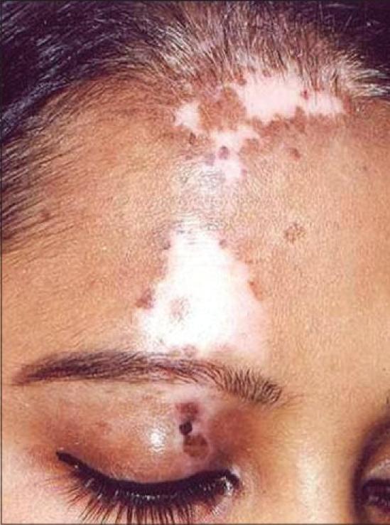
Clinical photograph of the forehead and periocular region of a patient with segmental vitiligo. The image demonstrates well-demarcated, irregularly shaped depigmented (white) macules and patches localized to the upper left quadrant of the face. A large, prominent patch of depigmentation is visible on the central-left forehead, extending toward the hairline. Scattered throughout and adjacent to the depigmented areas are smaller hyperpigmented macules, characteristic of repigmentation clusters following medical or surgical intervention (such as punch grafting). On the left upper eyelid, a cluster of dark, pigmented macules is evident within a localized area of skin alteration, possibly indicating the site of a therapeutic procedure or localized inflammation. The clinical presentation illustrates the dermatological manifestations of vitiligo and the resulting pigmentary variations during treatment monitoring in a dermatology setting.
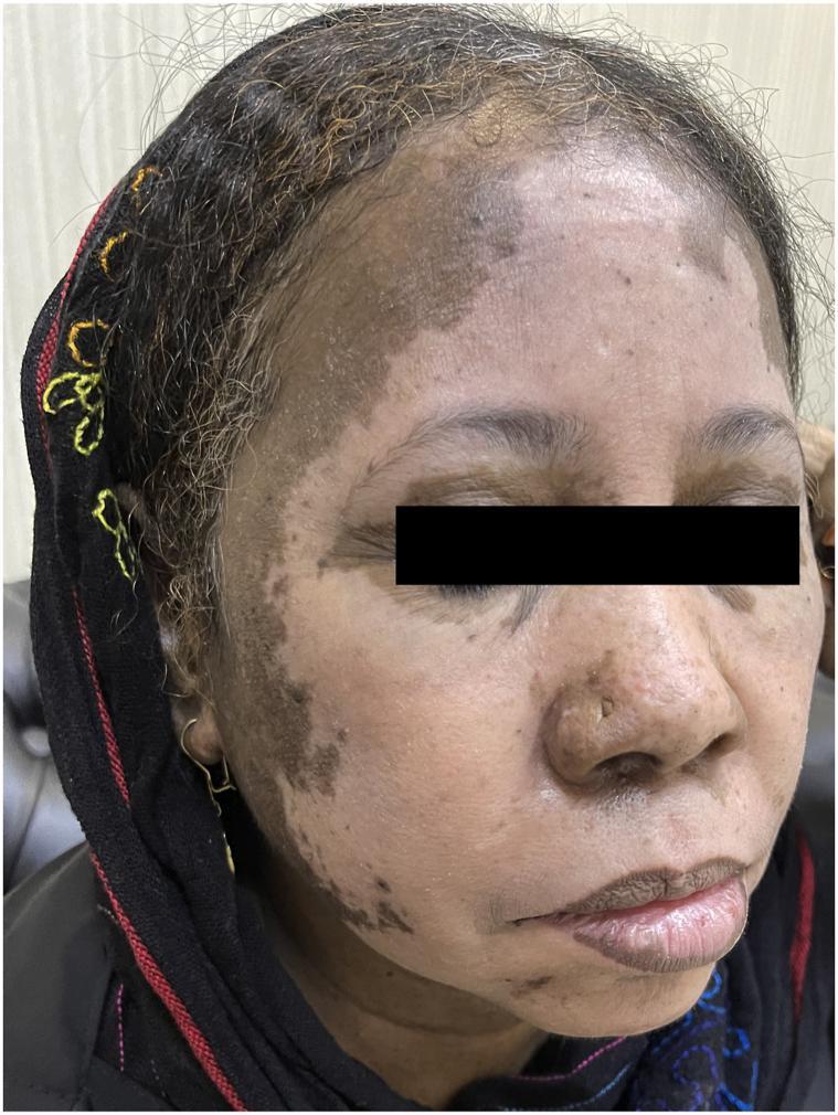
This clinical photograph shows a close-up frontal view of a patient's face, demonstrating extensive, well-demarcated patches of depigmentation. The condition is identified as drug-induced vitiligo secondary to carbamazepine. Large, milk-white depigmented macules and patches are most prominent on the forehead, right cheek, and extending along the preauricular region into the hairline. The distribution is asymmetrical, with notable involvement of the right temple and upper cheek, while the left side shows milder, mottled hypopigmentation. Perioral involvement is also visible, with depigmentation appearing at the vermilion border of the lower lip. The surrounding skin exhibits normal pigmentation, creating a sharp contrast with the affected areas. There is no evidence of secondary changes such as scaling, crusting, or ulceration. This image serves as a clinical example of chemically induced leucoderma, highlighting the importance of medication history in the evaluation of rapid-onset pigmentary disorders.
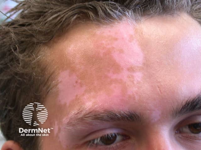
Clinical photography of a living patient displaying segmental vitiligo on the face. Frontal close-up of the forehead and periorbital/temporal region shows patches of depigmentation with sharply defined borders, ranging from pearl to porcelain white, and limited to a unilateral facial dermatomal distribution. Affected areas are hypopigmented to depigmented with uniform color loss and no visible scale or erythema. The surrounding skin shows mild hyperpigmentation at the lesion margins and subtle perifollicular pigment. The imaging modality is standard white-light dermatologic photography; no dermoscopic magnification or fluorescent staining is used. This presentation is typical for segmental vitiligo, an autoimmune melanocyte destruction pattern that often follows a dermatomal distribution and tends to progress rapidly before stabilization. The image demonstrates key diagnostic features including crisp lesion borders and absence of pigment in the epidermis on histology would be expected, but is not visible here. Clinically, vitiligo patches are often more conspicuous in darker skin types and are prone to Koebner phenomenon with friction or trauma. Relevance includes differential diagnosis against post-inflammatory hypopigmentation, pityriasis alba, and fungal hypopigmentation, and relevance for therapeutic planning such as topical corticosteroids, calcineurin inhibitors, photoprotection, and consideration of light therapy. Documented patch stability and follow-up imaging inform treatment decisions effectively.
Vitiligo
Why It Looks This Way
- Flat, white patches with sharp, well-defined borders
- No scaling, crusting, or skin texture change
- The surrounding normal skin has more pigment, creating strong contrast (especially noticeable in darker skin tones)
- Located on the forehead/hairline - one of the most commonly affected sites
Who Gets It
- Affects ~0.5-1% of the world population, but over 8% in some regions of India
- Can start at any age; about half of cases begin before age 20
- Has a genetic component - up to 30% of patients have an affected relative
- Associated with other autoimmune conditions: thyroid disease (most common), type 1 diabetes, and alopecia areata
Treatment Options
1. Topical Treatments (First Line)
| Treatment | Notes |
|---|---|
| Topical corticosteroids (e.g., mometasone, clobetasol) | First-line for localized vitiligo; use with caution on face (risk of skin thinning) |
| Topical calcineurin inhibitors (tacrolimus 0.1%, pimecrolimus) | Preferred on the face - no atrophy risk; effective for facial lesions |
| Ruxolitinib cream (Opzelura) | Newer JAK inhibitor cream; FDA-approved for non-segmental vitiligo (2022); very effective |
2. Phototherapy (Moderately Extensive or Non-responsive)
- Narrowband UVB (NB-UVB) phototherapy - most widely used and effective for widespread vitiligo
- Excimer laser (308 nm) - targeted therapy for focal patches like on the forehead; excellent for facial lesions
- PUVA (psoralen + UVA) - older option, less preferred due to side effects
3. Systemic Treatments (Rapidly Progressive Disease)
- Short course of oral corticosteroids - to halt rapid spread
- Oral JAK inhibitors (ritlecitinib, baricitinib) - emerging evidence for systemic use
4. Surgical Treatments (Stable, Resistant Lesions)
- Minigrafting
- Autologous epidermal cell suspension transplantation
- Ultrathin epidermal grafts
5. Camouflage & Sun Protection
- Sunscreen (SPF 30+) is important - depigmented skin burns easily and UV exposure can worsen patches
- Cosmetic camouflage creams or dihydroxyacetone-based self-tanning products can help match skin tone
Important Notes
- See a dermatologist for confirmation with a Wood's lamp (UV light) exam, which makes depigmented patches glow bright white, confirming the diagnosis
- Vitiligo is not contagious and not dangerous, but can have significant psychological impact - especially in children
- Thyroid function tests are recommended as a screening check, given the autoimmune association
- Early treatment generally gives better results