
Nocturnal enuresis
"nocturnal enuresis"[MeSH Terms] AND management
nocturnal enuresis bedwetting alarm treatment children
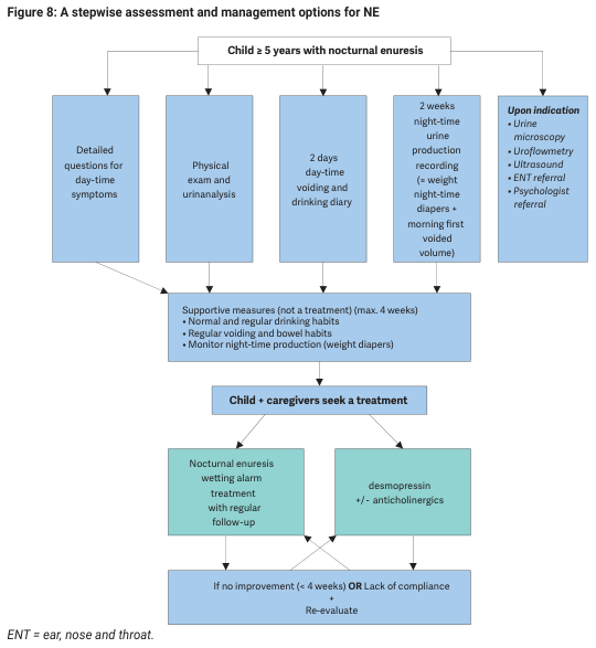
Summary : This flowchart outlines a stepwise assessment and management protocol for children aged 5 years or older with nocturnal enuresis (NE), detailing initial evaluation, supportive measures, treatment options, and re-evaluation steps. flowchart: # Nodes : • Child ≥ 5 years with nocturnal enuresis (rectangle) • Detailed questions for day-time symptoms (rectangle) • Physical exam and urinalysis (rectangle) • 2 days day-time voiding and drinking diary (rectangle) • 2 weeks night-time urine production recording (= weight night-time diapers + morning first voided volume) (rectangle) • Upon indication: Urine microscopy, Uroflowmetry, Ultrasound, ENT referral, Psychologist referral (rectangle) • Supportive measures (not a treatment) (max. 4 weeks): Normal and regular drinking habits, Regular voiding and bowel habits, Monitor night-time production (weight diapers) (rectangle) • Child + caregivers seek a treatment (rectangle) • Nocturnal enuresis wetting alarm treatment with regular follow-up (rectangle) • desmopressin +/- anticholinergics (rectangle) • If no improvement (< 4 weeks) OR Lack of compliance + Re-evaluate (rectangle) • ENT = ear, nose and throat. (footnote) # Connectors : • The initial node ("Child ≥ 5 years with nocturnal enuresis") branches into five parallel assessment nodes. • All five assessment nodes converge into the "Supportive measures" node. • "Supportive measures" leads to "Child + caregivers seek a treatment". • "Child + caregivers seek a treatment" branches into two treatment options: "Nocturnal enuresis wetting alarm treatment with regular follow-up" and "desmopressin +/- anticholinergics". • Both treatment options converge into the final node: "If no improvement (< 4 weeks) OR Lack of compliance + Re-evaluate". # Layout : • The flowchart is organized in a top-down manner, starting with the initial assessment, followed by supportive measures, then treatment options, and ending with re-evaluation. • Parallel nodes for initial assessment are arranged horizontally. • Treatment options are presented as parallel branches below the main treatment-seeking node. # Analysis : • The flowchart provides a clear, stepwise approach for managing nocturnal enuresis in children aged 5 and above. • Initial assessment is comprehensive, including history, physical exam, diaries, and targeted investigations. • Supportive measures are emphasized before initiating treatment. • Two main treatment options are presented, with a built-in mechanism for re-evaluation if initial interventions fail or compliance is poor. • The process is cyclical, allowing for repeated assessment and adjustment of management as needed.
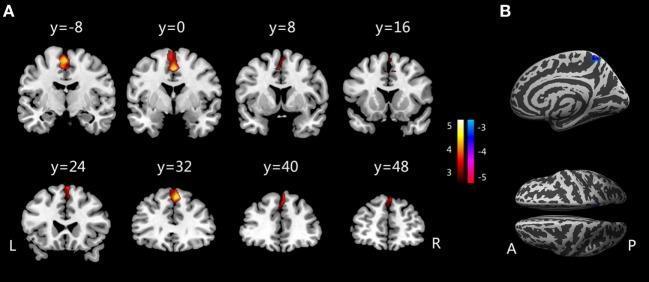
This diagnostic image displays neuroimaging findings comparing brain structures between children with primary monosymptomatic nocturnal enuresis (PMNE) and healthy controls. Section A presents a series of eight axial brain slices (labeled y=-8 to y=48) showing T1-weighted structural templates with heat map overlays. The orange-yellow clusters indicate regions of significantly increased gray matter volume (GMV), predominantly localized in the medial frontal regions, including the supplementary motor area (SMA) and medial prefrontal cortex (PFC). Section B illustrates a cortical surface rendering (gyrification index map) from a sagittal and dorsal perspective. A localized blue-shaded area on the right posterior hemisphere (right precuneus) represents a region of significantly decreased gyrification indices (GI) in the PMNE group. A color scale indicates statistical t-values, where positive values (red/yellow) denote increased GMV and negative values (blue/purple) denote decreased GI. The image provides clinical evidence of structural brain alterations in the central nervous system pathways potentially related to bladder control in pediatric enuresis.
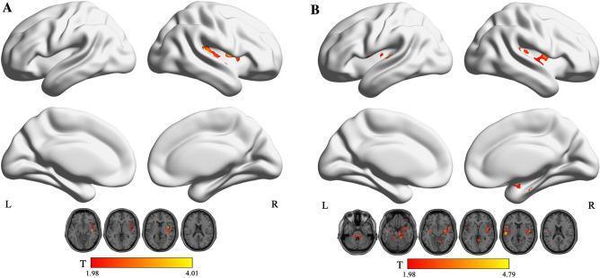
This diagnostic image displays functional magnetic resonance imaging (fMRI) surface renderings and axial slices illustrating abnormal fractional amplitude of low-frequency fluctuation (fALFF) in children with nocturnal enuresis (NE). The image is divided into two comparative panels: (A) shows increased fALFF within the typical low-frequency range (0.01–0.08 Hz), primarily localized to the left insula, with activity intensities reaching a T-value of 4.01. (B) displays results for the slow-5 band (0.01–0.027 Hz), revealing broader clusters of increased activity in the superior cerebellum, right rolandic operculum, and superior temporal gyrus, with higher peak T-values up to 4.79. Each panel includes lateral and medial cortical surface views and representative axial brain slices. Color-coded heatmaps (red to yellow) indicate the statistical significance of regional activity changes. This content is designed for medical researchers and neurology specialists studying the pathophysiology of pediatric urinary disorders and neuroimaging biomarkers.
Nocturnal Enuresis
Definition
Classification
| Type | Description |
|---|---|
| Primary NE | Never achieved a sustained dry period (≥6 months) |
| Secondary NE | Returns after ≥6 months of established dryness - signals possible organic pathology |
| Monosymptomatic NE (MNE) | NE without any other lower urinary tract symptoms |
| Non-monosymptomatic NE | NE + daytime symptoms (urgency, frequency, incontinence) |
Epidemiology
- ~10% of 7-year-olds are affected (Harriet Lane)
- More common in boys: ~7% of boys vs ~3% of girls at age 5
- Spontaneous remission rate: 5-10% per year after age 5
- By age 10: 3% boys, 2% girls
- By age 18: only ~1% boys still wet
- Persists into adulthood in 2-3% of cases
- Strong genetic tendency - if one parent had NE, risk is ~40%; if both parents, ~70%
Pathophysiology / Etiology
- Nocturnal polyuria - reduced nocturnal antidiuretic hormone (AVP/ADH) secretion leading to urine production that exceeds bladder capacity
- Reduced functional bladder capacity - the bladder cannot hold the volume produced overnight; often associated with overactive bladder
- Failure of arousal - the child does not wake up when the bladder contracts or is full (abnormal sleep arousal threshold)
- Genetics (autosomal dominant with variable penetrance in many families)
- Constipation and bowel dysfunction
- Obstructive sleep apnoea (ENT referral if suspected)
- Neurodevelopmental disorders (ADHD, autism) are associated
- Secondary NE: UTI, diabetes mellitus, diabetes insipidus, urologic anomalies, emotional stress, sexual abuse should be ruled out
Assessment
- Primary vs secondary classification
- Birth/developmental history
- Voiding and defecation history (constipation?)
- Daytime symptoms (urgency, frequency, incontinence)
- Family history of enuresis
- Sleep quality, snoring (OSA?)
- Fluid intake patterns
- Parental response / impact on child
- Neurologic exam including gait and anal reflex (wink)
- Lumbosacral examination (spinal dysraphism)
- Abdominal palpation (bladder, constipation)
- Genitalia
- Urinalysis - mandatory (UTI, glucosuria, low specific gravity)
- If history and exam are normal → no further workup needed
- Targeted: urine microscopy, renal ultrasound, uroflowmetry, ENT referral, psychology referral - as indicated
Management
1. Reassurance & Watchful Waiting
- Children < 7 years: reassurance is most appropriate - most will achieve dryness spontaneously
- Remove blame; educate parents that this is not willful behavior
2. General / Supportive Measures (all patients, max 4 weeks trial)
- Fluid restriction in the evening - reduce fluid intake from 1 hour before bedtime
- Regular timed voiding - void before sleep
- Bowel management - treat constipation if present
- Reward systems (star charts) - positive reinforcement, not punishment
- Lifting (waking the child 1.5-2 hrs after sleep onset) - may help reduce wet nights especially in younger children (4-5 yrs)
3. First-Line Treatment: Enuresis Alarm
- Mechanism: conditioning - the alarm wakes the child when wetting begins, training the bladder-arousal response
- Requires 6-12 weeks of consistent use
- Needs motivated child and family
- Success rate: ~60-70%; lower relapse rate than medications once dry
- Combine with dry-bed training for better outcomes
4. Pharmacological Treatment
Desmopressin (First-line pharmacotherapy)
- Synthetic ADH analogue - reduces nocturnal urine production
- Oral tablets (preferred over intranasal - intranasal no longer FDA-indicated for primary NE due to risk of severe hyponatremia and seizures)
- Dose (oral): Start 0.2 mg at bedtime; titrate by 0.2 mg every 3 days up to max 0.6 mg/24 hr (Harriet Lane)
- Fluid restriction mandatory: limit fluids from 1 hour before dose until next morning (at least 8 hrs)
- Effects are good but temporary - high relapse rate on discontinuation
- Key adverse effects: headache, hyponatremia, seizures (if fluid restriction not followed), hypertension
- Caution: hyponatremia risk, renal impairment (avoid if GFR < 50 mL/min), hypertension, CAD
Tricyclic Antidepressants (Second-line; use limited by safety profile)
- Imipramine most commonly used; nortriptyline is an alternative with fewer CNS/anticholinergic side effects
- Mechanism: anticholinergic + central (increased arousal?) + ADH-like effect
- Nortriptyline dosing (Harriet Lane):
- 6-7 yr (20-25 kg): 10 mg PO QHS
- 8-11 yr (26-35 kg): 10-20 mg PO QHS
-
11 yr (36-54 kg): 25-35 mg PO QHS
- Side effects limit use: anorexia, drowsiness, cardiac arrhythmias (QT prolongation)
- Contraindicated with Brugada syndrome, linezolid, IV methylene blue
- Monitor for suicidal ideation; do not discontinue abruptly
- High relapse rate on discontinuation
Anticholinergics (oxybutynin)
- Used as add-on to desmopressin when bladder overactivity is a contributing factor (non-monosymptomatic NE)
- Not effective as monotherapy for pure NE
5. Combination Therapy
- Alarm + desmopressin can be combined for refractory cases
- Desmopressin + anticholinergics for NE with overactive bladder features
Treatment Comparison Summary
| Treatment | Efficacy | Relapse on Stop | Best For |
|---|---|---|---|
| Enuresis alarm | High (60-70%) | Low | Motivated families; long-term cure |
| Desmopressin | Good (short-term) | High | Quick response needed; monosymptomatic NE |
| Tricyclics | Moderate | High | Second-line; limited by side effects |
| Anticholinergics | Limited alone | - | OAB component (add-on) |
Special Considerations
- Adults with NE: In men, may indicate high-pressure chronic urinary retention with risk of upper tract dilation and renal failure - warrants urologic evaluation (Campbell-Walsh-Wein Urology)
- Neurodevelopmental disorders: Higher prevalence of NE in ADHD and autism; management may need to be adapted
- Complementary therapies (acupuncture, hypnotherapy): uncertain/insufficient efficacy evidence
Recent Evidence (2025-2026)
- A 2025 systematic review on management of NE in children (PMID: 39964278) - confirms current management approach
- A 2025 network meta-analysis on pharmacological treatment (PMID: 40272475) - provides comparative data on drug therapies for pediatric NE