
Stripkinase mechanism how clot formed in detail easy word safe dose
Reading File
Now let me search the medical library for streptokinase mechanism, clot formation, and dosing - all in parallel.
Finding Sources
Finding Sources
Finding Sources
Excellent results. Now let me read the key sections from Harrison's (streptokinase mechanism with diagram) and Lippincott's (clot formation + streptokinase dose) in parallel.
Reading File
Reading File
Reading File
I have excellent content. Now let me get the dosing details and fetch the key diagram image.
Finding Sources
Loading Image
Loading Image
I now have all the information needed for a complete, well-illustrated answer. Let me compile it.
Streptokinase - Complete Guide in Easy Words
Part 1: How a Clot Forms (Before Understanding How Streptokinase Works)
Think of a clot like a dam built in a blood vessel after an injury. Here is how it happens step by step:
Step 1 - Blood vessel gets injured
When a vessel wall breaks, it exposes substances underneath (collagen and tissue factor) that were hidden from blood.
Step 2 - Platelets rush in (Primary plug)
Platelets (tiny blood cells) stick to the injured area and clump together. They release chemicals (ADP, serotonin, thromboxane A2) that call more platelets. They also switch on a receptor called GP IIb/IIIa, which grabs fibrinogen (a glue protein) and links platelets together - forming a soft platelet plug.
Step 3 - Coagulation cascade fires (Fibrin net)
The tissue factor triggers a chain reaction (the coagulation cascade) through two pathways - extrinsic and intrinsic. The end result of both is the same: thrombin is made. Thrombin then cuts fibrinogen into fibrin threads. These threads weave a mesh that traps platelets and blood cells.
Step 4 - Clot is stabilized
Factor XIII cross-links the fibrin threads tightly, making the clot strong and firm.
So the final clot = platelets + fibrin mesh + trapped blood cells = a solid plug blocking blood flow.
Part 2: How Streptokinase Breaks the Clot
Streptokinase is a clot-busting drug (thrombolytic/fibrinolytic). It comes from bacteria - beta-hemolytic streptococci.
Key point: Streptokinase is NOT an enzyme itself
It works in a clever indirect way:
Mechanism - Step by Step (Easy)
Step 1 - Streptokinase binds to Plasminogen
In the blood there is a dormant protein called plasminogen (like a sleeping scissors). Streptokinase latches onto plasminogen in a 1:1 ratio, forming a streptokinase-plasminogen complex.
Step 2 - Shape change unlocks the active site
When streptokinase binds, it bends/twists plasminogen into a new shape (conformational change). This reveals a hidden active site - like opening a folded knife.
Step 3 - The complex acts as a factory
This activated streptokinase-plasminogen(now plasmin) complex now attacks OTHER plasminogen molecules in the blood, converting them to plasmin - the actual clot-cutter.
Step 4 - Plasmin digests the fibrin
Plasmin chops up fibrin threads into small pieces called fibrin degradation products (FDPs). The clot dissolves.
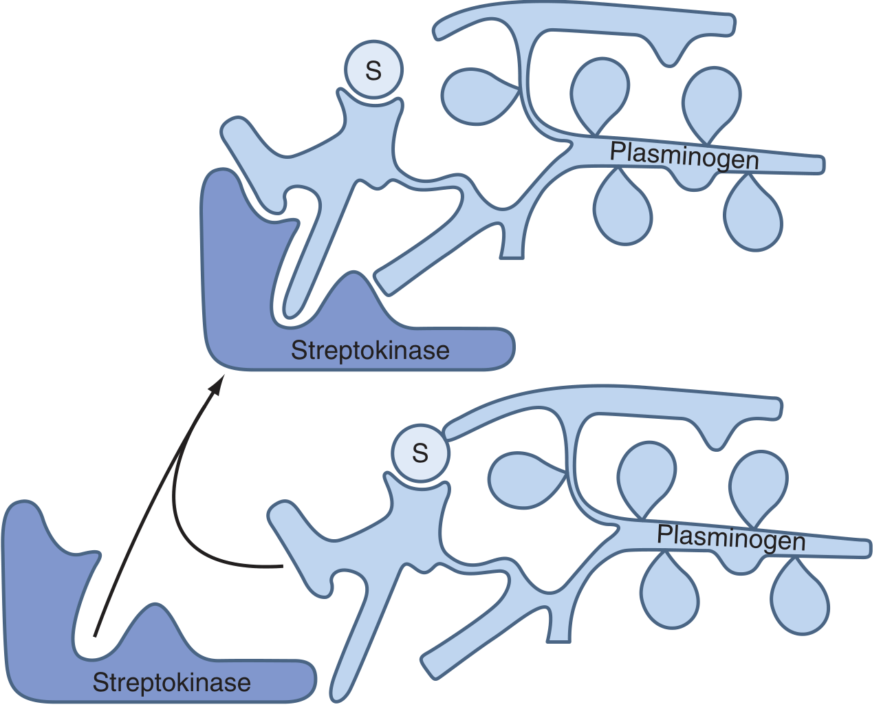
The diagram shows: streptokinase (bottom left, dark blue) binds to plasminogen (right side, light blue) → the complex changes shape (top) → this activated complex converts more plasminogen to plasmin.
The Fibrinolytic System - Big Picture
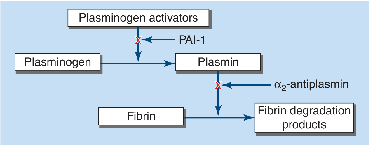
Two natural brakes exist in this system:
- PAI-1 (Plasminogen Activator Inhibitor-1) - blocks plasminogen activators
- α2-antiplasmin - grabs and destroys free plasmin
Streptokinase generates SO MUCH plasmin that it overwhelms α2-antiplasmin, causing a systemic lytic state - meaning fibrin is broken down everywhere in the body, not just at the clot. This is why bleeding is a major risk.
Why Streptokinase is "Non-Specific" (Important Exam Point)
| Feature | Streptokinase | tPA (alteplase) |
|---|---|---|
| Works directly on fibrin? | No - no affinity for fibrin | Yes - fibrin-specific |
| Activates circulating plasminogen? | Yes (both free + clot-bound) | Mostly clot-bound |
| Systemic lytic state? | Yes (high bleeding risk) | Less so |
| Enzyme itself? | No (allosteric modifier) | Yes |
Because streptokinase has no affinity for fibrin, it activates plasminogen floating freely in the blood too - not just the plasminogen sitting on the clot. This floods the body with plasmin, breaking down clotting factors everywhere.
Part 3: Safe Dose
| Indication | Dose | Route & Duration |
|---|---|---|
| Acute MI (STEMI) | 1.5 million IU (1,500,000 IU) | IV infusion over 60 minutes |
| Pulmonary embolism | 250,000 IU loading over 30 min, then 100,000 IU/hr for 24 hrs | IV infusion |
| Deep vein thrombosis | 250,000 IU loading, then 100,000 IU/hr for 72 hrs | IV infusion |
The standard and most commonly tested dose is 1.5 million IU IV over 1 hour for acute STEMI.
- Textbook of Family Medicine 9e - Table 27-16: "Streptokinase: 1.5 million IU IV, give infusion over 60 min"
Part 4: Important Safety Points / Contraindications
- Allergic reactions / anaphylaxis - streptokinase is a foreign bacterial protein; antibodies from prior streptococcal infection can cause reactions
- Cannot be re-used within 5 years - prior exposure creates antibodies that neutralize it
- Hypotension - give slowly; can cause BP drop
- Major bleeding risk - 20% risk of major hemorrhage; 3-5% risk of hemorrhagic stroke
- Contraindicated in: recent surgery, active bleeding, history of stroke, severe hypertension, pregnancy
Quick Memory Summary
Injury → Platelet plug + Fibrin mesh → CLOT
         ↑
Thrombin made from coagulation cascade
         ↑
Streptokinase → binds Plasminogen → conformational change
             → SK-Plasminogen complex → converts more Plasminogen → PLASMIN
                                                                    ↓
                                                          Fibrin → FDPs (clot dissolves)
Dose to remember: 1.5 million IU IV over 60 min (for STEMI)
Sources: Harrison's Principles of Internal Medicine 22E, p. 998 | Braunwald's Heart Disease | Lippincott Illustrated Reviews: Pharmacology | Textbook of Family Medicine 9e
Mechanism of Tramadol and how body feel pain and normal dose of Tramadol
Reading File
Now let me search all three topics in parallel - tramadol mechanism, pain physiology, and tramadol dosing.
Finding Sources
Finding Sources
Finding Sources
Good results. Now let me read the detailed tramadol section from Katzung and the pain pathway from Ganong, plus the dosing table from Goldman-Cecil - all in parallel.
Reading File
Reading File
Reading File
Excellent content from the textbooks. Now let me get the pain pathway details from Ganong and fetch the key opioid mechanism image.
Reading File
Loading Image
I now have all the information needed. Let me also quickly search for the pain pathway diagram and a tramadol-specific image.
Finding Sources
Finding Sources
Reading File
I now have all the content I need. Let me compile the complete, well-structured answer.
Tramadol - Complete Guide in Easy Words
Part 1: How the Body Feels Pain (Before Understanding Tramadol)
Think of the pain system like a fire alarm system in a building. Here is how it works step by step:
Step 1 - The Alarm is Triggered (Nociceptors)
When tissue is damaged - by a cut, burn, pressure, or inflammation - special nerve endings called nociceptors (pain receptors) are activated. They are found in skin, muscles, organs, and joints.
Two types of pain nerve fibers carry the signal:
- Aδ (A-delta) fibers - thin, myelinated, fast. They carry sharp, sudden, "first pain" (like when you first touch something hot).
- C fibers - very thin, unmyelinated, slow. They carry dull, burning, throbbing "second pain" that lingers.
These fibers fire when exposed to heat, pressure, chemicals (like bradykinin, histamine, ATP, prostaglandins) or acidic environments.
Step 2 - Signal Travels to the Spinal Cord
The pain signal travels from the nociceptor up the nerve → into the dorsal horn of the spinal cord. Here, the pain neuron releases chemicals called glutamate and neuropeptides (substance P) onto the next neuron (spinothalamic tract neuron).
This is the relay station - the signal is processed here and "decided" whether to go up to the brain.
Step 3 - Signal Goes to the Brain (Spinothalamic Tract)
The spinothalamic tract carries the signal upward to:
- Thalamus - the brain's relay center
- Somatosensory cortex - where you feel WHERE the pain is
- Limbic system - where you feel the EMOTIONAL suffering (fear, anxiety, distress)
This is why pain is both a sensation AND an emotion.
Step 4 - The Body's Own Pain Brake System
The brain also has a descending pain control system. From the brainstem (periaqueductal gray, PAG), signals travel back DOWN to the spinal cord and release:
- Endorphins / enkephalins - natural opioids that block pain signals at the dorsal horn
- Norepinephrine and serotonin - released from descending fibers to suppress pain transmission at the spinal cord level
This is exactly where tramadol works!
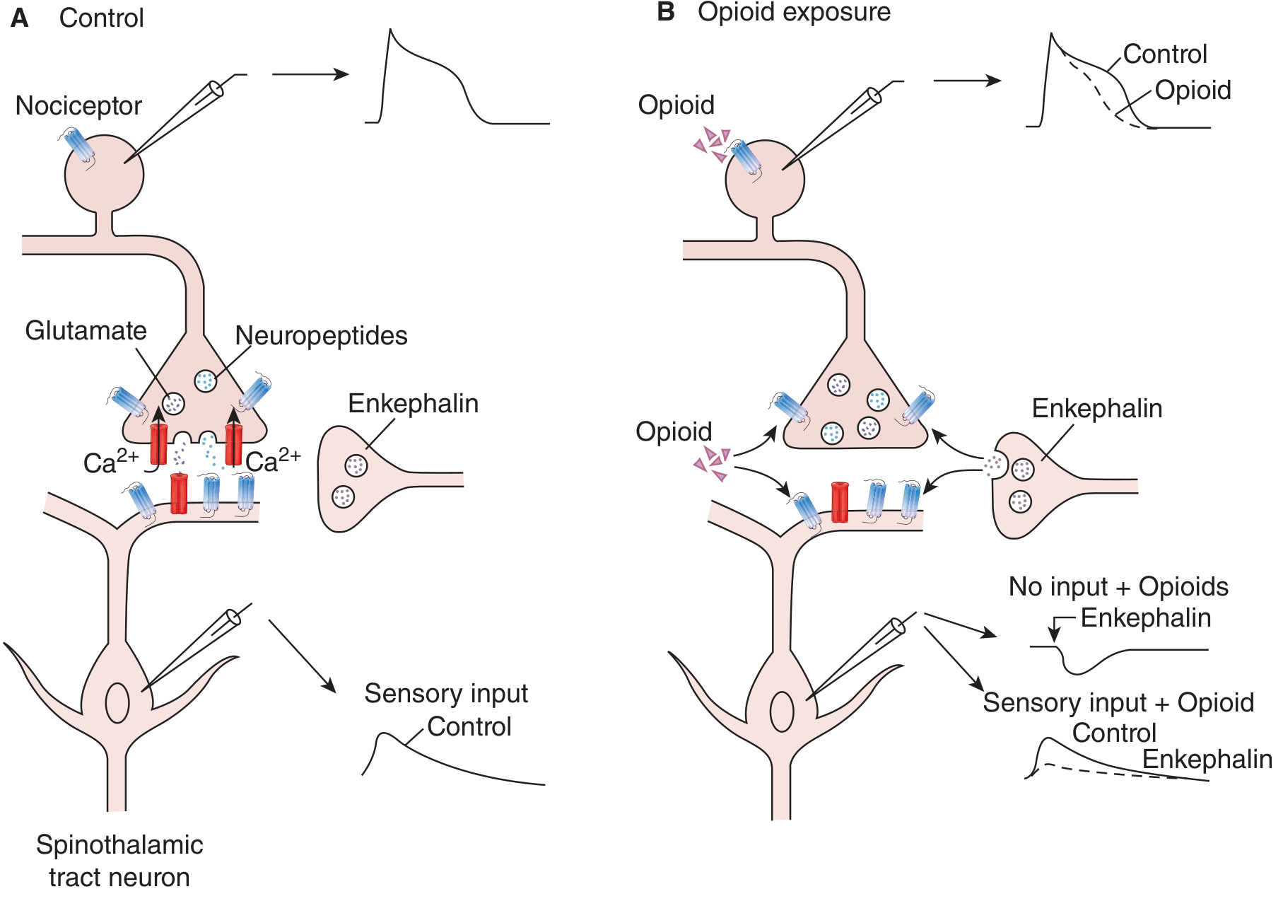
Left (A): Normal pain signal path. Right (B): Opioid/tramadol suppresses the signal - less calcium enters, less glutamate is released, and the spinal cord neuron is harder to activate.
Part 2: Mechanism of Tramadol (Dual Action)
Tramadol is a synthetic codeine analogue with a dual (two-pronged) mechanism - it works in two completely different ways at the same time.
Mechanism 1 - Weak Opioid Action (mu receptor)
Tramadol (and its active metabolite M1/O-desmethyltramadol) binds to mu-opioid receptors (MOR) in the brain and spinal cord.
When it binds:
- Calcium channels (Ca²⁺) close at the pain neuron's nerve ending → less glutamate and substance P are released → pain signal is not passed forward
- Potassium channels (K⁺) open at the receiving neuron → the cell hyperpolarizes (becomes harder to fire) → pain signal blocked
Important: Tramadol's affinity for the opioid receptor is much WEAKER than morphine. Its active metabolite M1 is 2-4x more potent than the parent drug and contributes significantly to pain relief.
Mechanism 2 - Reuptake Inhibition (like an antidepressant)
Tramadol blocks the reuptake of serotonin and norepinephrine back into nerve endings. This means more of these chemicals stay active in the synapse.
- More norepinephrine → strengthens the brain's descending pain-suppression system → blocks pain signals at the spinal cord
- More serotonin → also activates descending inhibitory pathways → reduces pain transmission
Tramadol comes as a racemic mixture (two mirror-image forms):
- (+) enantiomer - binds opioid receptor + blocks serotonin reuptake
- (−) enantiomer - blocks norepinephrine reuptake + activates α₂ receptors
Both forms together are MORE effective than either alone - that's why it's kept as a mixture.
Simple Summary of Dual Action:
TRAMADOL
    │
    ├──► Opioid receptor (μ) → blocks pain signal at spinal cord
    │         (Mechanism 1 - like a weak morphine)
    │
    └──► Blocks reuptake of Serotonin + Norepinephrine
              (Mechanism 2 - like an antidepressant SNRI)
              → activates the brain's own descending pain-control system
Part 3: Normal (Safe) Dose of Tramadol
| Form | Dose | Frequency | Maximum Daily Dose |
|---|---|---|---|
| Immediate-release (oral) | 50-100 mg per dose | Every 4-6 hours | 400 mg/day |
| Extended-release (ER) | 100-300 mg | Once daily | 300 mg/day |
| Acute pain (start low) | 50 mg | Every 4-6 hours | 400 mg/day |
| Chronic/neuropathic pain | 200-400 mg/day | Divided doses | 400 mg/day |
| Elderly (>75 years) | Max 300 mg/day | - | 300 mg/day |
- Onset of action: within 1 hour of oral dose
- Peak effect: 2-3 hours
- Duration: about 6 hours
- Half-life: 6 hours (tramadol), 7.5 hours (active metabolite M1)
- Bioavailability: 68% after single oral dose
The standard dose is 50-100 mg every 6 hours, not exceeding 400 mg per day.
Part 4: Side Effects and Safety Points
| Side Effect | Reason |
|---|---|
| Nausea, vomiting | Opioid effect on gut + CTZ in brain |
| Dizziness, sedation | CNS opioid effect |
| Constipation | Slowed gut motility (opioid) |
| Dry mouth, sweating | Serotonergic effect |
| Seizures | Lowers seizure threshold (risk if used with SSRIs/SNRIs/TCAs) |
| Serotonin syndrome | Too much serotonin - fever, agitation, muscle rigidity (DO NOT combine with MAOIs) |
Key Drug Interactions (Dangerous):
- MAO inhibitors - severe serotonin syndrome - CONTRAINDICATED
- SSRIs / SNRIs / TCAs - increased seizure risk + serotonin syndrome
- Triptans - serotonin syndrome risk
- Warfarin/Coumadin - tramadol exacerbates anticoagulant effect
Advantages over classic opioids:
- Lower respiratory depression risk
- Lower addiction/dependence potential (but NOT zero - abuse has been reported)
- Effective for neuropathic pain (due to SNRI component)
Quick Memory Trick:
TRAMADOL = T-wo mechanisms
 T - opioid receptor (μ) agonist (weak)
 R - euptake inhibitor of Serotonin + Norepinephrine

DOSE: 50-100 mg every 4-6 hours | MAX 400 mg/day
Sources: Katzung's Basic & Clinical Pharmacology 16e | Firestein & Kelley's Textbook of Rheumatology | Harrison's Principles of Internal Medicine 22e | Ganong's Review of Medical Physiology 26e
Mechanism of Tramadol and how body feel pain and normal dose of Tramadol
You asked the same question I just answered in detail! Here is a complete summary from the previous response (sourced from Harrison's, Katzung's, Ganong's, and Firestein's textbooks):
Tramadol - Complete Guide in Easy Words
Part 1: How the Body Feels Pain
Think of the pain system like a fire alarm - injury triggers it, nerves carry the signal, and the brain registers it.
Step 1 - Nociceptors Detect the Injury
Special pain receptors called nociceptors are activated by heat, pressure, chemicals (bradykinin, histamine, prostaglandins), or acidic pH. Two types of nerve fibers carry the signal:
- Aδ (A-delta) fibers - fast, sharp "first pain" (the sudden sting)
- C fibers - slow, dull "second pain" (the burning that lingers)
Step 2 - Signal Travels to the Spinal Cord
Pain fibers enter the dorsal horn of the spinal cord and release glutamate + substance P onto the next neuron. This is the key relay point.
Step 3 - Signal Goes Up to the Brain
The spinothalamic tract carries the signal to:
- Thalamus → relay center
- Somatosensory cortex → WHERE you feel the pain
- Limbic system → the EMOTIONAL suffering (fear, anxiety)
This is why pain is both a sensation AND an emotion.
Step 4 - The Brain's Own Brake System
The brain sends signals back DOWN (descending pathways from the periaqueductal gray) releasing endorphins, norepinephrine, and serotonin at the spinal cord - naturally suppressing pain. This is exactly where tramadol acts.
Part 2: Mechanism of Tramadol (Two Actions Together)
Tramadol is a synthetic codeine analogue with a dual mechanism - it hits the pain system in two different ways simultaneously.
Action 1 - Weak Opioid (mu receptor agonist)
Tramadol binds to mu-opioid receptors (μ-MOR) in the brain and spinal cord:
- Closes Ca²⁺ channels at the pain neuron terminal → less glutamate/substance P released
- Opens K⁺ channels at the receiving neuron → cell hyperpolarizes → harder to fire → pain signal blocked
Its active metabolite M1 (O-desmethyltramadol) is 2-4x more potent than tramadol itself and carries a large share of the opioid effect.
Action 2 - Reuptake Inhibition (SNRI-like)
Tramadol blocks reuptake of serotonin and norepinephrine → these accumulate in synapses → amplify the brain's own descending pain suppression system.
Tramadol is a racemic mixture (two mirror forms that work together):
- (+) enantiomer → binds opioid receptor + inhibits serotonin reuptake
- (−) enantiomer → inhibits norepinephrine reuptake + activates α₂ receptors
TRAMADOL
    │
    ├──► μ-Opioid receptor agonist (weak)
    │         → blocks pain at spinal cord level
    │
    └──► Serotonin + Norepinephrine reuptake inhibitor
              → boosts descending pain suppression from brain
Part 3: Normal (Safe) Dose
| Form | Dose Per Dose | Frequency | Max Daily Dose |
|---|---|---|---|
| Immediate-release (oral) | 50-100 mg | Every 4-6 hours | 400 mg/day |
| Extended-release | 100-300 mg | Once daily | 300 mg/day |
| Elderly (>75 yrs) | Lower doses | - | 300 mg/day |
- Onset: within 1 hour (oral)
- Peak effect: 2-3 hours
- Duration: ~6 hours
- Half-life: 6 hours (tramadol), 7.5 hours (M1 metabolite)
- Bioavailability: 68%
Standard dose: 50-100 mg orally every 6 hours. Never exceed 400 mg/day.
Part 4: Key Side Effects and Safety
| Side Effect | Why it Happens |
|---|---|
| Nausea, vomiting | Opioid effect on gut and brain |
| Dizziness, sedation | CNS opioid effect |
| Constipation | Slowed gut motility |
| Dry mouth, sweating | Serotonergic effect |
| Seizures | Lowers seizure threshold |
| Serotonin syndrome | Excess serotonin - fever, agitation, rigidity |
Dangerous Combinations (Avoid):
- MAO inhibitors - serotonin syndrome - CONTRAINDICATED
- SSRIs / SNRIs / TCAs - seizures + serotonin syndrome
- Triptans - serotonin syndrome
- Warfarin - enhanced anticoagulant effect
Advantages over classic opioids:
- Lower respiratory depression
- Lower addiction potential (though not zero)
- Works on neuropathic pain too (due to SNRI component)
Quick Memory Formula:
TRAMADOL = TWO mechanisms
  1. Weak opioid (μ receptor) → blocks spinal pain relay
  2. SNRI-like → boosts brain's own pain brake

DOSE: 50-100 mg every 4-6 hours | MAX 400 mg/day
Sources: Katzung's Basic & Clinical Pharmacology 16e | Firestein & Kelley's Textbook of Rheumatology | Harrison's Principles of Internal Medicine 22e | Ganong's Review of Medical Physiology 26e
How body get cramps and mechanism of levocratinine dose
Reading File
Searching all three topics in parallel - muscle cramp mechanism, levocarnitine mechanism, and levocarnitine dose.
Finding Sources
Finding Sources
Finding Sources
Let me broaden the searches.
Finding Sources
Finding Sources
Finding Sources
Good findings. Now let me read the detailed cramp section from Bradley & Daroff's, the carnitine transport diagram from Basic Medical Biochemistry, and levocarnitine dose details - all in parallel.
Reading File
Reading File
Reading File
Excellent content. Now let me get the carnitine diagram image, the levocarnitine dosing details, and the EMG cramp discharge details together.
Loading Image
Finding Sources
Finding Sources
Finding Sources
Reading File
I now have comprehensive content from the textbooks. Let me compile the full answer.
Muscle Cramps + Levocarnitine - Complete Guide in Easy Words
Part 1: How the Body Gets Cramps
What is a Cramp?
A cramp is a sudden, painful, involuntary contraction of a muscle or muscle group. The muscle becomes hard and tightly knotted. It lasts from seconds to minutes, then relaxes on its own. Stretching the muscle relieves it.
Normal Muscle Contraction (How Muscles Work - Background)
Think of muscle fibers like a rope that gets pulled tight. Normally:
- The brain sends a nerve signal → travels down the motor nerve to the muscle
- At the neuromuscular junction, the nerve releases acetylcholine
- This triggers calcium (Ca²⁺) to rush out of the muscle's storage tanks (sarcoplasmic reticulum)
- Calcium activates actin-myosin bridges → the muscle contracts
- When the signal stops, calcium is pumped back → muscle relaxes
This is a controlled, brief, coordinated process.
How a Cramp Happens (The Mechanism)
A cramp occurs when motor nerve terminals or motor axons fire spontaneously and repetitively without any instruction from the brain. On EMG (electrical test), cramp discharges show:
- Motor units firing at 40-150 times per second (Hz)
- Abrupt onset and cessation
The muscle fires uncontrollably - like a car alarm going off on its own. The key trigger is hyperexcitability of the motor nerve terminal.
Why does the nerve become hyperexcitable? Several reasons:
| Cause | How it triggers cramps |
|---|---|
| Dehydration | Blood volume drops → nerve endings become irritable |
| Hyponatremia (low sodium) | Disrupts nerve membrane potential - nerve fires easily |
| Hypocalcemia (low calcium) | Ca²⁺ normally stabilizes nerve membranes; when low, membranes fire spontaneously - classic tetany/cramp |
| Hypomagnesemia (low magnesium) | Mg²⁺ acts as a natural calcium channel blocker; when low, uncontrolled Ca²⁺ entry → hyperexcitability |
| Exercise/fatigue | Lactic acid buildup + local electrolyte shifts irritate nerve terminals |
| Pregnancy | Electrolyte shifts + compression of nerves |
| Dialysis/post-dialysis | Rapid fluid and electrolyte shifts |
| Motor neuron disease | Damaged/partially denervated muscles become spontaneously excitable |
| Carnitine deficiency | Energy failure in muscle → metabolic cramps |
Where Do Cramps Originate?
Cramps originate in motor axons or nerve terminals (peripheral), NOT in the muscle fiber itself. This is proven because:
- Cramps show motor unit action potentials on EMG (nerve-driven)
- Contractures (true muscle fiber contraction without nerve activity) show electrical silence on EMG
- CNS has only minor modulating influence on cramp threshold
Most Common Cramp Locations and Times
- Calf and foot muscles - most common (especially at night)
- Night cramps - very common, especially in elderly (up to 50%)
- Exercise-related cramps - during or immediately after exercise
- Pregnancy - due to electrolyte and fluid changes
Simple Summary of Cramp Mechanism
Normal: Brain → nerve signal → controlled Ca²⁺ release → contraction → relaxation

Cramp:  Motor nerve terminal fires spontaneously (no brain command)
              ↑
        Triggered by: low electrolytes (Na⁺, Ca²⁺, Mg²⁺)
                      dehydration, fatigue, nerve damage, carnitine deficiency
              ↓
        Uncontrolled repetitive firing at 40-150 Hz
              ↓
        Sustained involuntary muscle contraction = CRAMP
Part 2: Levocarnitine (L-Carnitine) - Mechanism
What is Levocarnitine?
Levocarnitine is the biologically active form of carnitine (also called L-carnitine). It is a vitamin-like compound naturally made in the body from amino acids (lysine and methionine). It is also found in red meat and dairy.
The Role of Carnitine in Energy Production - The Core Mechanism
The body gets energy from burning (oxidizing) fat inside mitochondria (the cell's power plants). But long-chain fatty acids cannot enter the mitochondria on their own - they need carnitine as a taxi/shuttle to carry them across the double mitochondrial membrane.
Here is the step-by-step process:
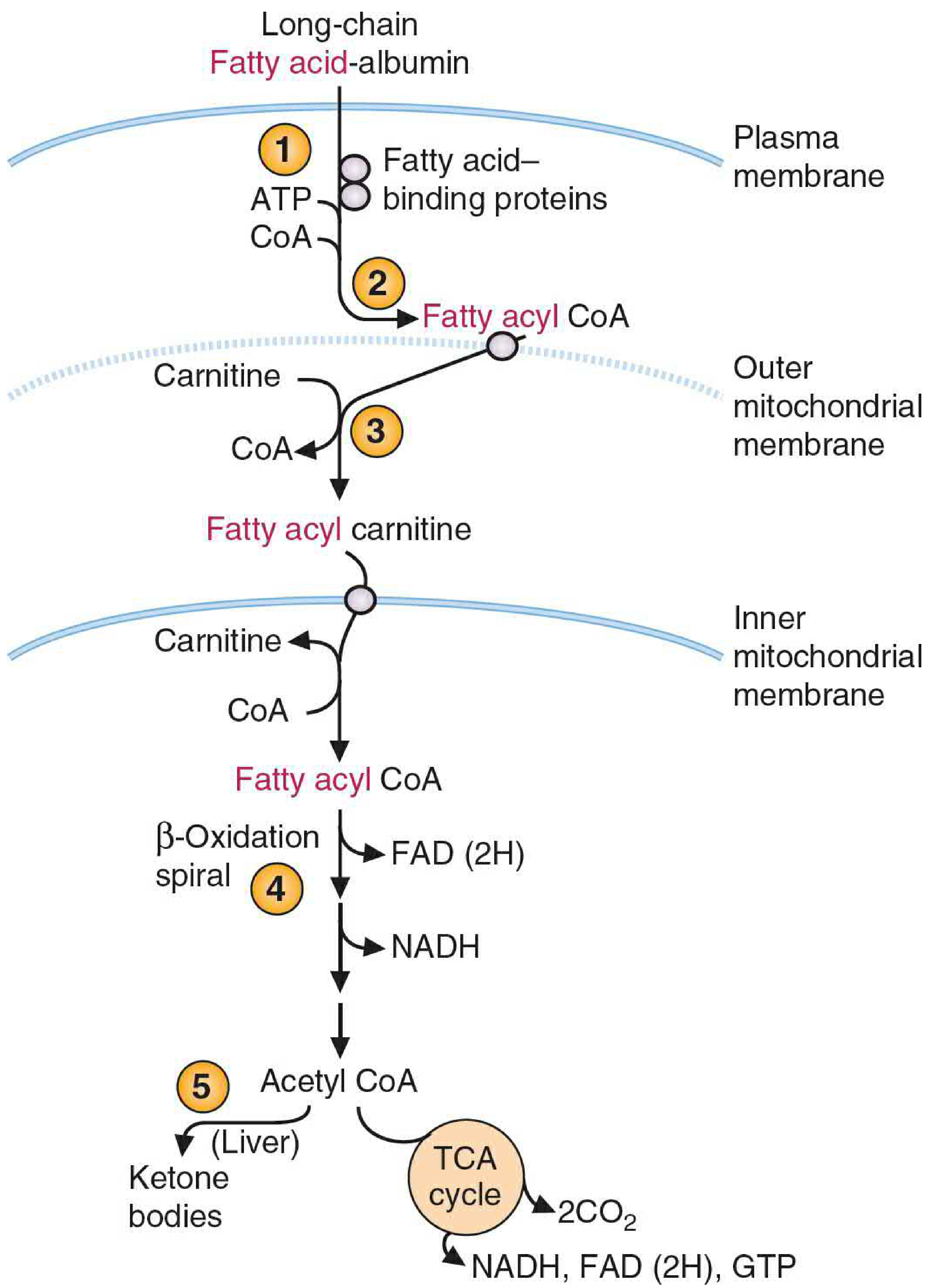
Step 1 - Long-chain fatty acids travel through blood bound to albumin and enter the cell via fatty acid-binding proteins.
Step 2 - Inside the cell, fatty acids are activated by attaching to CoA (Coenzyme A), forming Fatty Acyl-CoA.
Step 3 - Carnitine (the taxi) picks up the fatty acyl group from CoA at the outer mitochondrial membrane. The enzyme CPT-1 (Carnitine Palmitoyl Transferase-1) does this, forming Fatty Acyl-Carnitine.
Step 4 - The Fatty Acyl-Carnitine crosses the inner mitochondrial membrane via a transporter (CACT - carnitine-acylcarnitine translocase). Carnitine is released back to the outside for reuse.
Step 5 - Inside the mitochondria, the fatty acyl group reattaches to CoA (via CPT-2), becoming Fatty Acyl-CoA again.
Step 6 - Beta-oxidation begins: the fatty acid chain is cut up repeatedly, generating:
- NADH → enters electron transport chain → makes ATP
- FAD(2H) → enters electron transport chain → makes ATP
- Acetyl-CoA → enters the TCA (Krebs) cycle → makes more ATP
Result: Fat is burned to make energy (ATP). Without carnitine, this entire process stops. The muscle and heart cells starve for energy even though fat is available.
What Happens When Carnitine is Deficient?
| Effect | Reason |
|---|---|
| Muscle weakness and cramps | Muscles cannot burn fat for energy; they run out of ATP |
| Hypoglycemia | Body cannot use fat; over-relies on glucose |
| Cardiomyopathy | Heart muscle starves (heart mainly uses fat for fuel) |
| Liver dysfunction | Fat accumulates (steatosis - fatty liver) |
| Brain dysfunction | Energy failure, especially during fasting |
Carnitine deficiency commonly occurs in:
- Chronic kidney disease / dialysis patients - carnitine is lost in dialysis
- Genetic disorders (primary carnitine deficiency)
- Premature infants - cannot synthesize enough
- Valproic acid use - drug depletes carnitine
Levocarnitine Mechanism Summary
Without carnitine:
Fat (Fatty Acyl-CoA) → STUCK at outer mitochondrial membrane → NO energy → muscle/heart failure

With levocarnitine:
Fat (Fatty Acyl-CoA) + Carnitine → CPT-1 → Fatty Acyl-Carnitine
                                         → crosses inner membrane (CACT)
                                         → CPT-2 → Fatty Acyl-CoA inside
                                         → Beta-oxidation → NADH + FAD(2H) + Acetyl-CoA
                                         → TCA cycle + Electron Transport Chain
                                         → ATP (ENERGY) ✓
Part 3: Normal (Safe) Dose of Levocarnitine
| Indication | Form | Dose | Frequency |
|---|---|---|---|
| Primary carnitine deficiency (adults) | Oral | 990 mg - 3 g/day | 2-3 divided doses |
| Primary carnitine deficiency (children) | Oral | 50-100 mg/kg/day | Divided doses |
| End-stage renal disease / dialysis | IV (post-dialysis) | 10-20 mg/kg per dialysis session | After each session |
| Acute metabolic crisis (illness) | IV | 100 mg/kg/day | Every 6 hours |
| Valproic acid toxicity | IV | 100 mg/kg loading | Divided doses |
Key Pharmacokinetic Points:
- Oral bioavailability: ~15-25% (fairly low; IV is much more effective for severe deficiency)
- Half-life: ~17 hours
- Excess carnitine is excreted in urine
- Available as: oral solution (1 g/10 mL), tablets (330 mg), and IV solution (200 mg/mL)
Quick Memory Summary
CRAMPS = Nerve terminal fires on its OWN
  Trigger: low Na⁺, Ca²⁺, Mg²⁺ | dehydration | fatigue | dialysis | nerve damage
  On EMG: 40-150 Hz motor unit firing
  Relief: stretch the muscle

LEVOCARNITINE = Fatty acid TAXI into mitochondria
  Without it: fat cannot be burned → no energy → muscle weakness/cramps
  With it: Fat → Beta-oxidation → ATP (energy)

  DOSE: Adults: 990 mg - 3 g/day orally (divided)
        Dialysis: 10-20 mg/kg IV per session
        Children: 50-100 mg/kg/day
Sources: Bradley & Daroff's Neurology in Clinical Practice | Basic Medical Biochemistry 6e (Lippincott) | Guyton & Hall Textbook of Medical Physiology | Thompson & Thompson Genetics and Genomics in Medicine 9e
This is a shared conversation. Sign in to Orris to start your own chat.