
Development of axial skeleton system with diagram
Reading File
The axial skeleton develops from somites, paraxial mesoderm, and neural crest cells through a process called somitogenesis and chondrification/ossification. The vertebral column, skull, ribs, and sternum are the main components.
Searching sources...
Finding Sources
Finding Sources
Reading File
Reading File
Loading Image
Loading Image
Reading File
Loading Image
Reading File
Reading File
Loading Image
Reading File
Loading Image
Here is a comprehensive, textbook-based account of the development of the axial skeleton, drawn from The Developing Human: Clinically Oriented Embryology.
Development of the Axial Skeleton
The axial skeleton consists of the cranium (skull), vertebral column, ribs, and sternum. Its development begins in the third week of embryogenesis and continues into early adult life.
1. Overview: Somites and the Sclerotome
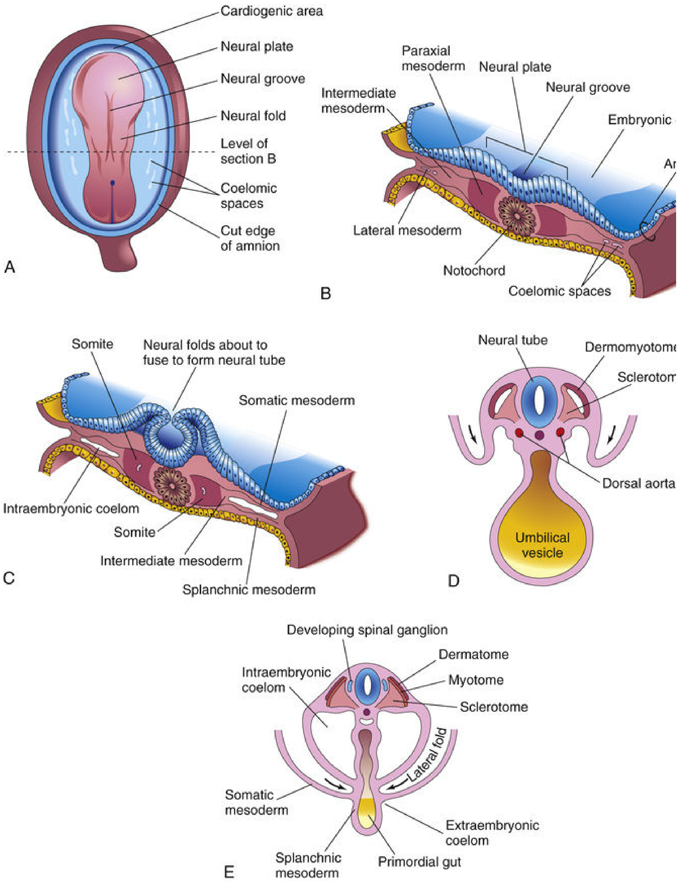
During the third week, intraembryonic mesoderm lateral to the notochord and neural tube thickens to form two longitudinal columns of paraxial mesoderm. These columns become segmented into blocks called somites (bead-like elevations along the dorsolateral embryo surface).
Each somite differentiates into two parts:
| Part | Region | Derivative |
|---|---|---|
| Sclerotome (ventromedial) | Surrounds neural tube and notochord | Vertebrae, ribs |
| Dermomyotome (dorsolateral) | - Myotome region - Dermatome region | Muscle (myoblasts) - Dermis (fibroblasts) |
2. Development of the Vertebral Column
Stages of Vertebral Development
Stage 1 - Mesenchymal (Precartilaginous) Stage
During the fourth week, sclerotomal cells migrate to three locations:
- Around the notochord (future vertebral bodies)
- Surrounding the neural tube (future vertebral arches)
- Into the body wall (future ribs)
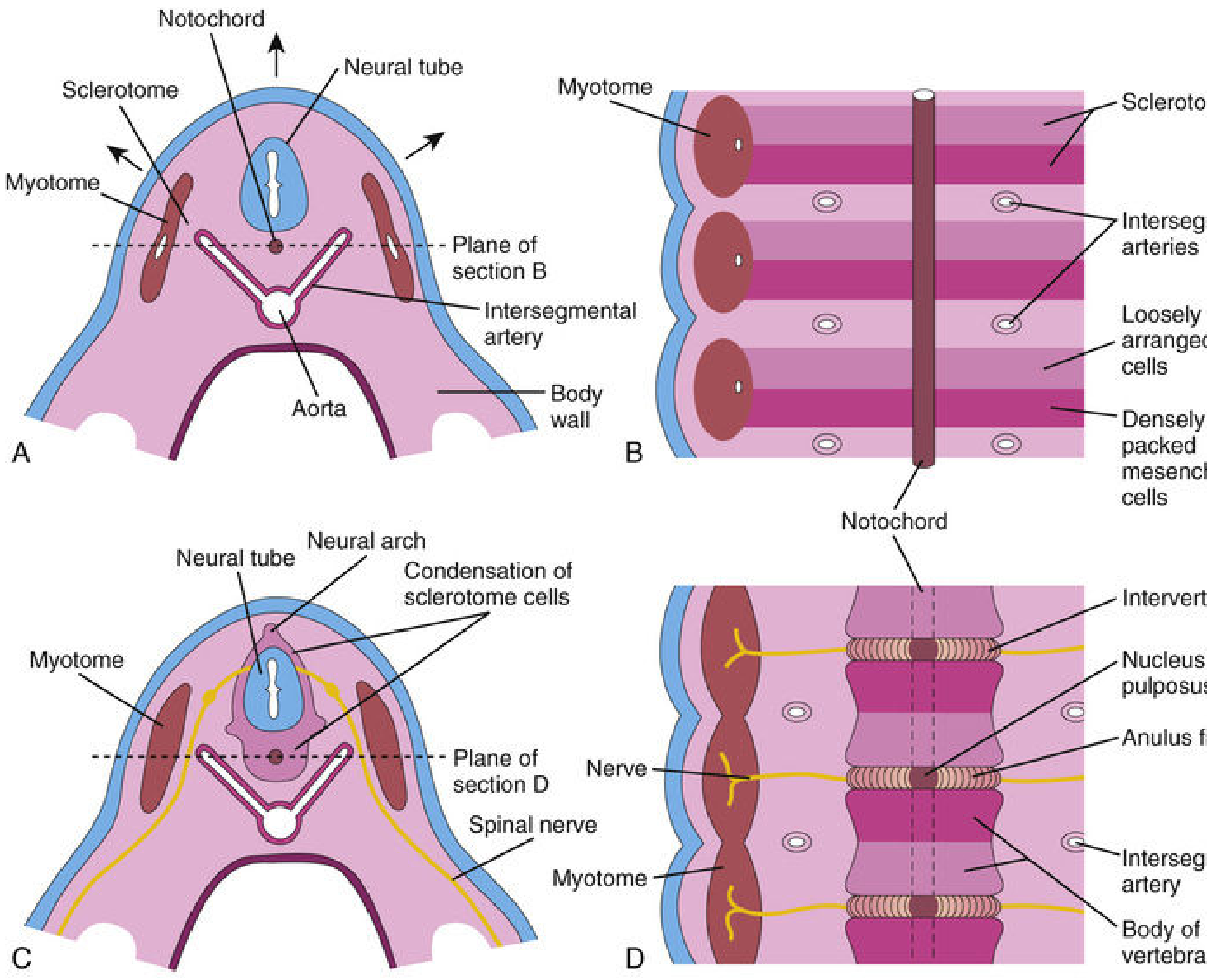
Each sclerotome has a cranial zone of loosely packed cells and a caudal zone of densely packed cells. The densely packed cells from one sclerotome fuse with the loosely arranged cells of the immediately caudal sclerotome to form the mesenchymal centrum (primordium of the vertebral body). This means each vertebral body is an intersegmental structure, derived from two adjacent sclerotomes - this is why spinal nerves lie between vertebrae and intersegmental arteries cross the vertebral bodies.
The notochord degenerates within the vertebral bodies but persists between them, expanding to form the nucleus pulposus of the intervertebral disc. The annulus fibrosus forms from surrounding mesenchyme. The neural arch (primordium of the vertebral arch) forms from mesenchyme surrounding the neural tube.
Key molecular regulators:
- TBX6, Hox, and PAX genes - regulate patterning along the anterior-posterior axis
- Fat4 and Dchs1 (protocadherins) - mediate planar cell polarity and early chondrogenesis
- Notch signaling - patterning of the vertebral column
Stage 2 - Cartilaginous Stage
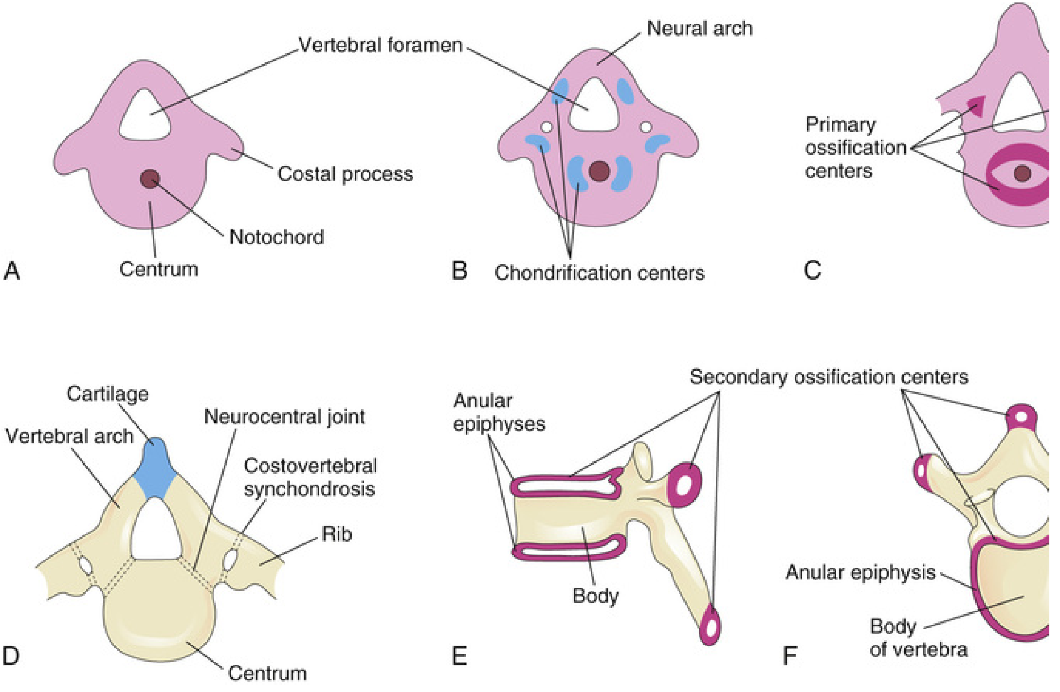
During the sixth week, chondrification centers appear in each mesenchymal vertebra. The two centers in each centrum fuse to form a cartilaginous centrum by the end of the embryonic period. Centers in the neural arches fuse with each other and with the centrum. Chondrification spreads until a complete cartilaginous vertebral column is formed.
Stage 3 - Bony (Ossification) Stage
Ossification begins in the seventh week and is not complete until age 25.
Primary ossification centers (3 per vertebra):
- 1 in the centrum (from fusion of ventral + dorsal centers)
- 1 in each half of the neural arch (present by 8th week)
Secondary ossification centers (5 per vertebra, appear after puberty):
- Tip of spinous process (1)
- Tip of each transverse process (2)
- Anular epiphyses - superior and inferior rims of vertebral body (2)
All secondary centers unite with the vertebra by approximately age 25.
Normal vertebral numbers: 7 cervical, 12 thoracic, 5 lumbar, 5 sacral (minor variations exist).
3. Development of the Ribs
- Ribs develop from the mesenchymal costal processes of thoracic vertebrae (visible in Fig. 14.7A as costal processes)
- They become cartilaginous during the embryonic period and ossify during the fetal period
- The original union site of costal processes with vertebrae is replaced by costovertebral synovial joints
Classification by attachment:
| Type | Pairs | Attachment |
|---|---|---|
| True ribs | 1-7 | Directly to sternum via own cartilage |
| False ribs | 8-10 | Via cartilage of another rib |
| Floating ribs | 11-12 | No sternal attachment |
4. Development of the Sternum
- A pair of vertical mesenchymal bands called sternal bars develops ventrolaterally in the body wall
- Chondrification occurs as they move medially
- By 10 weeks, they fuse craniocaudally in the median plane to form cartilaginous models of:
- Manubrium
- Sternebrae (segments of sternal body)
- Xiphoid process
- The manubrium also receives contributions from neural crest cells
- Centers of ossification appear craniocaudally before birth (except the xiphoid process, which appears during childhood and may never fully ossify)
5. Development of the Cranium (Skull)
The cranium develops from mesenchyme around the developing brain and is divided into two parts:
Neurocranium (braincase)
a) Cartilaginous Neurocranium (Chondrocranium)
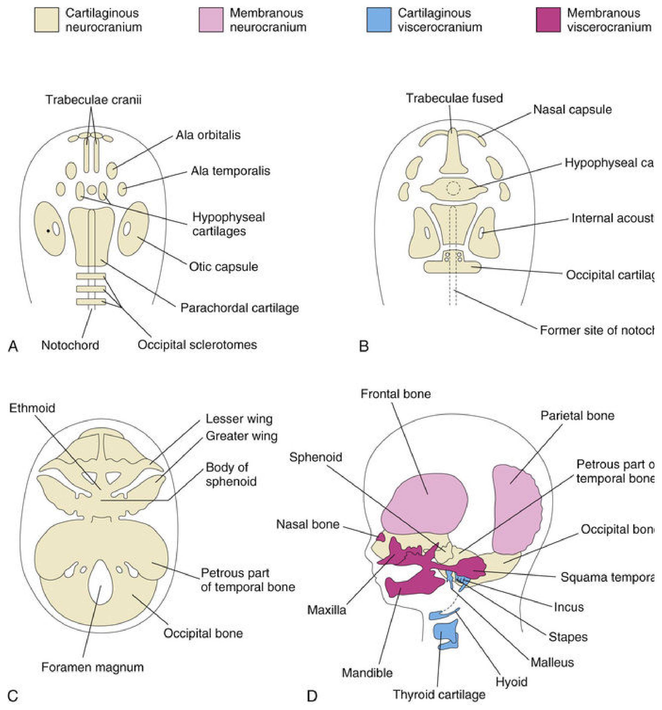
Forms the base of the skull via endochondral ossification. Multiple cartilages fuse sequentially:
| Cartilage | Fuses to form |
|---|---|
| Parachordal cartilage (basal plate) | Base of occipital bone + foramen magnum |
| Hypophyseal cartilage | Body of sphenoid |
| Trabeculae cranii | Body of ethmoid |
| Ala orbitalis | Lesser wing of sphenoid |
| Otic capsules | Petrous and mastoid parts of temporal bone |
| Nasal capsules | Contributes to ethmoid |
Ossification sequence: occipital bone → body of sphenoid → ethmoid bone
b) Membranous Neurocranium (Calvaria)
Forms the sides and top of the skull via intramembranous ossification in head mesenchyme. The flat bones (frontal, parietal, squamous temporal, squamous occipital) are separated in fetal life by:
- Sutures - fibrous joints between bones
- Fontanelles - 6 large fibrous areas at suture intersections
Fontanelles and their closure times:
| Fontanelle | Closure |
|---|---|
| Posterior | 2-3 months after birth |
| Anterolateral (sphenoid) | 2-3 months after birth |
| Posterolateral (mastoid) | End of first year |
| Anterior (largest) | End of second year |
| Frontal suture | Obliterated by ~8 years |
The fontanelles allow molding of the calvaria during birth and rapid postnatal brain growth.
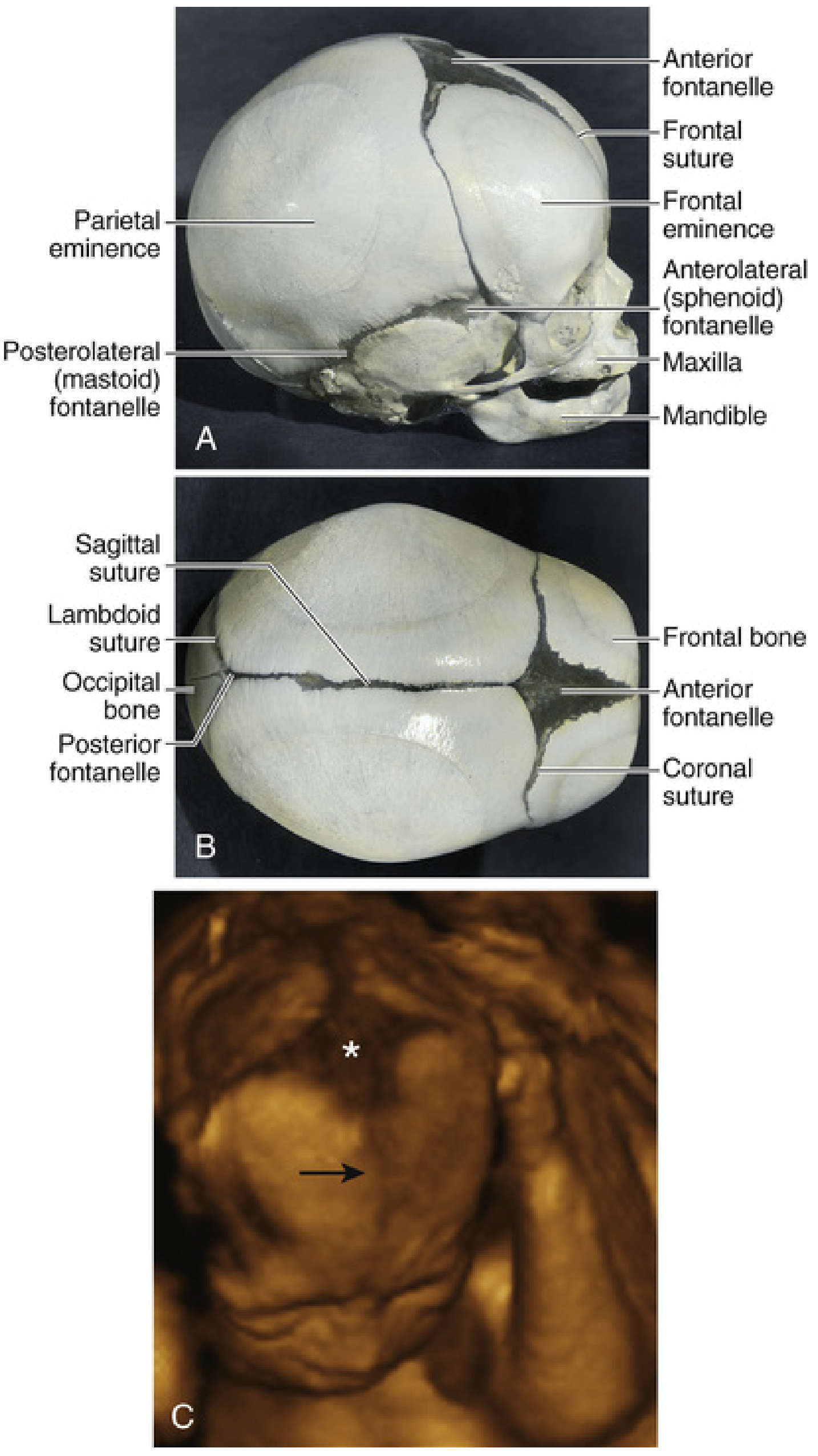
Viscerocranium (facial skeleton)
Derived largely from neural crest cells that migrate into the pharyngeal arches. Hox genes regulate neural crest migration and differentiation.
- First pharyngeal arch (Meckel's cartilage): Malleus and incus (middle ear ossicles)
- Second pharyngeal arch (Reichert's cartilage): Part of stapes, styloid process, lesser horn of hyoid
- Third pharyngeal arch: Greater horn and lower body of hyoid
Membranous Viscerocranium
Intramembranous ossification of neural crest mesenchyme forms: maxilla, zygomatic bone, squamous temporal bone, most of mandible, and the nasal and lacrimal bones.
Summary Diagram: Components and Origins of the Axial Skeleton
AXIAL SKELETON
├── VERTEBRAL COLUMN
│   └── Source: Sclerotome (paraxial mesoderm → somites)
│   └── Stages: Mesenchymal (Wk 4) → Cartilaginous (Wk 6) → Ossification (Wk 7 → Age 25)
│   └── Key: Each vertebral body is INTERSEGMENTAL (from 2 adjacent sclerotomes)
│   └── Notochord → Nucleus pulposus (persists between vertebrae)
│
├── RIBS
│   └── Source: Costal processes of thoracic vertebrae (sclerotome)
│   └── Ossify during fetal period
│   └── Types: True (1-7), False (8-10), Floating (11-12)
│
├── STERNUM
│   └── Source: Sternal bars (body wall mesenchyme + neural crest for manubrium)
│   └── Fuse craniocaudally by 10 weeks
│   └── Ossifies craniocaudally (xiphoid process last)
│
└── CRANIUM
    ├── NEUROCRANIUM
    │   ├── Cartilaginous (chondrocranium) → Endochondral ossification → Skull BASE
    │   └── Membranous (calvaria) → Intramembranous ossification → Skull VAULT
    └── VISCEROCRANIUM
        ├── Cartilaginous → Neural crest + pharyngeal arches → Middle ear ossicles, hyoid
        └── Membranous → Neural crest mesenchyme → Maxilla, mandible, zygomatic, etc.
Source: The Developing Human: Clinically Oriented Embryology, Chapter 14 (Skeletal System Development), pp. 901-926. All diagrams are from the same source.
Clinical note: Defects in Notch-pathway genes can cause severe vertebral anomalies, including VACTERL syndrome (vertebral, anal, cardiac, tracheal, esophageal, renal, and limb defects). Residual notochord tissue can give rise to a chordoma, a rare slow-growing malignant tumor occurring at the cranial base or lumbosacral region.
This is a shared conversation. Sign in to Orris to start your own chat.