
Course of sciatic nerve , compartments of thigh along with muscle , action, compartment of leg along with muscle and actions
Reading File
Finding Sources
Finding Sources
Finding Sources
Searching PubMed
"sciatic nerve" AND anatomy
Searching the Web
sciatic nerve thigh leg compartments muscles actions anatomy
Reading File
Reading File
Reading File
Searching Images
labeled diagram compartments of thigh and leg muscles anatomy
Course of the sciatic nerve
- Root value: L4-S3, from the sacral plexus.
- It is the largest nerve in the body.
- Leaves the pelvis through the greater sciatic foramen, usually inferior to piriformis.
- Enters the gluteal region deep to gluteus maximus, midway between the greater trochanter and ischial tuberosity.
- Descends in the posterior thigh:
- deep to the long head of biceps femoris
- superficial to adductor magnus
- lateral to semitendinosus and semimembranosus.
- Usually divides at the superior angle/apex of the popliteal fossa, often in the lower third of the thigh, into:
- Tibial nerve
- Common fibular (peroneal) nerve
Division can occur higher, including in the gluteal region. The sciatic nerve has no direct cutaneous supply to the posterior thigh, which is supplied by the posterior femoral cutaneous nerve.
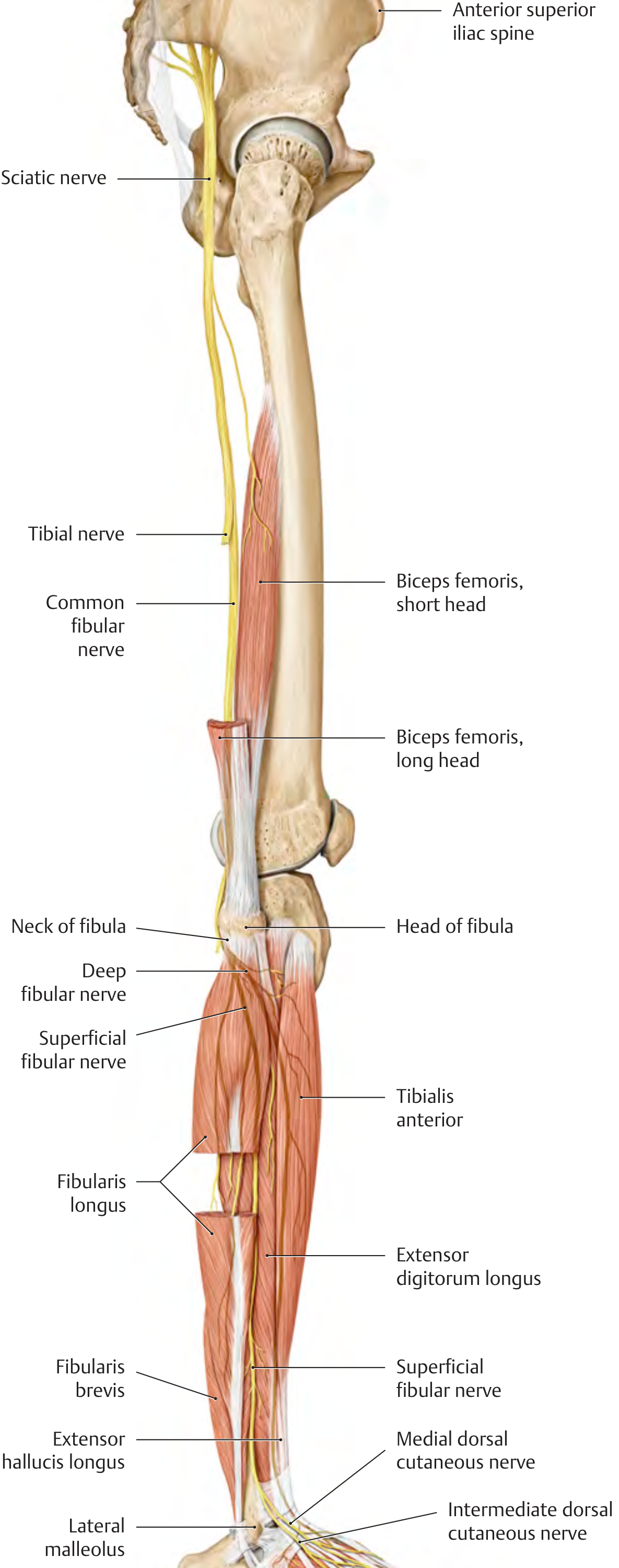
Motor supply in thigh
- Tibial part: semitendinosus, semimembranosus, long head of biceps femoris, hamstring part of adductor magnus.
- Common fibular part: short head of biceps femoris.
- Through its terminal branches, it supplies all muscles of the leg and foot.
General Anatomy and Musculoskeletal System (THIEME Atlas), p. 565.
Compartments of thigh
The thigh has three fascial compartments: anterior, medial, and posterior.
Gray's Anatomy for Students, p. 686.
Gray's Anatomy for Students, p. 686.
| Compartment | Main nerve | Muscles | Main actions |
|---|---|---|---|
| Anterior | Femoral nerve | Iliacus, sartorius, quadriceps femoris: rectus femoris, vastus lateralis, vastus medialis, vastus intermedius; articularis genus | Mainly extension of leg at knee. Rectus femoris also flexes thigh. Sartorius flexes, abducts and laterally rotates thigh, then flexes knee. Iliacus flexes thigh. |
| Medial | Mainly obturator nerve | Pectineus, adductor longus, adductor brevis, adductor magnus, gracilis, obturator externus | Mainly adduction of thigh at hip. Pectineus also flexes thigh. Adductor longus/brevis assist hip flexion. Adductor magnus: adductor part flexes and adducts thigh; hamstring part extends thigh. Gracilis also flexes knee and medially rotates leg. Obturator externus laterally rotates thigh. |
| Posterior | Sciatic nerve | Biceps femoris, semitendinosus, semimembranosus, hamstring part of adductor magnus | Mainly extension of thigh at hip and flexion of leg at knee. With knee flexed, biceps femoris laterally rotates the leg; semitendinosus and semimembranosus medially rotate it. |
High-yield: quadriceps actions
- Vastus lateralis, medialis, intermedius: extend knee.
- Rectus femoris: extends knee and flexes hip.
- Vastus medialis: helps stabilize patella, particularly against lateral displacement.
High-yield: hamstrings
“Hamstrings” are:
- Semitendinosus
- Semimembranosus
- Long head of biceps femoris
- Hamstring part of adductor magnus
They generally extend the hip and flex the knee. The short head of biceps femoris is not a true hamstring because it does not cross the hip joint.
Compartments of leg
The leg has four compartments: anterior, lateral, superficial posterior, and deep posterior.
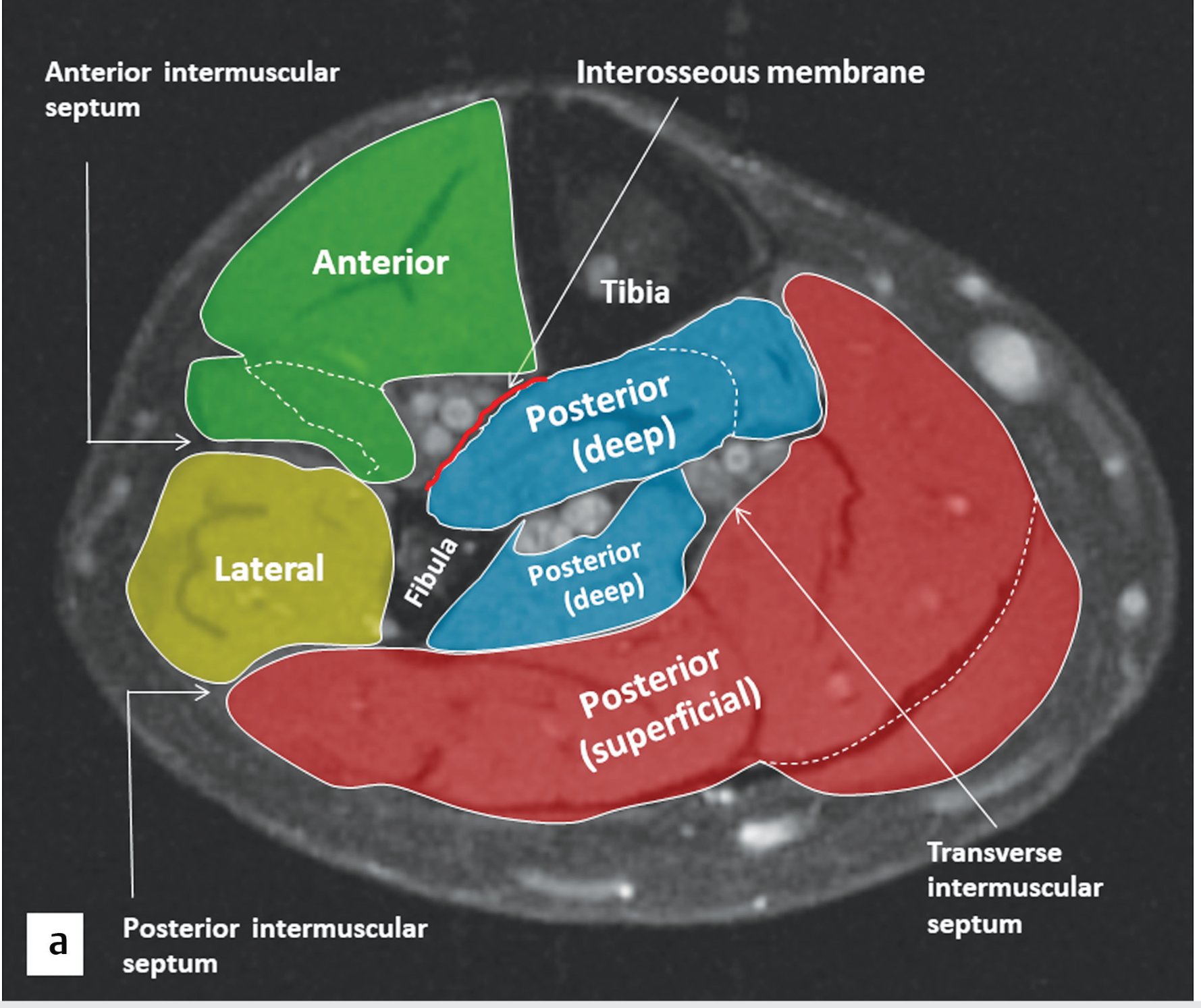
Imaging Anatomy Text and Atlas, p. 314.
| Compartment | Nerve supply | Muscles | Main actions |
|---|---|---|---|
| Anterior | Deep fibular nerve | Tibialis anterior, extensor hallucis longus, extensor digitorum longus, fibularis tertius | Dorsiflexion of ankle and extension of toes. Tibialis anterior also inverts foot. Fibularis tertius assists eversion. |
| Lateral | Superficial fibular nerve | Fibularis longus, fibularis brevis | Eversion of foot; weak plantarflexion. Fibularis longus also supports transverse arch of foot. |
| Superficial posterior | Tibial nerve | Gastrocnemius, soleus, plantaris | Mainly plantarflexion. Gastrocnemius also flexes knee. Soleus is important for postural standing and venous return. |
| Deep posterior | Tibial nerve | Popliteus, tibialis posterior, flexor digitorum longus, flexor hallucis longus | Plantarflexion and inversion; flexion of toes. Tibialis posterior supports medial longitudinal arch. Popliteus “unlocks” the extended knee. |
Individual muscle actions in leg
1. Anterior compartment
- Tibialis anterior: dorsiflexes and inverts foot.
- Extensor hallucis longus: extends great toe and dorsiflexes foot.
- Extensor digitorum longus: extends lateral four toes and dorsiflexes foot.
- Fibularis tertius: dorsiflexes and everts foot.
2. Lateral compartment
- Fibularis longus: everts and weakly plantarflexes foot; supports transverse arch.
- Fibularis brevis: everts and weakly plantarflexes foot.
3. Superficial posterior compartment
- Gastrocnemius: plantarflexes foot when knee is extended; flexes knee.
- Soleus: powerful plantarflexor, especially during standing.
- Plantaris: weak plantarflexion and weak knee flexion.
4. Deep posterior compartment
- Popliteus: unlocks knee by laterally rotating femur on fixed tibia, or medially rotating tibia on femur; weak knee flexor.
- Tibialis posterior: plantarflexes and inverts foot; supports medial longitudinal arch.
- Flexor digitorum longus: flexes lateral four toes; assists plantarflexion and supports longitudinal arches.
- Flexor hallucis longus: flexes great toe; assists plantarflexion and supports medial longitudinal arch, especially during push-off.
Nerve correlation
- Common fibular nerve supplies anterior and lateral leg compartments through its deep and superficial branches.
- Tibial nerve supplies both posterior compartments.
- Injury to the common fibular nerve at the neck of fibula can cause weak dorsiflexion and eversion, producing foot drop.
General Anatomy and Musculoskeletal System (THIEME Atlas), p. 565.
Recent anatomy literature warning: sciatic nerve division and its relation to piriformis show meaningful anatomical variation, relevant to injections and surgery (PMIDs: 41785245, 41612052).
This is a shared conversation. Sign in to Orris to start your own chat.