


Trigger 5 A 55–year–old man was brought under the influence of alcohol, in a state of drowsiness and a history of cough and fever with chills and rigors of 2 days duration Chest Xray Trigger 6 A 63-year-old man who is a long-standing [ 35 years] smoker attends the chest clinic with a 3-year history of worsening exertional dyspnea, associated with a productive cough. On examination, he is drowsy and has flapping tremors First photo trigger 5 , 2nd photo is trigger 6 Make one mcq and one saq question based on history , clinical examination , investigation , management . Make each for both triggers With explanation for its answer
TRIGGER 5 - Aspiration Pneumonia / Lobar Pneumonia
TRIGGER 5 - MCQ
- A. Mycoplasma pneumoniae
- B. Klebsiella pneumoniae
- C. Streptococcus pneumoniae
- D. Legionella pneumophila
- E. Staphylococcus aureus
- Alcoholics (impaired mucociliary clearance, aspiration risk, poor immunity)
- Diabetics and debilitated patients
- "Bulging fissure sign" (the weight of the exudate pushes down the horizontal fissure - look for downward bowing)
- Thick, mucoid ("currant jelly") sputum due to blood-stained mucoid exudate
- High mortality if untreated; tends to cavitate
TRIGGER 5 - SAQ
- Diagnosis: Lobar pneumonia - right upper lobe (most likely Klebsiella pneumoniae given the clinical context)
- Key predisposing factor: Chronic alcohol use - causes aspiration due to obtunded gag reflex, impaired mucociliary clearance, nutritional immunosuppression, and leucocyte dysfunction
- Inspection: Reduced chest expansion on the affected side; tachypnoea; use of accessory muscles
- Palpation: Reduced tactile vocal fremitus initially (pre-consolidation); increased TVF / vocal fremitus over consolidated lobe
- Percussion: Dullness to percussion over the right upper zone
- Auscultation: Bronchial breathing and crackles (crepitations) over the right upper lobe; whispering pectoriloquy; aegophony
- Sputum culture and Gram stain (before starting antibiotics)
- Expected finding: Klebsiella produces short, plump gram-negative rods; mucoid colonies on culture. Blood cultures should also be obtained (bacteraemia is common). Sputum Gram stain may show PMNs + gram-negative rods.
| Component | Detail |
|---|---|
| Supportive | Supplemental O2 to maintain SpO2 >94%; IV fluids (he is drowsy/possibly septic); analgesia for pleuritic pain |
| Positioning | Nurse semi-upright; aspiration precautions given alcohol intoxication |
| Antibiotics | Klebsiella: IV ceftriaxone (3rd-gen cephalosporin) OR piperacillin-tazobactam; if CAP uncertain: add a macrolide (e.g. azithromycin) for atypical coverage |
| Monitor | Pulse oximetry, RR, BP, urine output; repeat CXR at 48-72 h |
| Severity scoring | CURB-65 score (Confusion, Urea >7, RR >30, BP systolic <90, age >65) - this patient likely scores at least 1-2 (confusion present), indicating moderate-severe CAP requiring hospital admission |
| Follow-up CXR | At 6 weeks post-discharge to exclude underlying malignancy (especially important in smokers/alcoholics) |
TRIGGER 6 - COPD with Type II Respiratory Failure (CO2 Retention)
TRIGGER 6 - MCQ
| Option | pH | PaO2 | PaCO2 | HCO3- |
|---|---|---|---|---|
| A | 7.52 | 56 | 28 | 22 |
| B | 7.38 | 55 | 62 | 34 |
| C | 7.28 | 58 | 68 | 22 |
| D | 7.45 | 90 | 38 | 24 |
| E | 7.30 | 50 | 70 | 26 |
- Option A: Respiratory alkalosis with low CO2 - this is hyperventilation/Type I failure, not CO2 retention
- Option B (CORRECT): Near-normal pH (7.38) despite elevated PaCO2 (62 mmHg) and elevated HCO3- (34 mEq/L) - this represents chronic compensated respiratory acidosis. The kidneys have retained bicarbonate over time (metabolic compensation) to normalize the pH. PaO2 is low (55). This is the pattern of chronic CO2 retainer with acute decompensation.
- Option C: Uncompensated acute respiratory acidosis (low HCO3- indicates no metabolic compensation yet) - more acute, but asterixis and drowsiness usually accompany chronic retention
- Option D: Normal blood gas - does not fit the clinical picture
- Option E: Acute respiratory acidosis with partial compensation - possible but HCO3- of 26 shows minimal compensation, not the chronic retainer pattern
TRIGGER 6 - SAQ
- Diagnosis: Severe COPD (likely GOLD Stage III-IV) with acute exacerbation and Type II respiratory failure (hypercapnic respiratory failure)
- Asterixis (flapping tremor): Signifies CO2 narcosis / hypercapnic encephalopathy - CO2 retention causes cerebral vasodilation and metabolic encephalopathy, manifesting as a coarse, non-rhythmic tremor on wrist extension. It is a red flag indicating ventilatory failure.
- Hyperinflation - >10 posterior ribs visible above the diaphragm
- Flattened hemidiaphragms - loss of normal dome shape, diaphragm at or below the level of the 7th rib anteriorly
- Increased retrosternal airspace (barrel chest on lateral CXR)
- Bullae - focal areas of avascular hyperlucency in the upper lobes
- Peripheral vascular pruning - reduced peripheral vascular markings with oligaemia in outer lung fields
- Narrow, vertical "dripping" heart
- Horizontal rib orientation with widened intercostal spaces
| Parameter | Finding in COPD | Significance |
|---|---|---|
| FEV1/FVC ratio | < 0.70 (post-bronchodilator) | Confirms obstructive defect - diagnostic criterion |
| FEV1 % predicted | Determines GOLD severity: Stage I >80%, Stage II 50-79%, Stage III 30-49%, Stage IV <30% | This patient likely Stage III-IV given drowsiness |
| Post-bronchodilator reversibility | <12% improvement (or <200 mL) in FEV1 | Confirms fixed obstruction (distinguishes from asthma) |
| Step | Action |
|---|---|
| Oxygen | Controlled low-flow O2: 24-28% via Venturi mask - target SpO2 88-92% (NOT >94%). High-flow O2 removes the hypoxic drive in CO2 retainers, worsening hypercapnia and CO2 narcosis. |
| ABG | Urgent arterial blood gas to confirm Type II failure, assess pH and PaCO2 |
| Bronchodilators | Nebulised salbutamol (SABA) + ipratropium (SAMA) - back-to-back nebulisers driven by air (not O2, or low-flow O2) |
| Systemic corticosteroids | Prednisolone 30-40 mg oral (or IV hydrocortisone) for 5 days - reduces airway inflammation and accelerates recovery |
| Antibiotics | If purulent sputum/infective exacerbation: amoxicillin or doxycycline or co-amoxiclav |
| NIV (BiPAP) | If pH <7.35 and PaCO2 rising despite above measures - non-invasive ventilation (BiPAP) is the definitive treatment for Type II failure in COPD to rest the respiratory muscles and correct acidosis |
| Avoid intubation if possible | CPAP/BiPAP preferred; if refractory, discuss ITU with patient's prior wishes |
| Monitor | Repeat ABG at 30-60 min after starting O2/NIV; ECG for cor pulmonale arrhythmias |
| Feature | Trigger 5 (Pneumonia) | Trigger 6 (COPD + Type II Failure) |
|---|---|---|
| Diagnosis | Right upper lobe pneumonia (Klebsiella) | COPD exacerbation with hypercapnic encephalopathy |
| Cause of drowsiness | Systemic sepsis / encephalopathy | CO2 narcosis (asterixis is the clue) |
| CXR | Lobar consolidation (RUL) | Hyperinflation, flat diaphragms, pruning, PAH |
| Organism/trigger | Klebsiella (alcoholism) | Smoking-induced COPD, likely infective exacerbation |
| O2 target | SpO2 >94% (standard) | SpO2 88-92% (controlled - avoid hypoxic drive suppression) |
| Specific treatment | IV ceftriaxone +/- macrolide | Bronchodilators + steroids + NIV (BiPAP) if pH <7.35 |
I meant to create new question , mcq and meq
You mentioned MCQ and MEQ — what do you mean by MEQ? · Should each question (MCQ + MEQ) cover ALL four areas together, or one question per area? · When you say 'new question' — do you want completely different questions from what I already wrote, or the same format but reworded?
Make a new scenario thats like this. Thats exp after finish learning regarding the triggers, need to ask questions right. Haaa that one
📋 ASSESSMENT — Trigger 5 Topic: Pneumonia in a Compromised Host
MCQ
- A. Streptococcus pneumoniae
- B. Mycoplasma pneumoniae
- C. Klebsiella pneumoniae
- D. Pseudomonas aeruginosa
- E. Legionella pneumophila
MEQ
- Chronic alcoholism - impaired immunity, aspiration risk, malnutrition
- Confusion / altered GCS - CURB-65 point (C = 1); suggests severity
- Pleuritic chest pain - indicates pleural involvement/possible parapneumonic effusion
- Delayed presentation (3 days) - infection has progressed without treatment, risk of complications
| Parameter | Criteria | This Patient |
|---|---|---|
| C - Confusion | New confusion (GCS <15) | ✅ Yes = 1 |
| U - Urea | Blood urea >7 mmol/L | Unknown (pending bloods) |
| R - Respiratory rate | RR ≥30/min | ❌ RR 26 = 0 |
| B - Blood pressure | Systolic <90 or diastolic ≤60 | ✅ BP 100/70 = 1 |
| 65 - Age | Age ≥65 | ❌ Age 60 = 0 |
| Investigation | Expected Finding |
|---|---|
| Chest X-ray (PA) | Lobar consolidation (likely right lower lobe); possible parapneumonic effusion |
| ABG (Arterial Blood Gas) | Type I respiratory failure: low PaO2 (<8 kPa), low/normal PaCO2, mild respiratory alkalosis |
| Blood cultures (x2 sets) | May grow gram-negative rod (Klebsiella) - bacteraemia in ~30% of severe CAP |
| Sputum Gram stain + culture | Gram-negative short rods; mucoid colonies on MacConkey agar |
| FBC | Leukocytosis (WBC >12 x10⁹/L), neutrophilia |
| CRP / Procalcitonin | Significantly elevated, confirming bacterial infection |
- High-flow O2 via non-rebreather mask; titrate to target SpO2 94-98% (no CO2 retention risk here - this is Type I failure)
- Sit patient upright; aspiration precautions given alcohol intoxication
- IV access x2; IV fluid resuscitation with 500 mL normal saline bolus (BP 100/70, HR 112 - septic picture)
- IV co-amoxiclav (amoxicillin-clavulanate) PLUS azithromycin (macrolide) for atypical coverage
- OR IV ceftriaxone (preferred if Klebsiella suspected given alcoholism) + azithromycin
- Guided by local hospital antibiogram; de-escalate once cultures available
- Continuous SpO2, ECG, hourly urine output; repeat ABG at 1 hour; repeat CXR at 48-72 h
- Parapneumonic effusion / empyema - monitor for persistence or worsening of dullness; if effusion develops, diagnostic pleural tap (Light's criteria); empyema requires chest drain + prolonged antibiotics
- Also acceptable: septic shock, ARDS, lung abscess (especially with Klebsiella)
📋 ASSESSMENT — Trigger 6 Topic: COPD with Hypercapnic Respiratory Failure
MCQ
- A. Oxygen toxicity causing retinal damage
- B. Suppression of hypoxic respiratory drive leading to worsening CO2 retention
- C. Nitrogen washout causing absorption atelectasis
- D. Pulmonary oxygen toxicity causing ARDS
- E. Precipitation of pulmonary oedema
MEQ
- Long-standing smoking history (40 years) - established COPD risk factor; 3 triggers are the same presentations of a known chronic disease
- Pre-existing reliever inhaler use ("all the time") - indicates significant background airflow obstruction already present before this admission
- 4-day history of worsening dyspnoea + change in sputum (increased volume, purulent/green) - classic precipitant of COPD exacerbation, most commonly infectious (viral or bacterial), on a background of chronically impaired lung function
| Finding | Pathophysiological Basis |
|---|---|
| Barrel chest | Chronic air trapping → hyperinflation → increased AP diameter of thorax; ribs become horizontal and intercostal spaces widen |
| Pursed-lip breathing | Expiration against resistance creates intrinsic PEEP, preventing small airway collapse during expiration - a compensatory mechanism to slow expiratory flow |
| Flapping tremor (asterixis) | CO2 retention (hypercapnia) causes impaired cortical inhibition of movement - metabolic encephalopathy from CO2 narcosis |
| Prolonged expiratory wheeze | Diffuse airflow obstruction from bronchospasm, mucosal oedema, and secretions in narrowed airways; expiration requires active effort against collapsed small airways |
- pH 7.31 = acidaemia
- PaCO2 9.2 kPa (high) = respiratory acidosis (primary problem)
- PaO2 5.1 kPa (low) = hypoxaemia - Type II respiratory failure
- HCO3- 36 mEq/L (high) + BE +8 = metabolic alkalosis = renal compensation
- Conclusion: Partially compensated respiratory acidosis = acute-on-chronic hypercapnic (Type II) respiratory failure
- Spirometry (pulmonary function tests): Post-bronchodilator FEV1/FVC <0.70 confirms obstructive defect. FEV1 % predicted stages severity (GOLD I-IV). This patient likely has GOLD Stage III-IV (FEV1 <50% predicted) given drowsiness and degree of failure.
- Remove the high-flow O2 immediately if applied
- Apply 24% Venturi mask → titrate to 28% if SpO2 remains <88%
- Target SpO2 88-92% - NOT higher, to preserve hypoxic drive
- Recheck ABG in 30-60 minutes after starting controlled O2
- Nebulised salbutamol 2.5-5 mg (SABA) + ipratropium 500 mcg (SAMA)
- Drive nebuliser with air (not O2) or low-flow O2 at max 6 L/min
- Repeat every 20-30 minutes in first hour, then 4-hourly
- Prednisolone 30-40 mg oral (or IV hydrocortisone 200 mg if unable to swallow)
- Course: 5 days - reduces airway inflammation, shortens exacerbation duration
- Green/purulent sputum = likely bacterial trigger
- Amoxicillin 500 mg TDS OR doxycycline 200 mg stat then 100 mg OD (5 days)
- If resistant/severe: co-amoxiclav or levofloxacin
- Indications met: pH 7.31 (<7.35), PaCO2 9.2 kPa, drowsy
- Start BiPAP (IPAP 10-20 cmH2O / EPAP 4-5 cmH2O) - this is the single most important intervention to correct acidosis and rest respiratory muscles
- Recheck ABG 1-2 hours after BiPAP
- If pH worsens despite BiPAP (<7.25) or patient deteriorates → escalate to ITU / intubation (discuss with patient's advance wishes)
- Continuous SpO2, ECG (right heart strain pattern - P pulmonale, right axis deviation)
- Monitor for cor pulmonale - check for RV failure
- Daily weight; fluid balance
- VTE prophylaxis (LMWH)
- Smoking cessation counselling on discharge
Marking Tip for Examiners: The MEQ is weighted 25 marks total (4+4+4+9 for Trigger 6; 4+4+4+8 for Trigger 5). The MCQs each carry 1 mark. Both scenarios test integration of clinical reasoning across all four domains as required by the PBL trigger assessment framework.
So now najw make a new scenario like for both trigger. Make one true and false question and one short case question for both trigger baded on it but make new scenario
No too many I just one tru and false question and one short case question that simple with newly made sceanrio based on the 2 trigger. Means each tigget need one tru fals question and short case.
TRIGGER 5 TOPIC — Pneumonia in Compromised Host
True / False Question
| # | Statement | Answer |
|---|---|---|
| 1 | The most likely causative organism is Streptococcus pneumoniae | FALSE - Klebsiella is the classic organism in alcoholics with upper lobe consolidation and bulging fissure sign |
| 2 | Alcoholism impairs the cough and gag reflex, increasing aspiration risk | TRUE - alcohol suppresses CNS protective reflexes, promoting micro-aspiration of oropharyngeal flora |
| 3 | A CURB-65 score of 3 or more indicates the patient can be safely treated at home | FALSE - score ≥3 indicates severe CAP requiring hospital admission, consider HDU/ICU |
| 4 | The "bulging fissure sign" on CXR is characteristic of Klebsiella pneumonia due to its heavy mucoid exudate | TRUE - the voluminous inflammatory exudate adds weight, pushing the fissure downward |
| 5 | High-flow oxygen should be avoided in this patient as he is at risk of CO2 retention | FALSE - this is Type I respiratory failure (pneumonia); high-flow O2 targeting SpO2 94-98% is appropriate. CO2 retention precaution applies to chronic COPD retainers, not acute pneumonia |
Short Case Question
- C (Confusion) = 1 ✅ drowsy
- U (Urea) = unknown (pending)
- R (RR ≥30) = 0 (RR is 28)
- B (BP systolic <90) = 1 ✅ BP 95/65
- 65 (Age ≥65) = 0
- Minimum score = 2 → Hospital admission required; if urea >7 = score 3 → HDU consideration
- Antibiotic: IV Ceftriaxone (3rd generation cephalosporin) covering gram-negative organisms including Klebsiella
- Complication: Lung abscess - Klebsiella has a high tendency to cavitate; monitor with repeat CXR at 48-72 hours
TRIGGER 6 TOPIC — COPD with Type II Respiratory Failure
True / False Question
| # | Statement | Answer |
|---|---|---|
| 1 | The flapping tremor (asterixis) in this patient is caused by CO2 retention | TRUE - hypercapnia causes metabolic encephalopathy; asterixis is a hallmark of CO2 narcosis |
| 2 | This patient should receive 15L/min O2 via non-rebreather mask immediately | FALSE - he is a chronic CO2 retainer; high-flow O2 removes hypoxic drive, worsening hypercapnia. Target SpO2 88-92% via 24-28% Venturi mask |
| 3 | The elevated HCO3- of 38 mEq/L indicates acute metabolic alkalosis caused by vomiting | FALSE - the raised bicarbonate reflects chronic renal compensation for long-standing respiratory acidosis (CO2 retention over months to years) |
| 4 | Non-invasive ventilation (BiPAP) is indicated when pH falls below 7.35 despite initial treatment | TRUE - NIV/BiPAP is the treatment of choice for hypercapnic respiratory failure in COPD when pH <7.35 |
| 5 | Enlarged central pulmonary arteries on CXR in COPD indicate the development of cor pulmonale | TRUE - chronic hypoxia causes pulmonary vasoconstriction → pulmonary hypertension → right heart strain/cor pulmonale |
Short Case Question
- CXR finding 1: Hyperinflation - more than 10 posterior ribs visible, flat diaphragms (1 mark)
- CXR finding 2: Bullae / peripheral vascular pruning - avascular hyperlucent areas from emphysematous destruction (1 mark)
- Enlarged pulmonary arteries: Indicate pulmonary hypertension secondary to chronic hypoxic vasoconstriction, a sign of cor pulmonale (right heart failure) (1 mark)
- Oxygen target: SpO2 88-92% via 24-28% Venturi mask - controlled O2 to preserve hypoxic drive (1 mark)
- Ventilatory intervention: Non-invasive ventilation (BiPAP) - indicated when pH <7.35 despite initial bronchodilators, steroids, and controlled O2; corrects acidosis and rests respiratory muscles (1 mark)
Ish you don't understand my question
When you say 'new scenario' — do you mean the TRUE/FALSE and Short Case questions should each have their OWN brand new patient scenario written at the top (like a new vignette/story), separate from the original triggers? · What do you mean by 'short case question'? Is it:

This is how my dr want
- True or False
- Case Scenario
- Short Essay Question
🫁 TRIGGER 5 — Pneumonia in a Compromised Host
1. TRUE OR FALSE
| # | Statement | Answer |
|---|---|---|
| 1 | Klebsiella pneumoniae is the most likely causative organism in this patient | TRUE |
| 2 | High-flow oxygen at 15L/min should be given immediately targeting SpO2 >98% | FALSE |
| 3 | Alcoholism increases pneumonia risk by impairing the cough and gag reflex | TRUE |
| 4 | A CURB-65 score of ≥3 means the patient can be discharged with oral antibiotics | FALSE |
| 5 | The bulging fissure sign occurs because Klebsiella produces heavy mucoid exudate | TRUE |
2. CASE SCENARIO
3. SHORT ESSAY QUESTION
- Alcohol impairs mucociliary clearance - cilia function is suppressed, secretions accumulate
- Impaired gag and cough reflex due to CNS depression - promotes aspiration of oropharyngeal/gastric contents
- Alcohol causes neutrophil dysfunction - impaired chemotaxis and phagocytosis
- Malnutrition (common in alcoholics) reduces T-cell and B-cell immunity
- Liver dysfunction reduces opsonin and complement production - gram-negative bacteria less effectively cleared
- Airway/Breathing: O2 via face mask, target SpO2 94-98% (Type I failure); sit upright; aspiration precautions
- Circulation: IV access; fluid resuscitation if hypotensive (sepsis); blood cultures before antibiotics
- Antibiotics: IV Ceftriaxone ± azithromycin (atypical cover); guided by sputum Gram stain
- Investigations: CXR, FBC, CRP, ABG, sputum C&S, blood cultures
- Severity scoring: CURB-65 to guide admission level
- Follow-up CXR at 6 weeks post-discharge to exclude underlying malignancy
🫁 TRIGGER 6 — COPD with Hypercapnic Respiratory Failure
1. TRUE OR FALSE
| # | Statement | Answer |
|---|---|---|
| 1 | This patient has Type II (hypercapnic) respiratory failure | TRUE |
| 2 | Oxygen should be given at 15L/min to rapidly correct his SpO2 to 99% | FALSE |
| 3 | The elevated HCO3- of 37 mEq/L represents acute metabolic alkalosis from vomiting | FALSE |
| 4 | BiPAP (non-invasive ventilation) is indicated because his pH is below 7.35 | TRUE |
| 5 | Enlarged central pulmonary arteries on CXR indicate pulmonary hypertension and possible cor pulmonale | TRUE |
2. CASE SCENARIO
3. SHORT ESSAY QUESTION
- Hyperinflation + flat diaphragms: Chronic air trapping from loss of lung elastic recoil (emphysema) → increased residual volume → lungs push diaphragm flat and ribs horizontal
- Bullae/hyperlucency: Destruction of alveolar walls by chronic inflammation (proteases overwhelm antiproteases) → large air spaces with no vascular markings
- Peripheral pruning: Loss of pulmonary capillary bed from emphysema → fewer peripheral vessels visible
- Enlarged central pulmonary arteries: Chronic hypoxia → hypoxic pulmonary vasoconstriction → pulmonary hypertension → right ventricular hypertrophy (cor pulmonale)
- Vertical/narrow heart: Hyperinflated lungs push the heart inferiorly and narrow its silhouette
- Controlled O2: 24-28% Venturi mask, SpO2 target 88-92%
- Bronchodilators: Nebulised salbutamol + ipratropium (driven by air)
- Steroids: Prednisolone 30-40 mg oral or IV hydrocortisone
- Antibiotics: Amoxicillin/doxycycline (green sputum = infective exacerbation)
- ABG at 30-60 min: If pH <7.35 → start BiPAP immediately
- If BiPAP fails: Escalate to ITU/intubation discussion
Okay this for which trigger?
-
🫁 TRIGGER 5 (the top half) = Pneumonia in a compromised host - based on the 55-year-old alcoholic man with cough, fever, and right upper lobe consolidation on CXR
-
🫁 TRIGGER 6 (the bottom half) = COPD with Hypercapnic Respiratory Failure - based on the 63-year-old long-term smoker with worsening dyspnoea, drowsiness, and flapping tremor
- ✅ True or False
- ✅ Case Scenario
- ✅ Short Essay Question
Okay honeyy , can you explain each answer why true , why false. Why the answers like that
🫁 TRIGGER 5 — True or False: Full Explanations
- Chronic alcoholism - alcohol impairs the immune system and gag reflex, making alcoholics specifically vulnerable to Klebsiella
- Right UPPER lobe consolidation - Klebsiella classically affects the upper lobes (unlike most other pneumonias that affect lower lobes)
- Reddish-brown ("currant jelly") sputum - this is the classic description of Klebsiella sputum, caused by blood mixed with thick mucoid exudate
- Bulging fissure sign - the heavy mucoid exudate produced by Klebsiella pushes the fissure downward
- The target SpO2 in standard pneumonia is 94-98%, not >98%
- There is no benefit in pushing SpO2 above 98% and excessive O2 can cause oxygen toxicity with prolonged use
- The >98% target via 15L is the wrong approach that should be reserved for cardiac arrest only
- CNS depression - alcohol suppresses the brainstem reflexes including the cough reflex and gag reflex. When these are impaired, the patient cannot clear secretions or prevent food/liquid/bacteria from entering the airway
- Aspiration - a drunk person lying down can silently aspirate oral bacteria (including Klebsiella which colonises the mouth of alcoholics) into the lungs without coughing it back up
- Mucociliary dysfunction - alcohol paralyses the cilia lining the airways that normally sweep bacteria upward and out
- Immune suppression - neutrophil (white cell) function is impaired; they cannot kill bacteria effectively
- C = Confusion
- U = Urea >7 mmol/L
- R = Respiratory rate ≥30/min
- B = Blood pressure (systolic <90 or diastolic ≤60)
- 65 = Age ≥65
| Score | Meaning | Action |
|---|---|---|
| 0-1 | Low severity | Consider home treatment |
| 2 | Moderate severity | Hospital admission |
| ≥3 | HIGH severity | Hospital admission, consider HDU/ICU |
- It weighs down the affected lobe
- This weight pushes the fissure (the dividing line between lung lobes) downward or outward
- On CXR, you see the horizontal fissure bowing downward instead of its normal horizontal position
🫁 TRIGGER 6 — True or False: Full Explanations
| Type | O2 | CO2 | Example |
|---|---|---|---|
| Type I | LOW | Normal or LOW | Pneumonia, pulmonary oedema |
| Type II | LOW | HIGH | COPD, severe asthma, neuromuscular disease |
- PaO2 5.2 kPa = LOW (normal >10.6 kPa) ✅ hypoxaemia
- PaCO2 9.0 kPa = HIGH (normal 4.7-6.0 kPa) ✅ hypercapnia
- pH 7.30 = acidaemia from CO2 retention
- The hypoxia is corrected
- The peripheral chemoreceptors stop firing
- The patient has NO drive to breathe
- They breathe less and less
- CO2 rises even further
- The patient becomes more drowsy, stops breathing → respiratory arrest
- Give 24-28% O2 via Venturi mask
- Target SpO2 88-92% only
- Keep them slightly hypoxic ON PURPOSE to maintain their breathing drive
- pH 7.30 = acidaemia (NOT alkalotic - rules out primary metabolic alkalosis)
- PaCO2 9.0 = very HIGH = respiratory acidosis (this is the PRIMARY problem)
- HCO3- 37 = HIGH = this is the body's RESPONSE to the high CO2, not a primary problem
- IPAP (inspiratory pressure) - helps push air INTO the lungs, increasing tidal volume and blowing off CO2
- EPAP (expiratory pressure) - keeps airways open, preventing collapse
- pH <7.35 (acidaemia) despite initial treatment with controlled O2, bronchodilators, and steroids
- PaCO2 rising
- Patient drowsy/tiring
- Rests the exhausted respiratory muscles
- Increases ventilation, blowing off excess CO2
- Corrects the acidosis
- Avoids the need for invasive intubation in most cases
- Enlarged central pulmonary arteries (dilated from high pressure)
- Peripheral pruning (peripheral vessels disappear as resistance increases)
- Eventually signs of right heart failure (raised JVP, ankle oedema clinically)
Okay for the true false and short case for each trigger thats, got x ray noted in scenario give me the x ray pocture
Klebsiella pneumonia right upper lobe consolidation bulging fissure chest X-ray
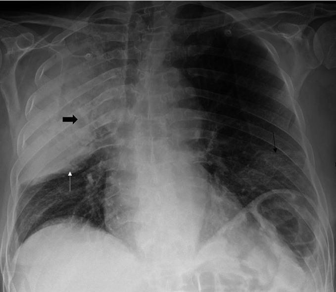
This chest X-ray in anteroposterior (AP) projection demonstrates multi-focal pulmonary pathology. The primary finding is a dense consolidation of the right upper lobe (RUL), characterized by a notable 'bulging fissure' sign where the horizontal fissure (marked by a thin white arrow) is displaced inferiorly, suggesting an expansive inflammatory process such as Klebsiella or Legionella pneumonia. Within the RUL consolidation, a thick black arrow points to an area of radiolucency representing pulmonary cavitation. Additionally, the image shows bilateral lower zone ground-glass opacities (indicated by a thin black arrow in the left lung base), manifesting as hazy increased density that does not fully obscure pulmonary vasculature. This combination of dense, cavitating lobar consolidation and diffuse ground-glass opacities is clinically significant for severe community-acquired or atypical pneumonia, potentially occurring in the context of co-infections such as SARS-CoV-2 and Legionella pneumophila. Educational value lies in identifying the bulging fissure sign, recognizing cavitary lesions on plain film, and distinguishing lobar consolidation from diffuse ground-glass patterns.
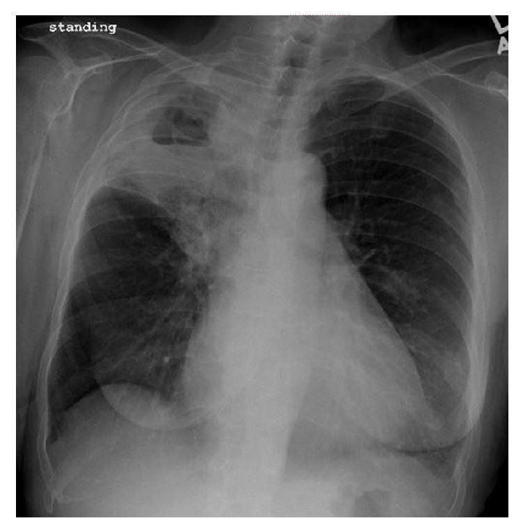
A posterior-anterior (PA) chest X-ray in a standing position demonstrating a significant pathological process localized to the right upper lobe. The primary finding is a dense, consolidated opacity involving the right upper lobe, clearly demarcated by the minor fissure. Within this consolidated region, there is a large, well-defined cavitary lesion containing a distinct horizontal air-fluid level, characteristic of necrotizing pneumonia or a lung abscess. The surrounding lung parenchyma in the right middle and lower lobes appears relatively aerated, though there is a subtle increase in interstitial markings. The left lung field remains clear with normal vascularity and no evidence of consolidation, effusion, or cavitation. The cardiac silhouette, mediastinal contours, and costophrenic angles appear within normal limits. This radiographic presentation is highly suggestive of cavitating pneumonia, potentially caused by virulent pathogens such as Pseudomonas aeruginosa, Klebsiella pneumoniae, or Staphylococcus aureus. The image serves as a clinical example of lung parenchyma destruction and abscess formation in the context of severe pulmonary infection.
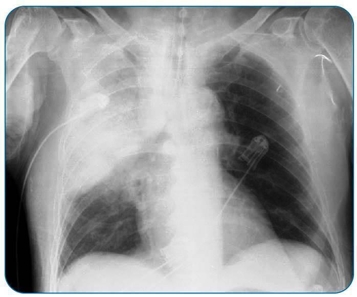
This diagnostic image is an anteroposterior (AP) chest X-ray of an adult patient. The primary finding is an extensive, dense homogeneous opacification occupying the right upper and middle lung zones, characteristic of lobar consolidation. Notable radiographic signs include the presence of air bronchograms—branching lucent tubular structures within the opaque area—indicating patent airways surrounded by alveolar exudate. There is a visible inferior displacement and convex bulging of the minor fissure (bulging fissure sign), suggesting an inflammatory process with significant volume expansion, often associated with aggressive bacterial infections such as Klebsiella or Acinetobacter pneumoniae. The left lung remains relatively clear with normal bronchovascular markings. Several medical devices are present, including an endotracheal tube, a right-sided jugular central venous catheter, and ECG leads. The diaphragmatic domes and costophrenic angles appear visible but partially obscured on the right side. This image serves as a classic educational example of acute lobar pneumonia with severe pulmonary involvement and signs of inflammatory pressure within the thoracic cavity.
COPD emphysema hyperinflation flat diaphragm enlarged pulmonary arteries chest X-ray
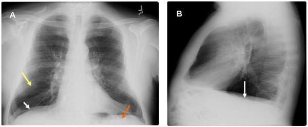
This diagnostic image displays a two-panel chest roentgenogram (X-ray) from a 65-year-old male, highlighting radiographic features of severe pulmonary emphysema and chronic obstructive pulmonary disease (COPD). Panel A presents a postero-anterior (PA) view, while Panel B provides a lateral view. Key findings include significant hyperinflation of the lung fields with marked flattening of the hemidiaphragms (indicated by white arrows), which loss their normal convex shape. In Panel A, a yellow arrow identifies areas of increased translucency and attenuation of vascular markings consistent with emphysematous parenchymal changes. A red arrow highlights the decreased zone of apposition, representing the diminished area where the diaphragm sits parallel to the inner rib cage. Panel B further illustrates the flattened diaphragmatic contour and an increased retrosternal clear space, reflecting an increased anterior-posterior thoracic diameter (barrel chest). These visual markers are clinically significant as they correlate with the mechanical basis of Hoover's sign, where the horizontal orientation of the diaphragm causes inward rib cage retraction during inspiration.
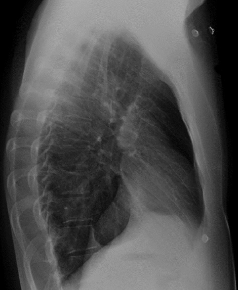
**Imaging Modality:** Lateral Chest Radiograph (X-ray). **Anatomical Region:** Thoracic cavity, including the mediastinum, lungs, and diaphragm. **Observed Pathology and Visual Features:** The image demonstrates signs of pulmonary hyperinflation. Key radiologic features include: * **Anterior Clear Space:** There is a notable increase in the retrosternal clear space (the lucent area between the posterior aspect of the sternum and the anterior wall of the ascending aorta), suggesting an increase in lung volume. * **Thoracic Configuration:** The anteroposterior (AP) diameter of the chest is increased, contributing to a "barrel chest" appearance. * **Diaphragmatic Appearance:** The hemidiaphragms appear flattened rather than normally domed. * **Lung Parenchyma:** Increased radiolucency is observed throughout the lung fields, consistent with air trapping. **Clinical Context:** These findings are classic radiographic markers for Chronic Obstructive Pulmonary Disease (COPD), specifically emphysema. **Key Diagnostic Features:** The combination of an enlarged retrosternal clear space (typically $>2.5$ cm), flattened diaphragms, and an increased AP diameter on a lateral projection are the primary diagnostic cues for identifying chronic lung hyperinflation.
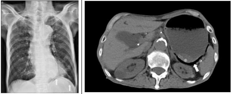
This dual-panel diagnostic image displays a posterior-anterior (PA) chest radiograph on the left and a non-contrast axial abdominal CT scan on the right, illustrating features of gastric emphysema. The chest X-ray demonstrates interstitial opacities in the left lower lung zone and a prominent air bubble located beneath the left hemi-diaphragm, indicated by white arrows. The cardiac silhouette appears slightly enlarged, and the lung fields show hyperinflation consistent with COPD. The axial CT scan on the right highlights a markedly dilated stomach. A white arrow points to pneumatosis gastrica (intramural gas), visualized as linear streaks and small pockets of air within the posterior stomach wall. Calcifications of the abdominal aorta and spinal osteophytes are also visible. This combination of findings is characteristic of gastric emphysema, an uncommon condition where gas collects within the gastric wall, often associated with gastric outlet obstruction or mucosal injury. These visuals serve as a clinical example of using multi-modal imaging to identify gastric distention and associated mural air in acute abdominal presentations.
🫁 TRIGGER 5 — Pneumonia (True/False & Case Scenario)
- Dense white opacity in the right upper lobe
- The horizontal fissure is pushed downward (bulging fissure sign) - due to heavy mucoid exudate from Klebsiella
- Area of dark lucency within the white opacity = early cavitation (Klebsiella tends to destroy lung tissue)
- Left lung is clear
🫁 TRIGGER 6 — COPD with Respiratory Failure (True/False & Case Scenario)
- Flat diaphragms (white arrows) - pushed down by hyperinflated lungs
- More than 10 posterior ribs visible above the diaphragm = hyperinflation
- Increased retrosternal clear space on lateral view = barrel chest (increased AP diameter)
- Reduced peripheral vascular markings = pruning from emphysema
- Ribs are horizontal instead of slanting downward normally
Umm but for true and false and for short case is it same x ray noted
- True/False scenario - mentions CXR with right upper lobe consolidation + bulging fissure ✅ same X-ray
- Short Case (Mr. Hassan) - mentions CXR with right upper lobe consolidation ✅ same X-ray
- True/False scenario - mentions CXR with hyperinflated lungs + enlarged pulmonary arteries ✅ same X-ray
- Short Case (Mr. Lim) - mentions CXR with hyperinflated lungs + flat diaphragms ✅ same X-ray
| X-ray | |
|---|---|
| Trigger 5 (both T/F and Short Case) | |
| Trigger 6 (both T/F and Short Case) | |
Different can aaa
lobar pneumonia upper lobe consolidation chest X-ray
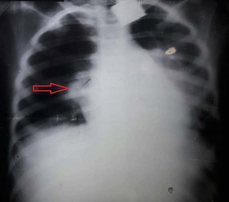
This diagnostic image is an anteroposterior (AP) chest X-ray of a pediatric patient demonstrating classic findings of lobar pneumonia. A prominent, dense, and homogenous opacification is localized to the right upper lobe, consistent with pulmonary consolidation. The opacification has a well-defined inferior border along the horizontal fissure, indicated by a red arrow, which is a hallmark of lobar involvement. Normal lung markings in this region are obscured, while the left lung field appears relatively clear and well-aerated. The cardiac silhouette and mediastinal structures appear within normal limits for this projection, with no significant shift or widening. The diaphragm and costophrenic angles are visible, though the right side is partially obscured by the adjacent consolidation. This image serves as an educational example of bacterial pneumonia presentation in a clinical setting, highlighting the radiographic appearance of alveolar space filling.

Multi-modal diagnostic imaging series illustrating community-acquired lobar pneumonia in a 30-year-old patient. (a) Posterior-anterior chest X-ray shows a clear area of consolidation in the left upper lobe. (b) B-mode lung ultrasound (LUS) reveals a complex, hypoechoic consolidation containing numerous hyperechoic punctate and linear structures, characteristic of a marked air bronchogram. (c-e) Contrast-enhanced ultrasound (CEUS) time-series demonstrates perfusion dynamics: (c) at 13 seconds, there is homogeneous pulmonary arterial enhancement; (d) by 1 minute, significant early washout occurs; (e) by 2 minutes, the washout is more pronounced. This specific CEUS pattern of rapid washout is indicative of pulmonary arterial vasoconstriction, often associated with the Euler–Liljestrand mechanism in response to local hypoxia in consolidated lung tissue. The series serves as an educational example of how multi-phase CEUS can supplement B-mode ultrasound and radiography in characterizing the hemodynamic response of pulmonary parenchyma during inflammatory processes.
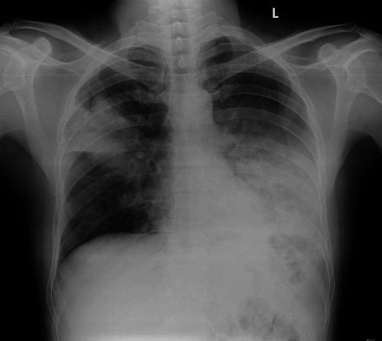
This diagnostic image is a posterior-anterior (PA) chest X-ray demonstrating multifocal pneumonia in a 49-year-old patient. The primary visual findings include significant areas of increased opacity, consistent with consolidation, located in the right upper lobe and the left lower lobe. The right upper lobe consolidation shows a relatively clear inferior margin along the minor fissure. In the left lung, a dense, patchy opacity is visible in the lower zone, obscuring the normal diaphragmatic contour and the left costophrenic angle, characteristic of lower lobe involvement. The heart size appears within normal limits (normal cardiothoracic ratio), and the trachea is midline without deviation. The mediastinal contours are preserved despite the adjacent pulmonary infiltrates. This radiographic presentation is typical for bacterial or community-acquired pneumonia, serving as an educational example of lobar consolidation patterns and the silhouette sign in clinical radiology.
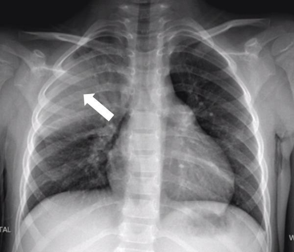
This diagnostic image is a posterior-anterior (PA) chest x-ray of a human patient. The primary finding is a dense, focal area of opacification located in the right upper-to-mid lung zone, indicated by a white arrow. This opacity is characteristic of lobar consolidation, exhibiting relatively well-defined borders and increased radiodensity compared to the more lucent left lung and right lower lung fields. The findings are clinically consistent with right-upper lobe pneumonia. The cardiac silhouette and mediastinal structures appear central and are within normal limits for size and morphology. The costophrenic angles are clear on this view, and the bony structures of the thorax, including the clavicles and ribs, appear intact. This radiological study serves as a classic educational example of community-acquired pneumonia and demonstrates the appearance of alveolar consolidation on plain film radiography.
COPD hyperinflation barrel chest flattened diaphragm chest X-ray PA view
This diagnostic image displays a two-panel chest roentgenogram (X-ray) from a 65-year-old male, highlighting radiographic features of severe pulmonary emphysema and chronic obstructive pulmonary disease (COPD). Panel A presents a postero-anterior (PA) view, while Panel B provides a lateral view. Key findings include significant hyperinflation of the lung fields with marked flattening of the hemidiaphragms (indicated by white arrows), which loss their normal convex shape. In Panel A, a yellow arrow identifies areas of increased translucency and attenuation of vascular markings consistent with emphysematous parenchymal changes. A red arrow highlights the decreased zone of apposition, representing the diminished area where the diaphragm sits parallel to the inner rib cage. Panel B further illustrates the flattened diaphragmatic contour and an increased retrosternal clear space, reflecting an increased anterior-posterior thoracic diameter (barrel chest). These visual markers are clinically significant as they correlate with the mechanical basis of Hoover's sign, where the horizontal orientation of the diaphragm causes inward rib cage retraction during inspiration.
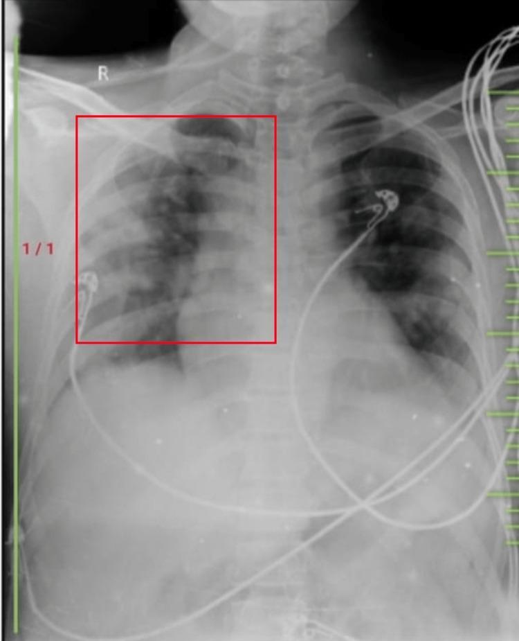
Diagnostic Imaging: This is a posteroanterior (PA) view chest X-ray demonstrating a clinical presentation of pulmonary hyper-translucency. The image captures the thoracic cavity with a red bounding box highlighting the right upper and middle lung zones. Distinctive visual features include increased radiolucency (hyper-translucency) across both lung fields, suggestive of pulmonary hyperinflation or increased air trapping. Despite the increased lucency, bronchovesicular markings remain visible throughout the parenchyma. A prominent hilar marking is identifiable on the right side within the highlighted region. Anatomical landmarks such as the clavicles, ribs, and cardiac silhouette are visible, though the diaphragm appears somewhat flattened, consistent with hyperinflation. Medical monitoring leads and cables are overlaid on the chest wall. This imaging is clinically significant for demonstrating radiographic signs of obstructive lung disease or other conditions involving alveolar over-distension, useful for students learning to identify variations in lung density and airway-related pathologies.
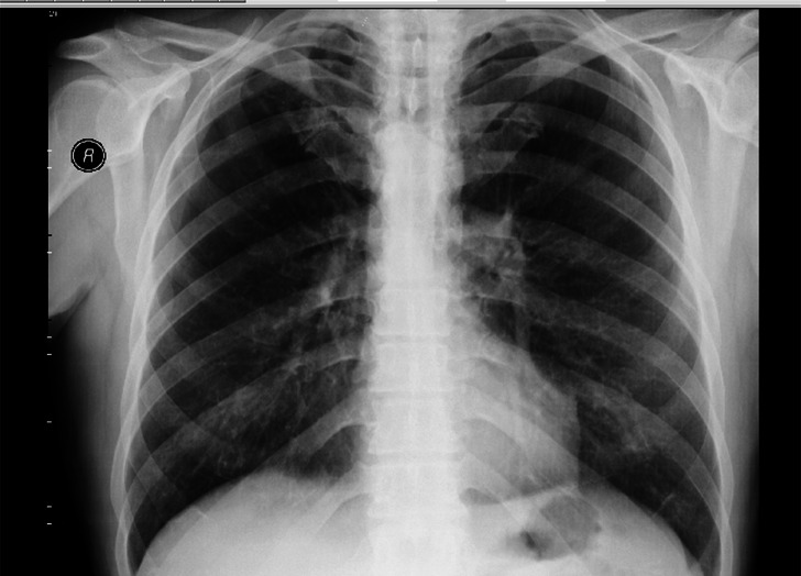
**Imaging Modality:** Posteroanterior (PA) Chest X-ray. **Anatomical Region:** Thoracic cavity, including the lungs, rib cage, mediastinum, and diaphragm. **Observed Pathology:** Bilateral pulmonary hyperlucency and lung overexpansion. The image demonstrates signs of chronic obstructive pulmonary disease (COPD) or emphysema. **Characteristic Visual Features:** * **Lungs:** Marked bilateral hyperlucency of the lung parenchyma with a noticeable reduction in vascular markings, particularly in the upper and mid-zones. * **Diaphragm:** Flattening of both diaphragmatic domes, indicating significant pulmonary hyperinflation. * **Rib Cage:** Increased width of the intercostal spaces and a more horizontal orientation of the ribs. * **Mediastinum:** The heart silhouette appears relatively narrow or "drop-shaped," a common secondary finding in hyperinflated lungs. * **Costophrenic Angles:** Blunting or widening due to the flattened diaphragm. **Key Diagnostic Features:** The combination of diffuse bilateral hyperlucency, flattened diaphragmatic contours, and increased retrosternal/intercostal space is highly characteristic of diffuse obstructive lung disease. No acute focal consolidations, large pleural effusions, or pneumothoraces are visualized.
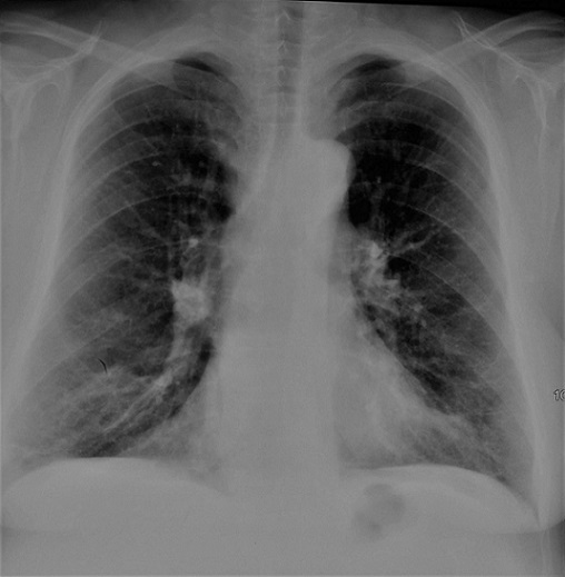
**Imaging Modality:** Posteroanterior (PA) chest X-ray. **Anatomical Region:** Thoracic cage, including lungs, mediastinum, and diaphragm. **Observed Pathology and Visual Features:** The radiograph demonstrates significant signs of pulmonary hyperinflation (thoracic distension). Key findings include: * **Diaphragmatic Flattening:** Bilateral depression and flattening of the hemidiaphragms, indicating increased lung volume. * **Increased Lucency:** Diffuse increase in lung radiolucency, consistent with air trapping. * **Bronchial Changes:** Visible peribronchial cuffing and thickening of the bronchial walls, particularly evident in the perihilar regions. * **Rib Configuration:** Horizontalization of the ribs and widening of the intercostal spaces. * **Mediastinal Profile:** A narrow, "vertical" heart configuration (drop heart) often associated with chronic obstructive pulmonary disease (COPD). **Clinical Context:** The combination of hyperinflation and bronchial wall thickening is highly characteristic of chronic obstructive airway disease, such as COPD or chronic bronchitis. **Key Diagnostic Features:** The presence of flattened diaphragms and peribronchial thickening serve as the primary visual cues for distinguishing obstructive lung pathology from restrictive patterns or acute consolidations. No acute pleural effusions or focal masses are identified.
🫁 TRIGGER 5 — Pneumonia
True/False X-ray
Short Case (Mr. Hassan) X-ray
🫁 TRIGGER 6 — COPD
True/False X-ray
Short Case (Mr. Lim) X-ray