
Describe the nucleus origin course branches of facial nerve add a note on bell’s palasy
facial nerve anatomy branches course diagram
facial nerve branches temporal zygomatic buccal marginal mandibular cervical
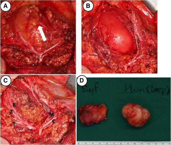
This composite of four clinical photographs illustrates intraoperative findings and gross pathology from a right parotidectomy for a deep-lobe tumor. Panel A shows the surgical field with a white arrow indicating a communicating nerve branch between the marginal mandibular and buccal branches of the facial nerve. Panel B demonstrates the tumor's location within the deep lobe of the parotid gland, adjacent to the masseter muscle. Panel C provides a comprehensive view of the preserved facial nerve branches following dissection, labeled as temporal (T), zygomatic (Z), buccal (B), marginal mandibular (M), and cervical (C). Panel D presents the gross surgical specimens against a green background with a metric scale; it differentiates the irregular superficial lobe parotid tissue ('Supf.') from the main, well-demarcated, multi-lobulated tumor mass ('Main (Deep)'). These images serve as educational material for head and neck surgery, specifically illustrating parotid gland anatomy, facial nerve preservation techniques, and surgical management of parotid neoplasms.
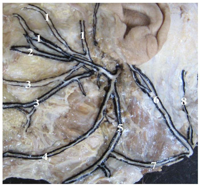
An anatomical dissection photograph illustrating the terminal branching patterns of the human facial nerve (CN VII) and its cervical connections. The image displays a 'Type I' branching variant characterized by a fan-like distribution over the facial musculature. Key structures labeled include the temporal branches (1) extending superiorly, zygomatic branches (2) coursing toward the midface, and buccal branches (3) distributed across the cheek area. Inferiorly, the marginal mandibular branch (4) follows the lower border of the mandible, while the cervical branch (5) descends toward the neck. The dissection also highlights the greater auricular nerve (6) and the lesser occipital nerve (8). Of clinical and surgical significance is the depicted connection (7) between the cervical branch of the facial nerve and the transverse cervical nerve of the cervical plexus. This anatomical resource serves to educate on neuroanatomical variations relevant to parotid surgery, facial reconstruction, and local anesthesia.
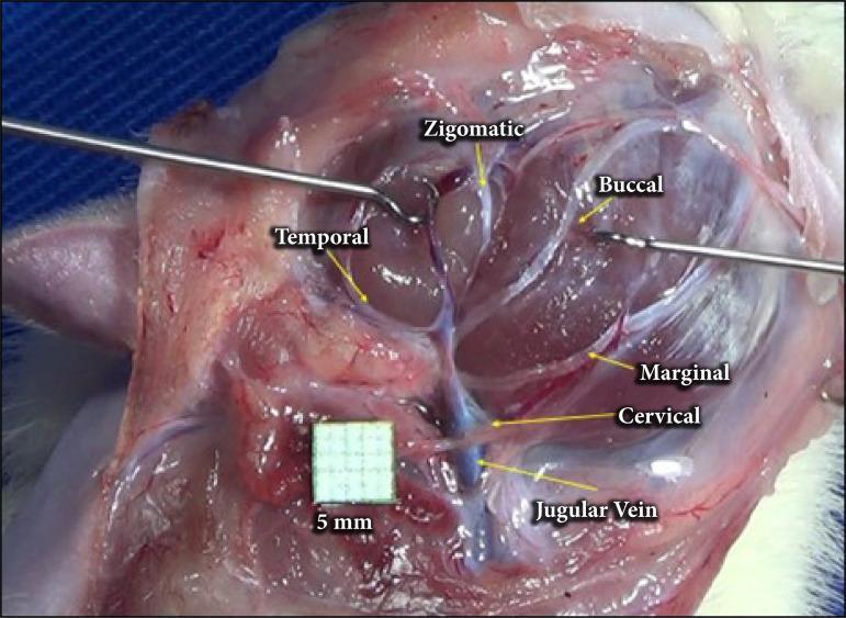
A clinical intraoperative photograph showing the anatomical arrangement of the facial nerve branches in a surgical dissection, following parotidectomy. The image provides a lateral view of the cervicofacial region where the parotid gland has been removed to expose the extratemporal facial nerve plexus. Labeled branches include the temporal branch extending superiorly, the zygomatic and buccal branches traveling toward the mid-face, and the marginal mandibular and cervical branches descending inferiorly. The cervical branch is depicted in close proximity to the jugular vein, which is visible in the lower surgical field. Surgical instruments (retractors) are positioned to maintain exposure of the deep tissues. A 5mm grid scale is included for anatomical measurement. This visual serves as an educational guide for surgical anatomy, identifying key landmarks for nerve preservation during parotid gland surgery or facial nerve decompression.
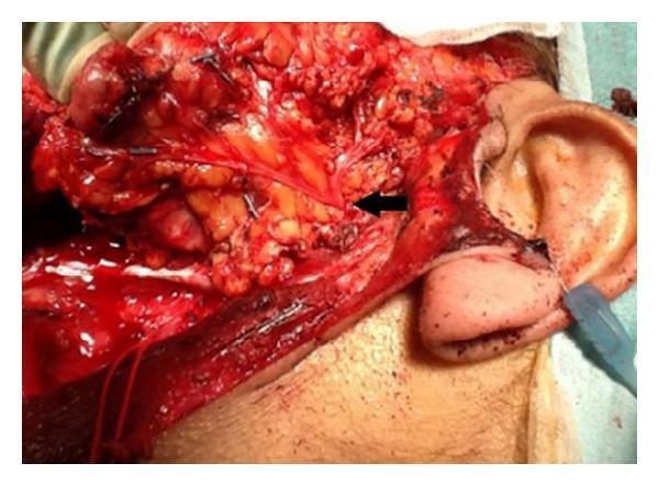
This clinical photograph displays a lateral view of an intraoperative surgical field following a subtotal parotidectomy. The image focuses on the preauricular and cervical regions, where the skin has been reflected to reveal the underlying anatomy. A black arrow indicates the pes anserinus of the facial nerve (Cranial Nerve VII) as it emerges from the parotid gland region. Clearly visible are the thin, yellowish-white, thread-like neural branches radiating from this central point, representing the temporal, zygomatic, buccal, marginal mandibular, and cervical branches. These nerves are preserved and meticulously dissected amidst the surrounding erythematous soft tissue and adipose deposits. The external ear is visible on the right as a landmark, with the neck and jawline extending inferiorly and anteriorly. The image demonstrates surgical anatomy crucial for preventing iatrogenic facial nerve injury during parotid gland surgery, highlighting the spatial relationship between the nerve branches and the deep salivary tissue.
Bell's palsy facial paralysis clinical appearance
The Facial Nerve (CN VII) - Nucleus, Origin, Course, and Branches
1. Nuclei and Fiber Components
| Fiber Type | Nucleus | Function |
|---|---|---|
| Special visceral efferent (SVE) | Motor nucleus of VII (pons) | Muscles of facial expression, stapedius, stylohyoid, posterior belly of digastric |
| General visceral efferent (GVE) | Superior salivatory nucleus | Lacrimal gland (via GSPN + pterygopalatine ganglion), submandibular & sublingual glands (via chorda tympani + submandibular ganglion) |
| Special afferent (taste) | Nucleus of the tractus solitarius | Taste from anterior 2/3 of tongue (via chorda tympani), palate & tonsillar fossa (via GSPN) |
| General somatic afferent | Spinal trigeminal nucleus | Skin of external auditory canal, conchal auricle, proprioception from facial muscles |
2. Origin and Intracranial Course
- The facial nerve exits the brainstem at the pontomedullary junction, between the pons and the olive, as two roots: the large motor root and the smaller nervus intermedius (which emerges between the pons and inferior cerebellar peduncle).
- The nerve traverses the cerebellopontine angle (CPA) cistern and enters the internal auditory meatus (IAM), lying anterosuperiorly alongside CN VIII. Within the IAM, it is positioned above the cochlear nerve, with vestibular nerves posteriorly.
- At the lateral end of the IAM, the nerve enters the fallopian canal (bony facial canal) - a tunnel through the temporal bone from the fundus of the IAC to the stylomastoid foramen. - Scott-Brown's Otorhinolaryngology, Vol. 2
3. Course Through the Temporal Bone (Intratemporal Segments)
A. Labyrinthine Segment
- First and narrowest segment - passes between the cochlea and vestibule (superior to cochlea)
- Opens into the geniculate fossa where the geniculate ganglion is located
- The overlying bone is dehiscent in ~25% of ears - making it surgically vulnerable
- This segment lacks epineurium and is a watershed vascular zone (between vertebrobasilar and ECA systems) - making it the most vulnerable site in Bell's palsy
- The First Genu is here: the nerve makes an acute posterior and slightly inferior turn
- Branch given off: Greater Superficial Petrosal Nerve (GSPN) - carries preganglionic parasympathetics to lacrimal and nasal glands via pterygopalatine ganglion; also carries taste from the palate
B. Tympanic (Horizontal) Segment
- Runs in the medial wall of the middle ear, above the promontory
- Passes over the cochleariform process and forms the superior wall of the oval window niche
- Most common site of congenital bony dehiscence (above the oval window), making it susceptible to toxic effects of suppurative otitis media
- Second Genu is at the pyramidal eminence (just anteroinferior to the lateral semicircular canal): the nerve turns downward into the vertical segment
C. Mastoid (Vertical) Segment
- Runs behind the external auditory canal (EAC)
- Branches given off:
- Nerve to stapedius - enters via a tiny canaliculus at the pyramidal eminence; supplies the stapedius muscle (loss = hyperacusis)
- Chorda tympani - carries taste from anterior 2/3 of tongue (via lingual nerve to CN V3) and preganglionic parasympathetics to submandibular/sublingual glands (via submandibular ganglion)
4. Exit from the Skull and Extracranial Course
- Posterior auricular nerve - supplies the occipital belly of occipitofrontalis and auricular muscles
- Branch to posterior belly of digastric and stylohyoid muscle
5. Five Terminal Branches (Emerge from Parotid Borders)
| Branch | Direction | Muscles Supplied |
|---|---|---|
| Temporal | Upper border → superiorly | Frontalis, orbicularis oculi (upper), corrugator supercilii |
| Zygomatic | Anterior border → midface | Orbicularis oculi (lower), zygomaticus |
| Buccal | Anterior border → cheek | Buccinator, orbicularis oris, muscles of upper lip |
| Marginal mandibular | Lower border → follows mandible | Depressor anguli oris, depressor labii, mentalis |
| Cervical | Lower border → neck | Platysma |
6. Summary of All Branches (Mnemonic Structure)
- Greater superficial petrosal nerve (at geniculate ganglion)
- Nerve to stapedius (at pyramidal eminence)
- Chorda tympani (in mastoid segment)
Note on Bell's Palsy
Definition
Pathophysiology
- The leading theory is reactivation of Herpes Simplex Virus type 1 (HSV-1) in the geniculate ganglion. HSV-1 DNA has been identified by PCR in endoneurial fluid from patients with Bell's palsy.
- Varicella-zoster virus (VZV) may account for up to one-third of cases (differentiating from Ramsay Hunt syndrome by absence of vesicles).
- The swollen, inflamed nerve becomes compressed within the narrow labyrinthine segment and bony fallopian canal, causing ischemia and axonal injury.
- Histopathology shows mononuclear cell infiltration consistent with viral or immune etiology.
Clinical Features
- Onset: Acute - maximum paralysis reached within 48 hours in most cases; essentially complete within 3-4 days
- Complete LMN palsy: All ipsilateral facial muscles affected, including the forehead (key distinction from UMN/supranuclear palsy, where the forehead is spared due to bilateral cortical representation)
- Pain behind the ear may precede paralysis by 1-2 days
- Hyperacusis - from stapedius involvement
- Loss of taste on anterior 2/3 of tongue (chorda tympani involvement)
- Reduced lacrimation if GSPN involved (proximal lesion)
- Incomplete eye closure - risk of exposure keratitis
Investigations
- Diagnosis is primarily clinical
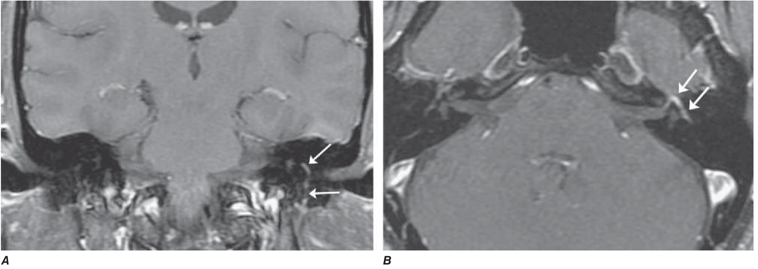
- MRI (gadolinium-enhanced): May show swelling and uniform enhancement of the geniculate ganglion and facial nerve within the temporal bone (see image below)
- EMG/nerve conduction: If denervation is found after 10 days, implies axonal degeneration with poorer prognosis and delayed recovery (3+ months)
- Bloods if atypical: ESR/CRP, blood glucose, Lyme titre, HIV, ACE level (sarcoidosis)
Treatment
| Intervention | Details |
|---|---|
| Corticosteroids (first-line) | Prednisone 60-80 mg/day for 5 days then tapered over 5 days - shortens recovery and improves outcome; most effective when started within 72 hours |
| Antivirals | Valacyclovir (1000 mg/day x 5-7 days) or acyclovir; evidence does not support adding antivirals to steroids alone for routine cases, but use if vesicular lesions are present (Ramsay Hunt) |
| Eye protection | Paper tape to close eyelid during sleep; artificial tears to prevent exposure keratitis |
| No surgical decompression | Evidence does not support decompression; may be harmful |
| Physical therapy | Facial muscle massage once recovery begins |
Prognosis
- 70% recover completely within 1-2 months
- 85% achieve near-normal function overall
- Favorable signs: Incomplete paralysis in the first week; return of taste in the first week; early motor recovery at 5-7 days
- Unfavorable: Complete paralysis; EMG denervation after 10 days (implies axonal degeneration; recovery may take 2+ years and be incomplete)
- Recurrence: ~7-8% of cases; interval averages ~10 years
- Aberrant reinnervation can produce synkinesis (e.g., involuntary eye closure on smiling) or crocodile tears (lacrimation during eating, from misdirected regenerating parasympathetic fibers)
Differential Diagnosis of Acute Facial Palsy
- Ramsay Hunt syndrome (VZV) - vesicular rash in external auditory canal/pinna/palate; worse prognosis than Bell's palsy
- Lyme disease - can cause bilateral facial palsy; Borrelia burgdorferi (>10% of facial palsies in endemic areas)
- Otitis media/cholesteatoma - middle ear disease eroding the fallopian canal
- Acoustic neuroma / CPA tumors - associated with CN VIII signs
- Sarcoidosis and Guillain-Barré syndrome - bilateral facial palsy
- Parotid malignancy - slow progressive palsy
- Melkersson-Rosenthal syndrome - recurrent facial palsy + facial edema + fissured tongue