
Ivdp vs lumbar spondylosis vs ankylosis spondylitis difference from history examination investigation and treatment
ankylosing spondylitis bamboo spine radiograph sacroiliitis
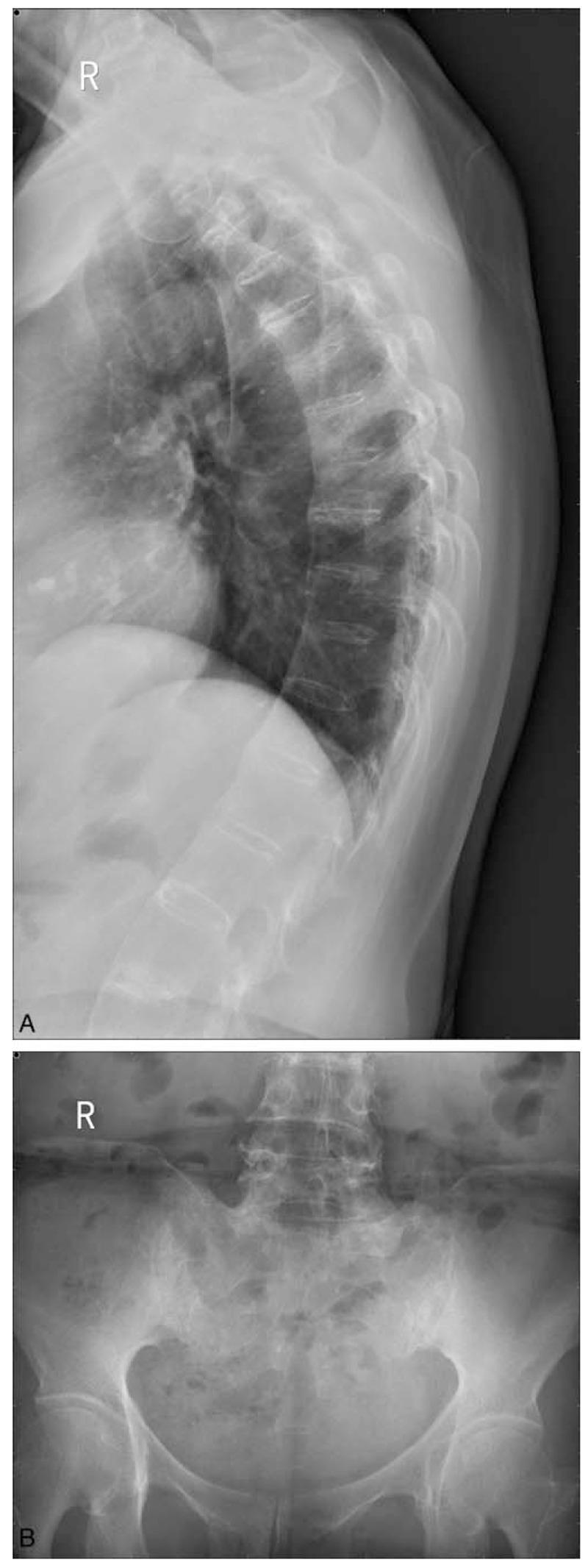
This composite of two X-ray images demonstrates classic radiographic signs of Ankylosing Spondylitis (AS). Panel A is a lateral chest/thoracic spine radiograph showing characteristic 'bamboo spine' appearance. This is characterized by extensive syndesmophyte formation, where bony bridges fuse the thoracic vertebral bodies together, creating a continuous, rigid vertical column. Notably, the intervertebral disc spaces remain relatively preserved in height despite the fusion. Panel B is an anteroposterior (AP) pelvic radiograph focusing on the sacroiliac joints. It shows Grade III bilateral sacroiliitis, evidenced by significant subchondral sclerosis (increased bone density) and narrowing with irregular joint margins. The sacrum and iliac wings exhibit focal areas of erosion and fusion. These findings are hallmarks of seronegative spondyloarthropathies and are used clinically to assess disease progression and staging in patients presenting with chronic inflammatory back pain and HLA-B27 positivity.
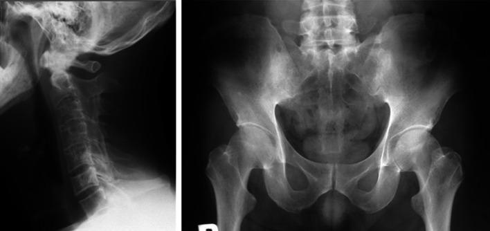
This composite diagnostic image features two X-ray radiographs illustrating the hallmark skeletal manifestations of advanced ankylosing spondylitis (SpA). The left panel is a lateral radiograph of the cervical spine showing 'bamboo spine' morphology. It demonstrates extensive marginal syndesmophytes and ankylosis leading to the fusion of vertebral bodies and complete ossification of the intervertebral disc spaces. The result is a continuous, rigid vertebral column with loss of the normal cervical lordosis. The right panel is an anteroposterior (AP) radiograph of the pelvis focusing on the sacroiliac (SI) joints. Both SI joints show Grade 4 sacroiliitis according to the Modified New York criteria, characterized by total ankylosis (fusion), significant subchondral sclerosis, and loss of visible joint space. These findings represent end-stage chronic inflammation in axial spondyloarthritis. The image is intended for medical education regarding the progression of rheumatological conditions and the interpretation of radiological signs of spinal fusion and sacroiliitis.
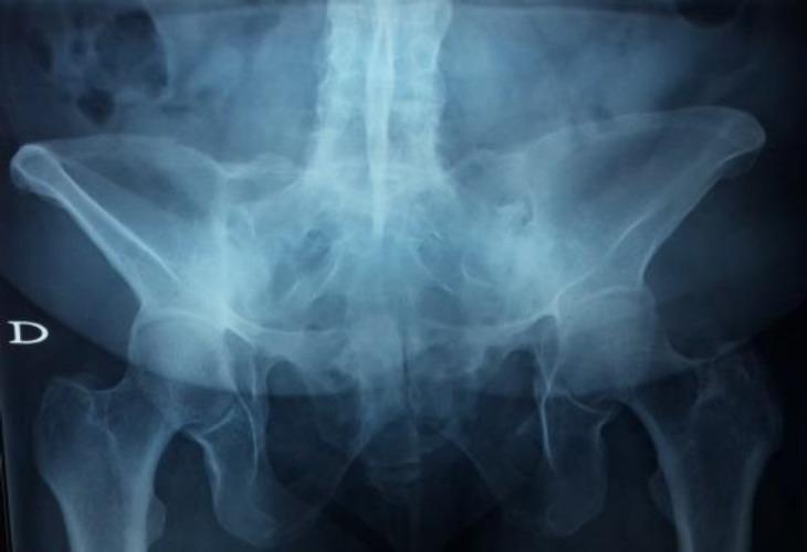
Anteroposterior (AP) pelvic radiograph demonstrating significant chronic changes of the axial skeleton, characteristic of advanced ankylosing spondylitis. The primary diagnostic finding is bilateral grade 4 sacroiliitis, visualized as complete bony ankylosis (fusion) of both sacroiliac joints. The normal joint spaces between the sacrum and iliac bones are entirely obliterated, replaced by continuous bony bridging. The lower lumbar spine shows signs of syndesmophyte formation and vertical fusion, contributing to the 'bamboo spine' appearance often associated with this pathology. The pelvic ring remains intact, and the iliac crests are well-defined. Both hip joints (femoroacetabular joints) are visualized, with the femoral heads well-seated within the acetabula, though the image focus remains on the sacroiliac and spinal involvement. This diagnostic image is used to teach the progression of seronegative spondyloarthropathies and the radiographic criteria for diagnosing advanced sacroiliitis.
lumbar disc prolapse herniation MRI L4 L5
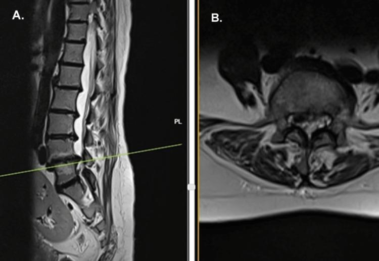
This diagnostic image consists of two MRI views of the lumbar spine: a sagittal section (A) and an axial section (B) at the L4-L5 level. In the sagittal view, the lumbar spine demonstrates preserved lordotic curvature, but multiple intervertebral discs exhibit signal hypointensity on T2-weighted imaging, consistent with widespread disc dehydration and degeneration. Notably, at the L4-L5 level, there is a significant posterior disc prolapse that is hypointense, suggesting potential calcification or dense fibrosis. The axial view (B) reveals a central and right paracentral disc herniation encroaching upon the spinal canal and the right lateral recess. This herniated material is causing visible mass effect and impingement on the neural structures, specifically the thecal sac and the right-sided nerve roots. These findings are characteristic of degenerative disc disease and may be associated with systemic metabolic conditions like alkaptonuria/ochronosis when presenting with such extensive disc calcification. The imaging serves as a clinical education tool for identifying lumbar disc herniation, spinal stenosis, and secondary neural impingement.
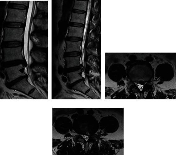
Diagnostic imaging series comprising sagittal and axial (labeled as coronal in clinical notes) T2-weighted MRI scans of the lumbar spine, specifically focusing on the L4-L5 intervertebral disc level. The series functions as a pre- and post-operative comparison for a prolapse poroscopy (percutaneous endoscopic lumbar discectomy). The preoperative sagittal view demonstrates a significant posterior disc herniation (prolapse) causing narrowing of the spinal canal and mechanical compression of the thecal sac. Corresponding axial sections show a focal disc protrusion resulting in lateral recess stenosis. Post-operative images illustrate the results of nucleus pulposus extraction, characterized by a visible reduction in the size of the herniated disc material, improved canal dimensions, and decompression of the neural elements. A minor signal intensity change is noted at the surgical site. This comparison is used to evaluate the efficacy of minimally invasive endoscopic spinal decompression and to assess for any residual disc material or nerve root impingement.
IVDP vs Lumbar Spondylosis vs Ankylosing Spondylitis - Complete Comparison
Overview at a Glance
| Feature | IVDP (Disc Prolapse) | Lumbar Spondylosis | Ankylosing Spondylitis |
|---|---|---|---|
| Nature | Mechanical / acute | Degenerative | Inflammatory / autoimmune |
| Age group | 30-50 years | >40 years | 15-35 years (young adults) |
| Sex | M > F | M = F | M >> F (3:1) |
1. HISTORY
IVDP (Intervertebral Disc Prolapse)
- Onset: Sudden, acute onset - classically while lifting weight from a bent/stooped position or during strenuous activity
- Site of pain: Low back (L4-L5 or L5-S1 most common), radiating to buttock, posterior thigh, leg, and down to toes (sciatica)
- Nature of pain: Sharp, stabbing, shooting - radiates along the affected nerve root distribution
- Aggravating factors: Movement, coughing, sneezing, defaecation (increased intradural pressure), forward bending
- Radiation: Unilateral - follows one nerve root (L4, L5, or S1)
- Relieving factors: Rest, recumbency
- History of recurrence: Subsequent attacks may follow trivial injury such as coughing
- No morning stiffness (pain is positional, not inflammatory)
- Bladder/bowel dysfunction if central disc prolapse compresses cauda equina (emergency)
Lumbar Spondylosis
- Onset: Insidious, chronic, gradual worsening over years
- Age: Invariably over 40 years; degenerative condition
- Site of pain: Diffuse low back pain, may radiate to legs if osteophytes compress nerve roots
- Nature of pain: Dull aching, constant, worsens with activity and prolonged standing/walking
- Aggravating factors: Sustained postures, movement, prolonged activity; better with rest
- Morning stiffness: Mild and brief (< 30 min) - unlike inflammatory causes
- No alternating sciatica - if present, is consistent on one side
- History of heavy physical work or obesity is common
Ankylosing Spondylitis
- Onset: Insidious onset, onset in adolescents/young adults (15-35 years); can precede by buttock/sacroiliac pain for months
- Site of pain: Low back + buttock pain (alternating sides - classic!); sacroiliac joint area
- Nature of pain: Intermittent, dull aching; "worse in the morning, improves with activity/exercise" (classic inflammatory pattern)
- Morning stiffness: Prominent, lasting >1 hour - hallmark feature; improves with movement
- Night pain: Wakes patient in second half of night (not relieved by rest)
- Radiation: Sciatica may occur but alternates from side to side - distinguishes it from disc prolapse
- Systemic features: Fatigue, malaise, weight loss; eye involvement (anterior uveitis/iritis ~30%), plantar fasciitis, Achilles tendinitis (enthesopathy)
- Family history of HLA-B27-associated diseases (AS, IBD, psoriasis, reactive arthritis)
- No clear precipitating injury
2. EXAMINATION
IVDP
- Typical lumbar scoliosis with convexity to the affected side
- Kyphosis, slight flexion of hips and knees
- Antalgic gait (leans away from painful side)
- Local deep tenderness on or slightly lateral to the affected spinous process
- Paraspinal muscle spasm
- Flexion and extension greatly restricted and painful
- Lateral flexion on the affected side is very painful
- Rotation may be relatively free
- Straight Leg Raise (SLR/Lasegue's test): Positive at < 60° - most important test for L4/L5/S1 disc
- Crossed SLR: Positive (suggests large disc herniation)
- Femoral Nerve Stretch Test (FNST): Positive for L2-L3 disc (pain in front of thigh)
- Naffziger's test: Jugular vein compression induces back/leg pain
| Level | Weakness | Sensation loss | Reflex lost |
|---|---|---|---|
| L3-L4 disc | Quadriceps (knee extension) | Medial lower leg | Knee jerk reduced |
| L4-L5 disc | Extensor hallucis longus (great toe extension) | Dorsum of foot | No significant reflex |
| L5-S1 disc | Plantar flexion, peroneals | Lateral foot/sole | Ankle jerk absent |
Lumbar Spondylosis
- Posture: May show loss of lumbar lordosis; no characteristic scoliosis
- Gait: Normal or slightly stiff
- Palpation: Diffuse paraspinal tenderness; spinous process tenderness
- Spinal movements: All lumbar movements limited and painful, but often less dramatically than acute disc prolapse
- SLR: May be mildly positive if nerve root compression by osteophyte
- Neurological: May show mild deficit in lower extremities (motor, sensory, reflexes) - usually less dramatic than acute IVDP
- "Paucity of physical findings" relative to reported pain - characteristic feature
- Back pain > leg pain (unlike IVDP where leg pain may dominate)
- No inflammatory signs; no sacroiliac tenderness
Ankylosing Spondylitis
- Loss of lumbar lordosis, increased thoracic kyphosis
- Compensatory neck hyperextension and flexion of hips/knees ("question mark posture")
- Reduced chest expansion
- Modified Schober's test: Marks 5 cm below and 10 cm above PSIS; < 5 cm expansion on maximal forward flexion = abnormal (normal >5 cm)
- Occiput-to-wall distance: Patient cannot touch wall with back of head (increased with cervical involvement)
- Chest expansion: Measured at 4th intercostal space; < 2.5 cm = abnormal (costovertebral joint involvement)
- BASMI (Bath Ankylosing Spondylitis Metrology Index): Composite of above mobility measures
- Sacroiliac joint tests:
- FABER test (Patrick's test): Hip flexion, ABduction, External Rotation - pain in SI joint
- Direct compression of SI joints: Pain
- Gaenslen's test: Positive for sacroiliitis
- Tenderness at entheses: Achilles tendon insertion, plantar fascia, iliac crests, pubic symphysis
- Anterior uveitis (red, painful eye)
- Aortic regurgitation murmur (late)
- Reduced chest expansion from costovertebral joint fusion
3. INVESTIGATIONS
IVDP
| Investigation | Finding |
|---|---|
| X-ray spine (AP/Lateral) | Normal in early attacks; later shows narrowing of intervertebral disc space, lipping of vertebral bodies |
| MRI spine | Investigation of choice - shows disc herniation, nerve root compression, thecal sac impingement; T2-weighted shows decreased signal ("dark disc") |
| CT scan | Good for bony detail; less preferred over MRI for soft tissue |
| Myelography | Filling defect at level of prolapse; largely replaced by MRI |
| Discography | Concordant pain reproduction + dye leakage through annular tear - used pre-operatively |
| Epidurography | Helpful in diagnosis |
| EMG/NCS | Shows denervation pattern in affected myotome - helps confirm radiculopathy level |
| Blood tests | Normal CBC, ESR, CRP |
Lumbar Spondylosis
| Investigation | Finding |
|---|---|
| X-ray spine | Disc space narrowing, vertebral body lipping/osteophytes, facet joint narrowing and sclerosis, loss of lumbar lordosis |
| MRI spine | Decreased T2 signal in disc (disc degeneration), annular tears (HIZ - High Intensity Zone), osteophyte formation, facet joint hypertrophy, foraminal stenosis |
| CT scan | Best shows osteophytes and facet joint degeneration; facet arthrosis |
| Discography | Can identify pain-generating discs pre-operatively |
| Blood tests | Normal ESR, CRP, CBC; no HLA-B27 association |
Ankylosing Spondylitis
| Investigation | Finding |
|---|---|
| X-ray sacroiliac joints (AP pelvis) | Bilateral, symmetrical sacroiliitis - erosions, subchondral sclerosis, joint space narrowing, eventual fusion (Grade 0-IV) |
| X-ray spine (lateral) | Romanus lesions (sclerotic "shiny corners"), syndesmophytes (marginal, vertical), bamboo spine (late - complete fusion), squaring of vertebral bodies |
| MRI sacroiliac joints | Earliest diagnosis - subchondral bone marrow oedema (STIR sequence) before X-ray changes; more sensitive than plain X-ray |
| ESR, CRP | Elevated (active disease) |
| HLA-B27 | Positive in ~90% of AS (positive in ~8% general population); not diagnostic alone |
| CBC | Normocytic normochromic anaemia of chronic disease |
| Rheumatoid factor (RF) | Negative (seronegative spondyloarthropathy) |
| ANA | Negative |
| Slit-lamp examination | For uveitis |
| Echo/CXR | Aortic regurgitation, apical pulmonary fibrosis (late) |
| BASFI, BASDAI | Disease activity/functional indices |
- Bilateral sacroiliitis ≥ Grade 2 OR Unilateral Grade 3-4, plus at least one clinical criterion (IBP, reduced lumbar mobility, reduced chest expansion)
4. TREATMENT
IVDP
- Rest - bed rest in acute phase, on firm mattress; avoid provocative activities
- Analgesics/NSAIDs: Ibuprofen, diclofenac, naproxen - first-line for pain
- Muscle relaxants: Diazepam, cyclobenzaprine for paraspinal muscle spasm
- Physiotherapy: Short-wave diathermy, traction (pelvic traction), McKenzie exercises after acute phase
- Epidural steroid injection: Methylprednisolone epidurally - for radicular pain
- Lumbar corset/brace - supportive
- Cauda equina syndrome - absolute emergency, immediate decompression
- Failed conservative treatment > 6-12 weeks with persistent radiculopathy
- Progressive neurological deficit
- Laminectomy/discectomy - removal of herniated disc fragment
- Microdiscectomy - minimally invasive, preferred
- Percutaneous endoscopic discectomy
- Spinal fusion if recurrent disease or instability
Lumbar Spondylosis
- NSAIDs: Ibuprofen, naproxen, diclofenac - mainstay
- Analgesics: Paracetamol, tramadol for breakthrough pain
- Physiotherapy: Core strengthening, lumbar stabilisation exercises, postural correction, hydrotherapy
- Weight loss if obese
- Epidural steroid injections for radicular symptoms
- Facet joint injections for facet arthropathy
- TENS, heat/cold therapy
- Lumbar support
- Decompressive laminectomy for spinal stenosis
- Foraminotomy for foraminal stenosis/osteophyte impingement
- Lumbar fusion (PLIF, TLIF) for instability or spondylolisthesis
- TLDR: Surgery reserved for neurological compromise or incapacitating pain after 3-6 months conservative therapy
Ankylosing Spondylitis
- Physiotherapy is MANDATORY - the single most important non-drug treatment; maintains spinal mobility, prevents deformity
- Daily exercises: Extension exercises, deep breathing, swimming
- Patient education, posture training, sleep on firm mattress without pillow
- NSAIDs (first-line): Indomethacin (drug of choice historically), naproxen, celecoxib - relieves pain AND may slow radiographic progression if taken continuously
- Analgesics for pain control
- Sulfasalazine: For peripheral joint involvement (NOT effective for axial disease)
- Methotrexate: For peripheral arthritis only; not for axial disease
- Anti-TNF-α agents: Infliximab (Remicade), etanercept (Enbrel), adalimumab (Humira), golimumab (Simponi) - decrease disease progression, especially when started early; first choice biologic
- IL-17A inhibitors: Secukinumab (Cosentyx), ixekizumab - alternative to anti-TNF
- IL-23 inhibitors: Risankizumab, guselkumab (for peripheral disease)
- Note: Anti-TNF agents carry increased risk of infection and lymphoma
- Intra-articular corticosteroid injections into sacroiliac joints (under imaging guidance)
- Systemic steroids - limited use; short courses for acute flares
- Total hip replacement (THA) - for hip joint ankylosis/destruction (not uncommon in AS)
- Corrective osteotomy - for severe fixed kyphotic deformity
- Spinal surgery rarely needed; fracture fixation for complications
Quick Summary Comparison Table
| Feature | IVDP | Lumbar Spondylosis | Ankylosing Spondylitis |
|---|---|---|---|
| Age | 30-50 | >40 | 15-35 |
| Gender | M > F | M = F | M >> F |
| Onset | Acute/sudden | Insidious | Insidious |
| Pain character | Sharp, shooting | Dull aching | Dull, inflammatory |
| Morning stiffness | Absent | < 30 min | > 1 hour |
| Activity effect | Worse with activity | Worse with activity | Improves with activity |
| Radiation | Unilateral sciatica | May radiate | Alternating sciatica |
| SLR | Positive | Mildly positive | Negative (usually) |
| Schober's test | Normal | Normal | Reduced |
| Chest expansion | Normal | Normal | Reduced |
| SI joint tenderness | Absent | Absent | Present |
| HLA-B27 | Negative | Negative | Positive (~90%) |
| ESR/CRP | Normal | Normal | Elevated |
| RF/ANA | Negative | Negative | Negative |
| X-ray | Disc space narrowing | Osteophytes, disc narrowing | Bamboo spine, bilateral sacroiliitis |
| MRI | Disc herniation | Dark disc, osteophytes | Bone marrow oedema (SI joints) |
| Key treatment | Rest, NSAIDs, discectomy | NSAIDs, PT, decompression | NSAIDs + physiotherapy, anti-TNF |