
Can you give information about Ayurveda diseases
- Ayurveda classifies diseases (called Rogas or Vikaras) based on the dosha involved, the affected tissue (dhatu), and the body channel (srotas) disturbed.
- Common categories include digestive disorders, skin diseases, fever (Jwara), metabolic conditions, and nervous system disorders.
- Treatment focuses on restoring dosha balance through diet, herbs, Panchakarma (detox therapies), and lifestyle.
Ayurveda diseases classification Vata Pitta Kapha Rogas overview
Ayurveda doshas Vata Pitta Kapha body types diagram
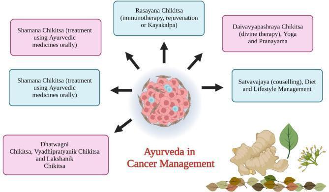
An educational infographic titled 'Ayurveda in Cancer Management' that illustrates a multidisciplinary integrative medicine approach. The central graphical element is a cross-sectional illustration of a tumor microenvironment, depicting a cluster of irregularly shaped pinkish-red neoplastic cells interspersed with small cyan-blue immune or signaling components. Black directional arrows radiate from this central mass toward surrounding rectangular boxes, signifying various therapeutic interventions. The described treatments include: 'Rasayana Chikitsa' (immunotherapy and rejuvenation), 'Shamana Chikitsa' (oral medications), 'Dhatwagni and Vyadhipratyanik Chikitsa' (antitumor therapies), 'Daivavyapashraya Chikitsa' (spiritual/divine therapy including Yoga and Pranayama), and 'Satvavajaya' (dietary and lifestyle counseling). At the bottom right, botanical illustrations including a ginger rhizome (Zingiber officinale) and medicinal leaves are shown, representing the phytochemical basis of these treatments. The diagram serves to categorize Ayurvedic cancer management procedures for healthcare providers and students interested in integrative oncology and complementary therapies.
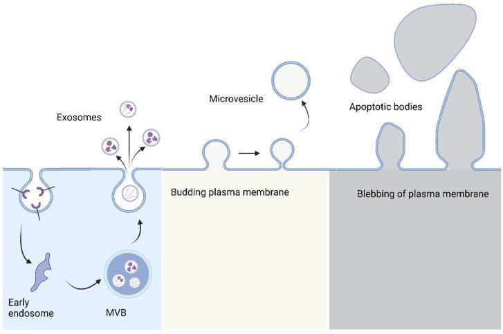
This pathophysiology diagram illustrates the biogenesis and release mechanisms of three major types of extracellular vesicles (EVs): exosomes, microvesicles, and apoptotic bodies. The diagram is divided into three sections from left to right. The first section shows the endosomal pathway for exosome formation, beginning with plasma membrane invagination to form an early endosome, which matures into a multivesicular body (MVB) containing intraluminal vesicles; these are released as exosomes via exocytosis when the MVB fuses with the cell surface. The second section depicts the formation of microvesicles through the direct outward budding and pinching off of the plasma membrane. The third section illustrates the formation of apoptotic bodies, which are larger, irregularly shaped remnants resulting from the extensive blebbing and fragmentation of the plasma membrane during programmed cell death (apoptosis). The illustration highlights key morphological differences in size and origin, identifying exosomes as the smallest vesicles derived from internal compartments, microvesicles as intermediate-sized surface buds, and apoptotic bodies as the largest, heterogeneous fragments of a dying cell.
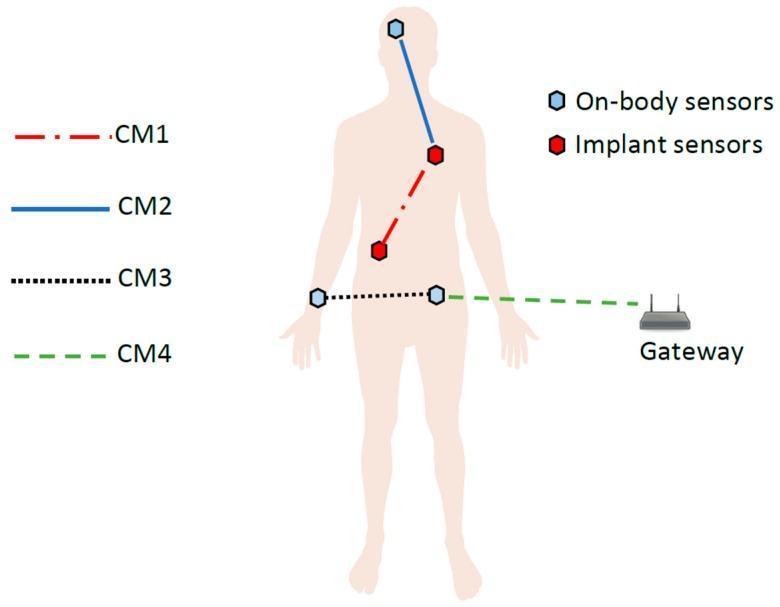
This medical technology diagram illustrates the IEEE 802.15.6 Channel Models (CM1-CM4) within a Wireless Body Area Network (WBAN) for remote patient monitoring. The visual features a human silhouette with two distinct sensor types: On-body sensors (represented by blue hexagonal icons) located on the head and waist, and Implant sensors (red hexagonal icons) situated within the torso and abdominal regions. The diagram identifies four communication pathways: CM1 (red dash-dot line) represents implant-to-implant or implant-to-body surface communication; CM2 (solid blue line) depicts on-body sensor-to-implant communication, spanning from the head to the chest; CM3 (dotted black line) shows on-body to on-body communication across the waist; and CM4 (dashed green line) illustrates off-body communication between a waist-mounted central node and an external gateway device. This figure is used in medical informatics and biomedical engineering to conceptualize data transmission architectures for real-time physiological monitoring and chronic disease management.
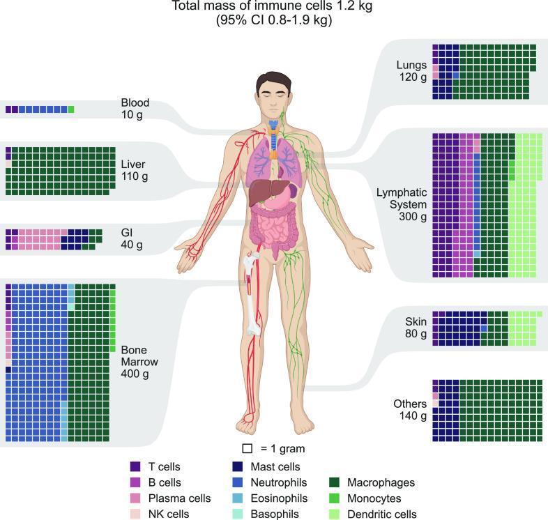
This medical infographic illustrates the distribution of immune cell mass across the human body, totaling approximately 1.2 kg. A central anatomical diagram is surrounded by waffle charts for specific organs and systems, where each square represents 1 gram of immune cells. The charts are color-coded to identify various cell types including T cells, B cells, Plasma cells, NK cells, Mast cells, Neutrophils, Eosinophils, Basophils, Macrophages, Monocytes, and Dendritic cells. Key findings highlighted include the Bone Marrow (400g) as the largest reservoir, dominated by Neutrophils, followed by the Lymphatic System (300g), which is primary composed of T and B Lymphocytes. Significant macrophage populations are visualized in the Lungs (120g) and Liver (110g). Other categorized regions include the Skin (80g), Gastrointestinal (GI) tract (40g), Blood (10g), and 'Others' (140g). This diagram serves as an educational tool for understanding quantitative immunology and the specialized tissue residency of different leukocyte lineages.
Ayurveda: Diseases, Doshas, and Healing
What is Ayurveda?
- Dermatology textbook (Griffiths et al.), p. 2804
- Essential Family Medicine (Rakel), p. 132-141
The Three Doshas
| Dosha | Elements | Governs | When Imbalanced |
|---|---|---|---|
| Vata | Air + Space | Movement - blood flow, nerve impulses, breathing, elimination | Dryness, scaling, loss of tissue; mental/nervous/digestive disorders; atopic dermatitis, photoaging |
| Pitta | Fire + Water | Metabolism - digestion, transformation, body temperature, inflammation | Blood toxicity, inflammation, infection; acne, psoriasis, rosacea, fever |
| Kapha | Water + Earth | Structure - cohesion, lubrication, collagen, tissue hydration | Excess mucus, weight gain, oedema, fibrosis; hypertrophic scars, scleroderma, lung disease |
Disease Classification in Ayurveda
1. By Dosha Involved
- Air-filled with pain = Vata imbalance
- Reddish with burning sensation = Pitta imbalance
- Pitting oedema with dull/no pain = Kapha imbalance
- A combination = multiple doshas deranged
2. By Affected Tissue (Dhatu)
| Dhatu | Tissue |
|---|---|
| Rasa | Plasma/lymph |
| Rakta | Blood/red blood cells |
| Mamsa | Muscle |
| Meda | Fat/adipose tissue |
| Asthi | Bone |
| Majja | Bone marrow/nerve tissue |
| Shukra | Reproductive tissue |
3. By Body Channel (Srotas)
Major Ayurvedic Disease Categories
Jwara (Fever)
- Vataja Jwara: irregular fever with bodyache, dryness
- Pittaja Jwara: high fever with burning, sweating
- Kaphaja Jwara: low-grade fever, heaviness, lethargy
Prameha / Madhumeha (Diabetes / Urinary Disorders)
Amavata (Rheumatoid Arthritis)
Kushtha (Skin Diseases)
- Maha Kushtha (7 major types): includes conditions resembling leprosy (Shwitra = vitiligo, Dadru = ringworm-like)
- Kshudra Kushtha (11 minor types): papules, eczema-like, urticaria-like conditions
- Psoriasis: Pitta imbalance; topical and oral curcumin (from Curcuma longa/turmeric) has shown evidence of down-regulating inflammatory kinases
- Atopic Dermatitis: Vata imbalance (dryness, scaling, tissue loss)
- Acne/Rosacea: Pitta imbalance (inflammation)
- Hypertrophic Scars/Scleroderma: Kapha imbalance (overproduction of fibrosis/collagen)
Arshas (Hemorrhoids / Piles)
Gulma (Abdominal Tumors / Masses)
Rajayakshma (Tuberculosis / Wasting Disease)
Unmada (Mental Disorders)
- Vatika Unmada: anxiety, restlessness, incoherent speech
- Paittika Unmada: rage, hallucinations, violence
- Kaphaja Unmada: depression, excessive sleep, withdrawal
Apasmara (Epilepsy)
Ayurvedic Diagnosis
- Prakriti (constitutional body type)
- Pulse diagnosis (Nadi Pariksha) - one of the most important diagnostic tools
- Examination of eyes, tongue, urine, feces, skin, voice/speech
- Factors considered: genetics, trauma, diet, habits, seasonal effects, climate, age, emotions, and karma
Ayurvedic Treatment (Chikitsa)
1. Panchakarma (Five Purification Therapies)
| Therapy | Method |
|---|---|
| Vamana | Therapeutic vomiting (clears Kapha) |
| Virechana | Purgation/laxatives (clears Pitta from intestines) |
| Basti | Medicated enemas (the most important, clears Vata) |
| Nasya | Nasal administration of medications (clears head/neck) |
| Raktamokshana | Blood purification (bloodletting or herbal teas) |
2. Shamana (Palliative Therapy)
3. Rasayana (Rejuvenation Therapy)
4. Diet and Lifestyle (Ahara & Vihara)
- Vata imbalance: warm, oily, grounding foods; regular routine; avoiding cold/dry environments
- Pitta imbalance: cool, non-spicy foods; avoiding overwork and heat
- Kapha imbalance: light, warm, stimulating foods; vigorous exercise
5. Herbal Formulations
- Turmeric (Curcuma longa) - anti-inflammatory, used in psoriasis
- Ashwagandha (Withania somnifera) - adaptogen, stress/fatigue
- Neem (Azadirachta indica) - skin diseases, antimicrobial
- Triphala - digestive/bowel regulator
- Brahmi (Bacopa monnieri) - cognitive/neurological conditions
Note on herb safety: Ayurvedic herbal products are regulated as dietary supplements rather than drugs in most Western countries. Heavy metal contamination has been a concern historically, though reputable companies now perform third-party testing. Herb-drug interactions are possible and must be assessed before combining with conventional medicines.
Ayurveda and Modern Diseases (GI Context)
- IBS (Irritable Bowel Syndrome): Triphala, Basti enemas
- Functional Dyspepsia: Digestive herbs (Trikatu, ginger)
- IBD (Inflammatory Bowel Disease): Curcumin, Boswellia
- Nausea/Vomiting: Ginger (Zingiber officinale)
- Liver Disease: Bhumiamalaki (Phyllanthus niruri)
Ayurveda and the Ancient Texts
- Charaka Samhita - focuses on internal medicine (Kayachikitsa)
- Sushruta Samhita - focuses on surgery (Shalya Tantra)
- Ashtanga Hridayam - a synthesis of both
Summary Table: Dosha Imbalance and Common Diseases
| Dosha | Key Diseases |
|---|---|
| Vata | Arthritis, constipation, nerve disorders, anxiety, insomnia, skin dryness (atopic dermatitis), osteoporosis |
| Pitta | Fever (Jwara), acid reflux, liver disease, skin inflammation (acne, psoriasis, rosacea), anger/irritability |
| Kapha | Obesity, diabetes (Prameha), respiratory conditions, depression, edema, scleroderma |
| Ama (toxins) | Rheumatoid arthritis (Amavata), digestive disorders, autoimmune conditions |
- Dermatology textbook (Griffiths et al.) - "Ayurveda in Dermatology," discussing dosha-based skin disease classification
- Essential Family Medicine (Rakel) - Ayurveda philosophy, diagnostics, and Panchakarma
- Sleisenger & Fordtran's Gastroenterology - Ayurveda as CAM for GI disorders
- AAPEC Journal - Ayurveda: a distinctive approach to health and disease