
give me arterial blood supply of heart Peer proper diagram and flashcard
arterial blood supply of heart coronary arteries anatomy diagram
Medical flashcard titled "Arterial Blood Supply of the Heart" with two sections: FRONT shows a clean labeled diagram of the heart's anterior view with the following arteries clearly marked: Right Coronary Artery (RCA) in the right atrioventricular groove, Left Coronary Artery (LCA) originating from left aortic sinus, Left Anterior Descending (LAD) artery in the anterior interventricular sulcus, Left Circumflex Artery (LCx) in the left AV groove, Right Marginal Artery, Left Marginal Artery, Posterior Interventricular Artery (from RCA). BACK section lists key supply areas in bullet points: RCA - right atrium, right ventricle, SA node (55%), AV node (80%), posterior 1/3 IVS; LAD - anterior 2/3 IVS, anterior LV, anterior RV; LCx - left atrium, posterior LV, SA node (45%). Include a "Dominance" note: Right dominant 67%, Left dominant 15%, Co-dominant 18%. Use a professional anatomy textbook style with clean white background, color-coded arteries (red for RCA, blue for LAD, green for LCx).
Arterial Blood Supply of the Heart
Diagrams
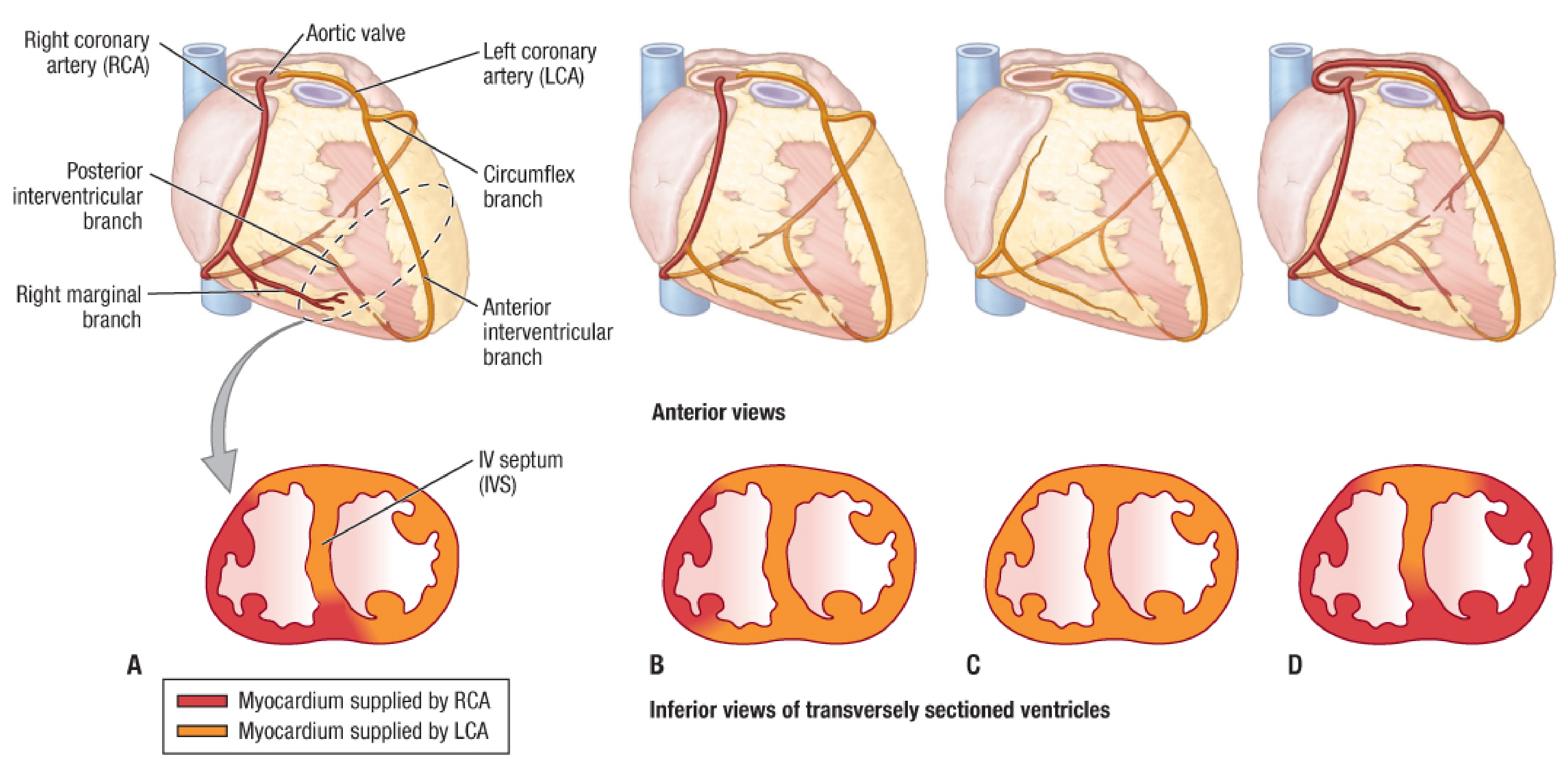
Red = Myocardium supplied by RCA | Orange = Myocardium supplied by LCA. Panels A-D show the 4 main dominance patterns.
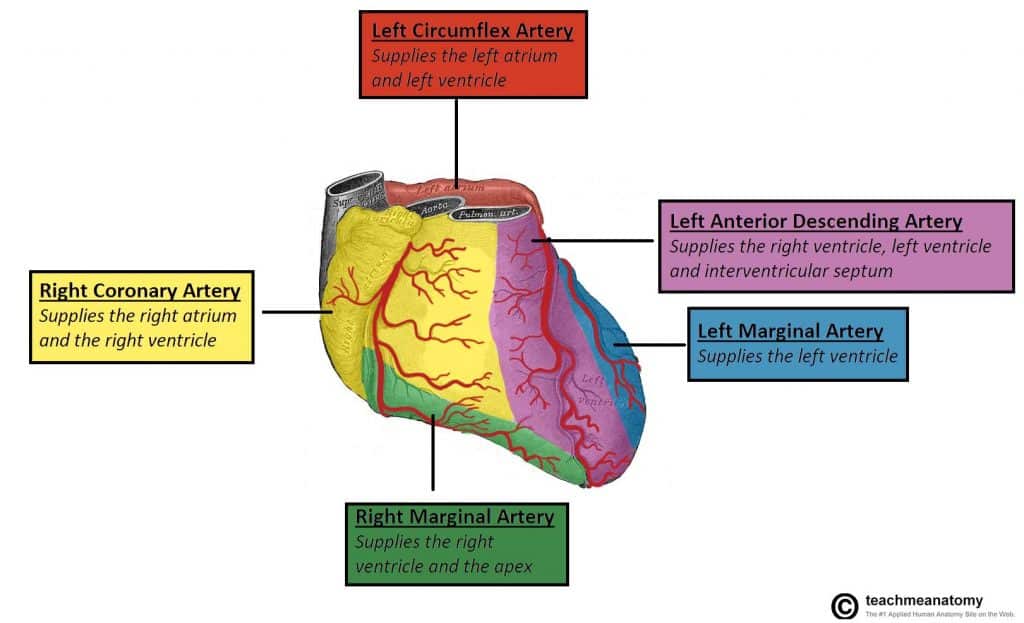
Flashcard
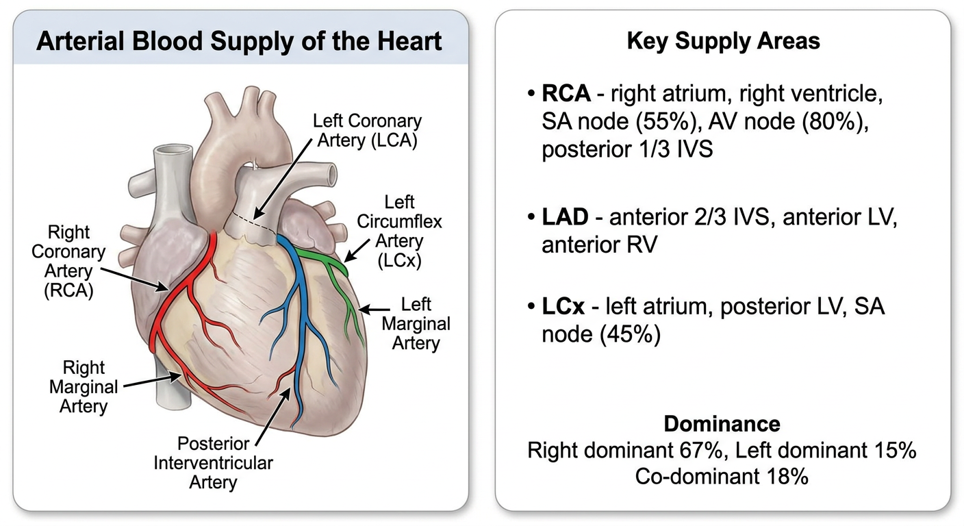
Full Explanation
Overview
1. Right Coronary Artery (RCA)
| Branch | Location | Supply |
|---|---|---|
| Sinu-atrial (SA) nodal branch | Passes around the superior vena cava | SA node (in ~55% of people) |
| Right marginal artery | Along the inferior (acute) margin | Right ventricle, apex |
| AV nodal branch | Near the crux of the heart | AV node (in ~80% of people) |
| Posterior interventricular (PDA) | Posterior interventricular sulcus | Posterior IVS, posterior LV |
- Right atrium
- Right ventricle
- SA node (55%), AV node (80%)
- Interatrial septum
- Posterior 1/3 of interventricular septum (IVS)
- Portion of the posterior left ventricle
2. Left Coronary Artery (LCA)
a) Left Anterior Descending (LAD) - Anterior Interventricular Branch
- Descends in the anterior interventricular sulcus toward the apex
- Curves around the apex to anastomose with the posterior interventricular branch (PDA)
- May give off diagonal branches across the anterior LV surface
- Supplies: Anterior left ventricle, anterior right ventricle, anterior 2/3 of IVS (including the bundle of His and bundle branches)
- Called "the artery of sudden death" - most commonly blocked in MI
b) Left Circumflex Artery (LCx)
- Courses in the coronary sulcus around the left border of the heart
- Gives off the left marginal artery (obtuse marginal)
- Usually terminates before the posterior interventricular sulcus (in right-dominant hearts)
- Supplies: Left atrium, posterior/lateral left ventricle, SA node (in ~45% of people)
Coronary Dominance
| Type | Frequency | PDA from |
|---|---|---|
| Right dominant | ~67% | RCA |
| Left dominant | ~15% | LCx |
| Co-dominant | ~18% | Both RCA and LCx |
Clinically Important Notes
| Vessel | Area at Risk in Occlusion | ECG Changes |
|---|---|---|
| LAD (proximal) | Anterior LV, IVS, bundle branches | Anterior ST elevation (V1-V4), LBBB |
| RCA (proximal) | Inferior LV, RV, SA/AV nodes | Inferior ST elevation (II, III, aVF), bradyarrhythmias |
| LCx | Lateral/posterior LV | Lateral ST elevation (I, aVL, V5-V6) |