
Hyperreflexia
Reading File
Now let me search the medical library for hyperreflexia content in parallel.
Finding Sources
Finding Sources
Searching PubMed
hyperreflexia upper motor neuron
Good results. Let me now read the most relevant sections - the Adams & Victor neurology section on hyperreflexia/spasticity and the Bradley & Daroff section on pathological hyperreflexia.
Reading File
Reading File
Reading File
Let me now read more of the Adams & Victor section (it had 9 paragraphs covering the topic well) and also search for the autonomic hyperreflexia / dysreflexia content.
Reading File
Finding Sources
Let me read the autonomic dysreflexia section from Campbell Walsh Urology, and also get the serotonin syndrome hyperreflexia context, and fetch the Babinski sign image.
Reading File
Reading File
Loading Image
I now have comprehensive content to provide a thorough answer on hyperreflexia. Let me compile the full response.
Hyperreflexia
Definition
Hyperreflexia refers to exaggerated (overactive) deep tendon/stretch reflexes. It is a cardinal sign of upper motor neuron (UMN) lesions - damage anywhere above the anterior horn cell, including the cerebral cortex, internal capsule, brainstem, or descending spinal cord pathways. It must be distinguished from the rare autonomic form (see below).
Pathophysiology
The stretch reflex arc is normally modulated by descending inhibitory signals from supraspinal centers. When these are lost:
- Disinhibition of spindle efferents - increased tonic gamma motor neuron activity amplifies the response to muscle stretch
- Loss of reticulospinal and vestibulospinal inhibition - these pathways normally dampen alpha motor neuron excitability
- The dorsal reticulospinal tract provides inhibitory effects on stretch reflexes; when injured (as in spinal cord lesions), hyperreflexia results
- Gamma neuron hyperactivity - blocking gamma neurons selectively abolishes spasticity and hyperactive tendon reflexes without affecting voluntary power, confirming they are separate mechanisms
The heightened tendon jerks are thus a "release" phenomenon - the result of interrupting descending inhibitory pathways. - Adams and Victor's Principles of Neurology, 12th Edition
Clinical Features of UMN Hyperreflexia
The Full UMN Syndrome
| Feature | Description |
|---|---|
| Hyperreflexia | Brisk, easily elicited tendon jerks (biceps, triceps, knee, ankle) |
| Spasticity | Velocity-dependent increase in muscle tone ("clasp-knife" phenomenon) |
| Babinski sign | Extension of great toe + fanning of other toes on plantar stimulation |
| Clonus | Rhythmic 5-7 Hz contractions on sustained stretch |
| Weakness | Predominantly anti-gravity muscles (flexors of arm, extensors of leg) |
| Loss of superficial reflexes | Absent abdominal and cremasteric reflexes |
- Bradley and Daroff's Neurology in Clinical Practice
Clasp-Knife Phenomenon
When muscles are stretched briskly, there is initial free movement followed by an abrupt catch with rapidly increasing resistance, then relaxation. This velocity-dependent tone is characteristic of spasticity. By contrast, if the limb is moved slowly, tone may be minimally changed. - Adams and Victor's Principles of Neurology
Clonus
Hyperreflexia may manifest as clonus - a series of rhythmic involuntary muscular contractions at 5-7 Hz in response to abruptly applied and sustained stretch. Common at the ankle and patella. It requires:
- Appropriate muscle relaxation
- Intact spinal stretch reflex mechanisms
- Sustained hyperexcitability of alpha and gamma motor neurons (suprasegmental influence)
- Synchronization of the contraction-relaxation cycle of muscle spindles
Babinski Sign
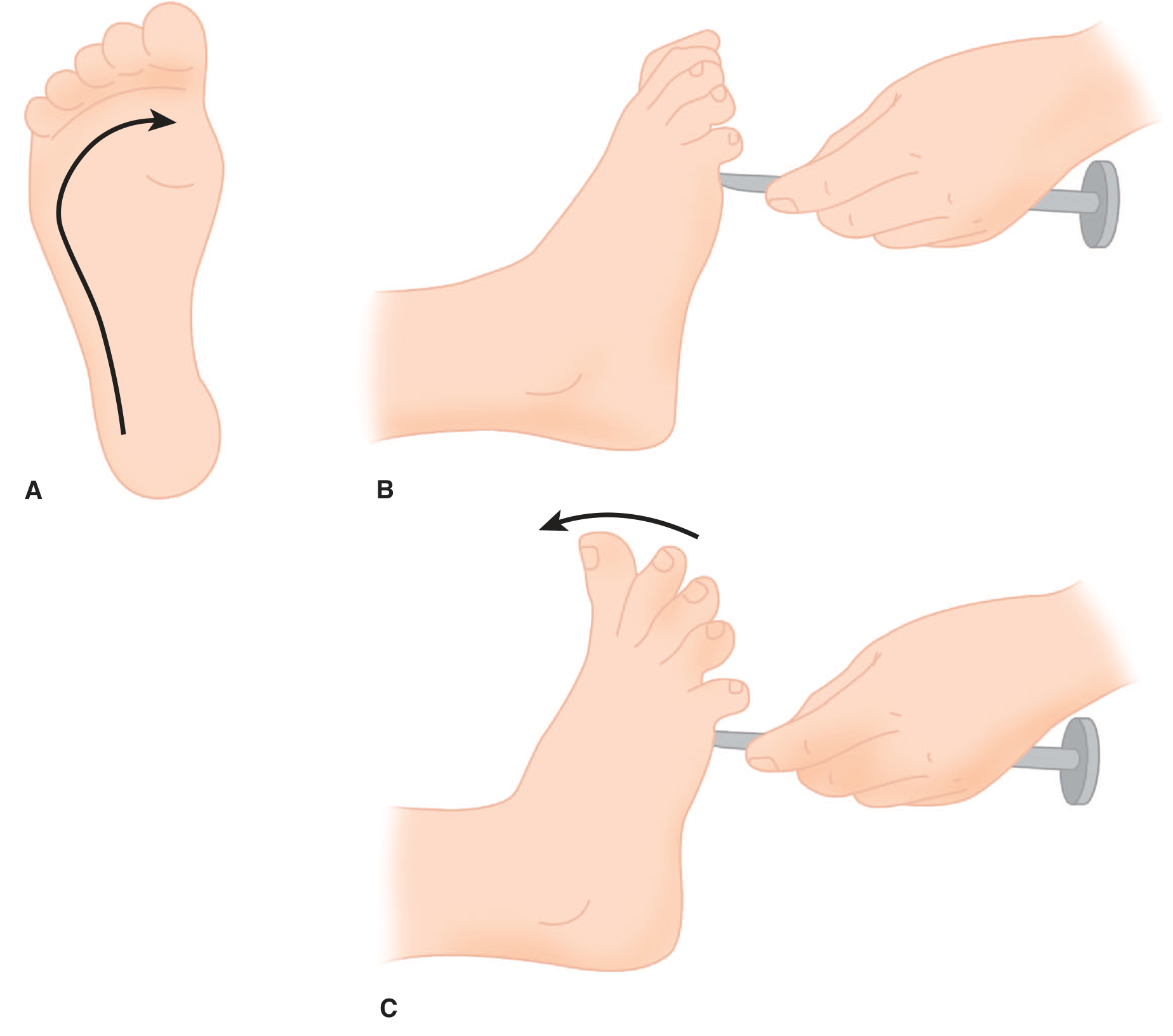
Figure: A. The plantar response is elicited by stroking from the heel upward across the ball of the foot. B. Normal response: toe flexion. C. Babinski sign (extensor plantar response): great toe extension + fanning of other toes.
The Babinski sign is the most reliable indicator of a UMN lesion. Physiologically, it is part of the "triple flexion response" - a nocifensive/protective withdrawal reflex of spinal origin that is normally suppressed by corticospinal input. It is present in normal infants until corticospinal tract myelination is complete. - Adams and Victor's Principles of Neurology
Spinal Shock vs. Hyperreflexia
After complete spinal cord transection, there is a biphasic course:
- Spinal shock (immediate) - all reflexes below the lesion are suppressed or abolished
- Hyperreflexia (weeks to months later) - reflexes gradually return and become greatly exaggerated, e.g., a light touch to the foot may trigger strong flexion withdrawal of the entire leg
This sequence is important to recognize - a patient with an acute cord injury may be areflexic initially and falsely appear to have an LMN lesion. - Kandel's Principles of Neural Science, 6th Edition
Important Causes of Hyperreflexia
| Category | Examples |
|---|---|
| Cerebral | Stroke, tumor, ALS, MS, TBI |
| Spinal cord | Cervical myelopathy, cord compression, MS, transverse myelitis, SCI |
| Metabolic/toxic | Serotonin syndrome, hyperthyroidism, hypocalcemia |
| Pre-eclampsia/eclampsia | Hyperreflexia with sustained clonus is a warning sign |
| Neonatal abstinence syndrome | Hyperreflexia, hypertonia (opioid withdrawal in newborns) |
Note on severity vs. weakness: The degree of spasticity and weakness are not proportional. Severe weakness may coexist with only mild spasticity. Extreme spasticity, as seen in cervical cord disease, may be disproportionate to weakness - these depend on separate mechanisms.
Autonomic Hyperreflexia (Autonomic Dysreflexia)
This is a distinct and potentially life-threatening syndrome, different from reflex hyperreflexia.
Definition: An acute, massive disordered autonomic (primarily sympathetic) response triggered by stimuli below the level of a spinal cord injury (SCI) above T6-T8 (the sympathetic outflow level). First described by Guttmann and Whitteridge in 1947.
Epidemiology: More common in cervical (60%) than thoracic (20%) SCI.
Common triggers (usually from bladder or rectum):
- Bladder distension, urinary catheter obstruction, clot retention
- Rectal distension, constipation
- LUT instrumentation, calculi
- Long bone fractures, pressure ulcers, sexual activity, electrocoagulation
Clinical features:
- Severe pounding headache
- Hypertension (can be life-threatening - may cause cerebral hemorrhage or seizures)
- Flushing and sweating above the lesion
- Bradycardia (reflex, though tachyarrhythmia may occur)
- Piloerection below the lesion
Pathophysiology: Nociceptive afferent impulses ascend the cord and trigger reflex sympathetic outflow causing arteriolar/pilomotor/pelvic visceral spasm. Normally the medulla would inhibit this, but the SCI prevents that descending control below the lesion. The splanchnic vascular bed is likely involved, contributing to the massive BP elevation. - Campbell Walsh Wein Urology
Anesthetic management: Regional anesthesia or deep general anesthesia effectively prevents hyperreflexia. Vasodilators must be immediately available. Succinylcholine should be avoided (risk of hyperkalemia from denervation). - Morgan and Mikhail's Clinical Anesthesiology
Serotonin Syndrome
Hyperreflexia is a key feature distinguishing serotonin syndrome from neuroleptic malignant syndrome (NMS):
| Feature | Serotonin Syndrome | NMS |
|---|---|---|
| Reflexes | Hyperreflexia, clonus | Hyporeflexia, rigidity |
| Onset | Hours | Days |
| Cause | Serotonergic excess | Dopamine antagonist |
| Tone | Myoclonus | "Lead-pipe" rigidity |
- Tintinalli's Emergency Medicine; Rosen's Emergency Medicine
Key Distinctions: UMN vs. LMN
| Feature | UMN (Hyperreflexia) | LMN (Hyporeflexia) |
|---|---|---|
| Reflexes | Increased (hyperreflexia) | Decreased/absent |
| Tone | Spastic (velocity-dependent) | Flaccid |
| Weakness | Pyramidal pattern | Focal/segmental |
| Atrophy | Minimal (disuse only) | Prominent |
| Fasciculations | Absent | Present |
| Babinski | Extensor (positive) | Flexor (negative) |
| Clonus | Present | Absent |
This is a shared conversation. Sign in to Orris to start your own chat.