
Myxomatous mitral valve echocardiography assesment
myxomatous mitral valve prolapse echocardiography
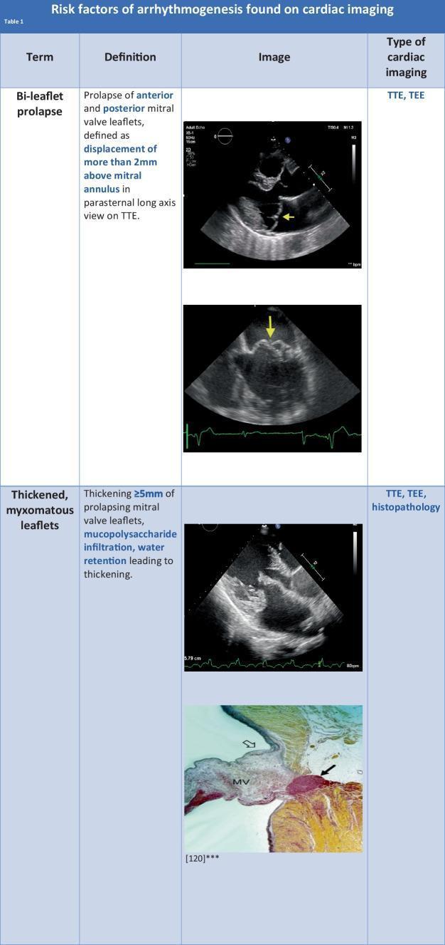
A comparison chart titled 'Risk factors of arrhythmogenesis found on cardiac imaging' illustrating diagnostic features of Mitral Valve Prolapse (MVP). The content is organized into categories covering bi-leaflet prolapse and thickened, myxomatous leaflets. For bi-leaflet prolapse, transthoracic echocardiogram (TTE) images in the parasternal long-axis view demonstrate the displacement of both anterior and posterior leaflets more than 2mm above the mitral annulus (indicated by yellow arrows). For thickened, myxomatous leaflets, the chart includes a TTE image showing mitral valve thickening of 5mm or more, alongside a histopathology slide (labeled MV) demonstrating mucopolysaccharide infiltration and structural expansion of the valve leaflet tissue. The visual material serves as a diagnostic guide for cardiologists and medical students to identify arrhythmogenic substrates in MVP using echocardiography and histopathological correlation. Key modalities mentioned include TTE, TEE (transesophageal echocardiography), and histopathology.
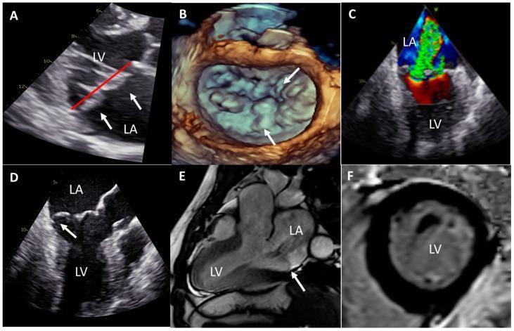
This multi-panel figure demonstrates multimodal clinical imaging of Barlow's mitral valve disease and associated cardiac remodeling. (A) Transthoracic echocardiography (parasternal long-axis) shows thickened, bileaflet mitral valve prolapse (white arrows) into the left atrium (LA) beyond the annular plane (red line). (B) 3D transesophageal echocardiography (TEE) 'surgeon's view' provides a detailed anatomical surface map of the multi-scallop, redundant leaflets characteristic of Barlow's disease. (C) TEE with color Doppler in a bicommissural view highlights a severe, multi-jet eccentric mitral regurgitation. (D) 2D TEE three-chamber view identifies posterior leaflet prolapse and the presence of mitral annular disjunction (MAD). (E) Cardiac magnetic resonance (CMR) three-chamber cine view confirms bileaflet prolapse, mild MAD, and a dilated left ventricle (LV). (F) Short-axis CMR late gadolinium enhancement (LGE) at the papillary muscle level shows no macroscopic myocardial fibrosis (black myocardium). The collection illustrates the diagnostic features of myxomatous valve degeneration, annular disjunction, and secondary ventricular dilatation across ultrasound and magnetic resonance modalities.
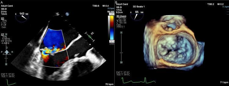
Preoperative transesophageal echocardiography (TEE) images demonstrating severe mitral valve regurgitation in a patient with Barlow’s disease. Panel A is a 2D TEE with color Doppler overlay showing a large blue-colored jet within the left atrium, indicating blood flow moving away from the transducer, with a high-velocity, turbulent mosaic of red and yellow at the valvular orifice representing significant regurgitant flow. Panel B is a 3D volume-rendered TEE reconstruction of the mitral valve from the surgical atrial perspective ('surgeon's view'). This image depicts the complex anatomy of the mitral valve leaflets, characterized by irregular surface texture, excessive tissue, and multiple bulging segments consistent with bileaflet prolapse and a flail posterior leaflet (P2/P3 segments). The anatomical visualization highlights severe left atrial enlargement and the distinctive bulky, thickened morphology associated with myxomatous degeneration of the mitral valve apparatus.
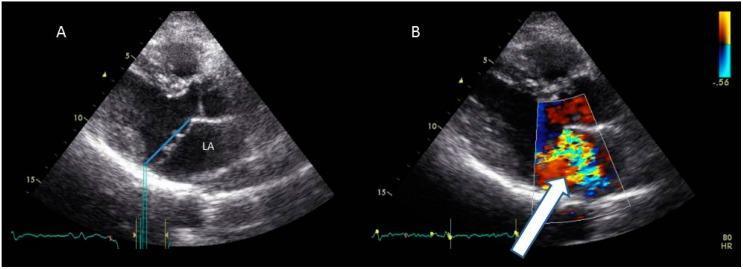
This diagnostic image consists of two panels (A and B) showing transthoracic echocardiography (TTE) in a parasternal long-axis (PLAX) view, demonstrating mitral valve prolapse (MVP). Panel A is a 2D grayscale image showing the anatomical relationship between the mitral valve leaflets and the left atrium (LA). A blue reference line marks the mitral annular plane; systolic billowing of the mitral leaflets exceeds this line by >2 mm, diagnostic of MVP. Panel B displays color Doppler imaging during systole. A large, eccentric, multi-colored (mosaic) jet originates from the mitral orifice and extends deep into the left atrium (indicated by a white arrow), signifying severe mitral regurgitation. The image illustrates key diagnostic criteria for myxomatous valve disease, emphasizing the displacement of leaflet tissue beyond the saddle-shaped annulus and the resultant functional hemodynamic impairment. This material is suited for cardiovascular education regarding valvular heart disease and echocardiographic interpretation.
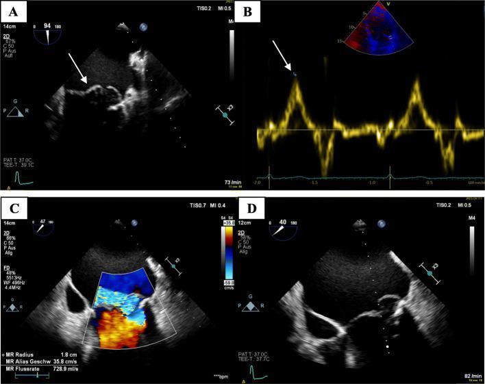
This composite of four transesophageal echocardiography (TEE) images illustrates the diagnostic findings of myxomatous mitral valve disease and its hemodynamic consequences. Panel A (2D TEE) demonstrates a classic bileaflet mitral valve prolapse (MVP), with the leaflets bulging superiorly into a significantly dilated left atrium (LA) during systole. Panel B utilizes Tissue Doppler Imaging (TDI) at the lateral annulus to show a high-velocity mid-systolic spike (Pickelhaube sign) reaching 18 cm/s, which is a characteristic marker associated with arrhythmogenic MVP. Panel C employs color Doppler flow mapping to visualize a severe, eccentric mitral regurgitation (MR) jet. The mosaic color pattern indicates high-velocity, turbulent retrograde flow into the LA, with quantitative metrics showing an MR radius of 1.8 cm and a flow rate of 728.9 ml/s. Panel D shows a detailed 2D view of the myxomatous degeneration, characterized by marked thickening and redundant tissue of the mitral leaflets. This collection is intended for cardiology education, highlighting the integration of anatomical 2D imaging with advanced Doppler techniques to assess valvular pathology and associated arrhythmic risk.
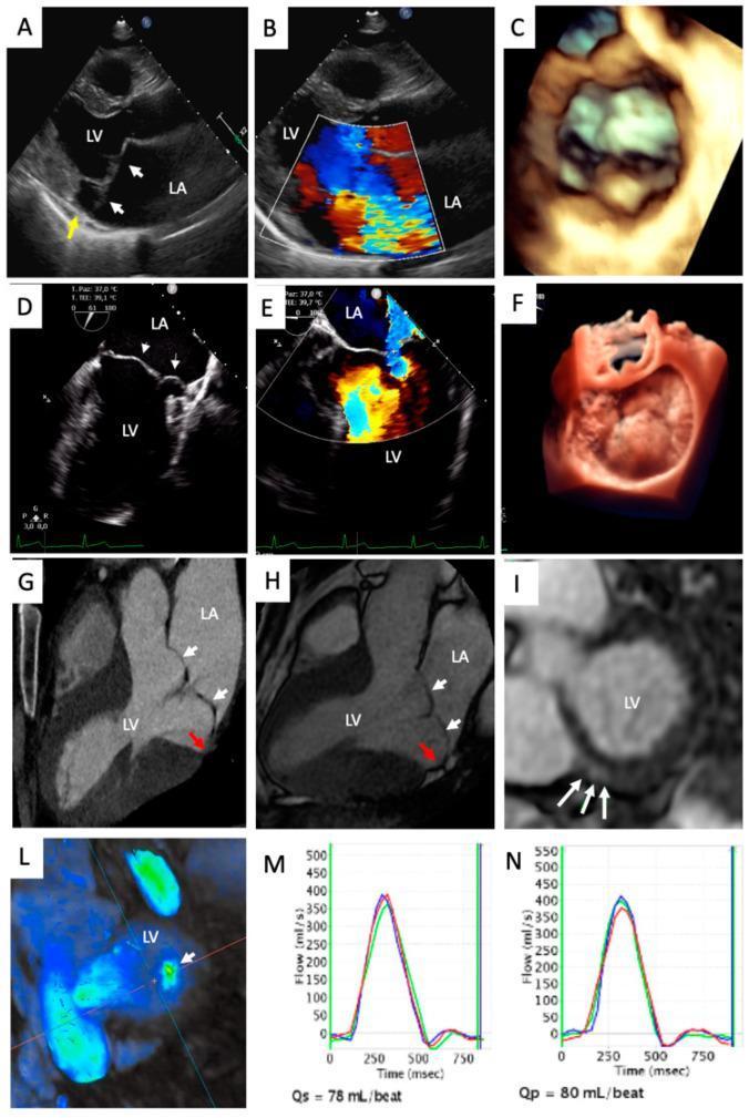
A multi-panel medical illustration demonstrating multimodality imaging of myxomatous mitral valve disease in a 37-year-old patient. Panels A, B, D, and E showcase Transthoracic (TTE) and Transesophageal Echocardiography (TEE), including parasternal long-axis (PLAX) and 4-chamber views. These images highlight bileaflet mitral valve prolapse (MVP), where leaflets (white arrows) are displaced superiorly into the left atrium (LA), accompanied by mitral annular disjunction (MAD, yellow arrow) and a centrovalvular mitral regurgitation jet on color Doppler. Panels C and F provide 3D-TTE and 3D-TEE reconstructions, visualizing the volumetric extent of the prolapse. Panels G and H represent Cardiac Computed Tomography (CCT) and Cardiac Magnetic Resonance (CMR) confirming bileaflet MVP and MAD (red arrows). Panel I (CMR Late Gadolinium Enhancement) reveals focal fibrosis in the left ventricle (LV) inferobasal wall. Panel L shows 4D-flow CMR visualizing the regurgitant jet, while panels M and N display phase-contrast flow measurements for aortic (Qs) and pulmonary (Qp) quantification. This composite facilitates comprehensive assessment of valvular morphology, regurgitation severity, and myocardial consequences.
myxomatous mitral valve echocardiography assessment
Myxomatous Mitral Valve Disease: Echocardiographic Assessment
1. Background and Pathology
2. Goals of Echocardiographic Assessment
- Confirming the diagnosis of MVP
- Identifying the mechanism and affected leaflet segments
- Quantifying mitral regurgitation (MR) severity
- Assessing LV/LA consequences
- Detecting high-risk features (mitral annular disjunction, papillary muscle changes)
- Surgical/transcatheter procedural planning
3. Imaging Modalities Used
3a. Transthoracic Echocardiography (TTE)
-
Diagnostic criterion: systolic displacement of the belly of the mitral leaflet(s) ≥2 mm above the mitral annular plane (saddle-shaped annulus reference - displacement must be measured in PLAX, not apical 4-chamber, to avoid overcalling)
-
Identifies whether prolapse is anterior leaflet, posterior leaflet, or bileaflet (Barlow's)
-
Leaflet thickening ≥5 mm indicates myxomatous disease (not FED)
-
Doppler (color, pulsed-wave, continuous-wave) quantifies associated MR
-
Harrison's Principles of Internal Medicine 22E, p. 2101-2102
-
Textbook of Clinical Echocardiography, pp. 400-401
3b. 3D Echocardiography (3D-TTE/3D-TEE)
-
"En face" surgical view (looking at the valve from the LA perspective) - enables intuitive description of the Carpentier segmental anatomy (P1, P2, P3; A1, A2, A3)
-
Identifies complex lesions often missed on 2D: commissural involvement, clefts, multi-scallop disease
-
3D-based software models measure: annular dimensions, leaflet surface area, prolapse height/volume, mitro-aortic angle - essential for surgical/TEER planning
-
Improves feasibility of pre-procedural planning
-
Fuster and Hurst's The Heart, 15th Edition, pp. 943-944
3c. Transesophageal Echocardiography (TEE)
- TTE is non-diagnostic or provides insufficient detail
- Pre-operative surgical planning (complex/anterior/bileaflet prolapse)
- Intraoperative monitoring during repair or TEER
- Suspected flail leaflet with eccentric jet making TTE quantification unreliable
4. Leaflet Segmentation (Carpentier Classification)
- Posterior leaflet: P1 (anterolateral), P2 (middle - most commonly prolapses in FED), P3 (posteromedial)
- Anterior leaflet: A1, A2, A3
5. Specific Echocardiographic Features
| Feature | Finding | Significance |
|---|---|---|
| Leaflet displacement | ≥2 mm above annulus in PLAX systole | Diagnostic of MVP |
| Leaflet thickness | ≥5 mm | Myxomatous (Barlow's), not FED |
| Bileaflet prolapse | Both AML + PML | Barlow's phenotype, higher regurgitation burden |
| Flail leaflet | Leaflet tip inverts into LA | Severe/eccentric MR, urgent surgery marker |
| Chordal rupture | Free chord visible | Acute/severe MR |
| Annular dilation | AP diameter >35 mm (women), >40 mm (men) | Contributes to MR severity |
| Mitral Annular Disjunction (MAD) | Hinge point of posterior leaflet displaced ≥3-5 mm from LA wall in systole | Arrhythmogenic substrate; associated with sudden cardiac death risk |
| Systolic anterior motion (SAM) | Post-repair complication | LVOTO after repair |
6. Quantification of Mitral Regurgitation Severity
| Parameter | Mild | Moderate | Severe |
|---|---|---|---|
| Vena contracta width | <0.3 cm | 0.3-0.69 cm | ≥0.7 cm |
| Regurgitant volume | <30 mL | 30-59 mL | ≥60 mL |
| Regurgitant fraction | <30% | 30-49% | ≥50% |
| Effective Regurgitant Orifice Area (EROA) | <0.20 cm² | 0.20-0.39 cm² | ≥0.40 cm² |
| Angiographic grade | 1+ | 2+ | 3-4+ |
- Textbook of Clinical Echocardiography, p. 400
- Fuster and Hurst's The Heart, 15th Ed., p. 944
7. ACC/AHA Disease Staging (Primary MR)
| Stage | Definition | Key Echo Findings |
|---|---|---|
| A | At risk | Mild MVP with normal coaptation; no/trivial MR jet (vena contracta <0.3 cm) |
| B | Progressive MR | Moderate-severe MVP with normal coaptation; central jet 20-40% LA or late-systolic eccentric jet; EROA <0.40 cm²; RVol <60 mL |
| C | Asymptomatic severe MR | Flail or loss of coaptation; vena contracta ≥0.7 cm; EROA ≥0.40 cm²; RVol ≥60 mL; RF ≥50%; Stage C1: LVEF >60%, LVESD <40 mm; Stage C2: LVEF ≤60% or LVESD ≥40 mm |
| D | Symptomatic severe MR | Same hemodynamic criteria as C; symptoms present |
- Textbook of Clinical Echocardiography, Table 12.7
8. LV and LA Consequences to Assess
- LV dimensions: LVESD ≥40 mm and LVEF ≤60% trigger surgery recommendations even in asymptomatic patients (Stage C2)
- LA enlargement: progressive LA dilation; severe enlargement associated with AF
- Pulmonary artery pressure: estimated by tricuspid regurgitation jet velocity (>50 mmHg at rest or with exercise = poor prognostic marker)
- LV strain (GLS): a 2024 systematic review (PMID 38976111) found that LV global longitudinal strain (GLS) is impaired in MVP even when LVEF appears preserved, serving as an early marker of subclinical LV dysfunction
9. High-Risk Echocardiographic Markers for Sudden Cardiac Death
- Bileaflet prolapse with myxomatous thickening
- Mitral Annular Disjunction (MAD): atrial displacement of the posterior leaflet hinge point ≥3 mm from the LA wall in TEE/TTE; associated with excessive mitral annular curling and mechanical stress on the papillary muscles
- Pickelhaube sign (TDI): high-velocity mid-systolic spike ≥16 cm/s at the lateral mitral annulus on tissue Doppler - a marker of abnormal annular motion linked to ventricular arrhythmias
- LGE on CMR: papillary muscle or inferobasal LV wall fibrosis in arrhythmic MVP
10. Role of 3D TEE in Procedural Planning
- Precise segment-level anatomy of prolapse
- Annular dimensions for ring selection
- Assessment of anterior leaflet height (for repair feasibility)
- Detection of calcification at posterior annulus
- Commissural integrity
- Real-time intraoperative guidance
Key Echocardiographic Images
Summary: Practical Echo Assessment Checklist
| Step | What to Do |
|---|---|
| 1. Confirm diagnosis | PLAX view - leaflet displacement ≥2 mm in systole |
| 2. Identify segment | Carpentier notation (A1-3, P1-3); single vs. bileaflet |
| 3. Leaflet morphology | Thickness, redundancy, FED vs. Barlow's |
| 4. Chordal status | Intact, elongated, ruptured → flail? |
| 5. Annulus | Measure AP and intercommissural diameters |
| 6. Detect MAD | PLAX/TEE - hinge point displacement ≥3 mm |
| 7. Quantify MR | Vena contracta + PISA EROA + RVol + RF (multiparametric) |
| 8. LV consequences | LVEF, LVESD, GLS |
| 9. LA size | Volume index; AF? |
| 10. PA pressure | TR jet velocity → RVSP |
| 11. Arrhythmic risk | MAD, leaflet thickness, Pickelhaube sign |
| 12. Procedural planning | 3D-TEE if repair/TEER planned |