
Nerve supply of git
Reading File
The GIT (gastrointestinal tract) receives both extrinsic nerve supply (sympathetic and parasympathetic from the CNS/ANS) and intrinsic nerve supply (the enteric nervous system within the gut wall).
Searching sources...
Finding Sources
Finding Sources
Finding Sources
Reading File
Reading File
Reading File
Reading File
Finding Sources
Reading File
Reading File
Reading File
Reading File
Loading Image
Loading Image
Here is a comprehensive overview of the nerve supply of the gastrointestinal tract (GIT), drawn from Costanzo Physiology, Ganong's Review, Sleisenger & Fordtran's, and Goldman-Cecil Medicine.
Nerve Supply of the GIT
The GIT has two levels of nervous control: extrinsic (from the CNS via the autonomic nervous system) and intrinsic (the enteric nervous system, or ENS, within the gut wall itself).
1. Extrinsic Innervation
A. Parasympathetic Innervation
Nerve fibers: Long preganglionic fibers that synapse in ganglia located within the gut wall (myenteric and submucosal plexuses).
| Region | Nerve | Origin |
|---|---|---|
| Esophagus (upper 1/3 - striated muscle), stomach, small intestine, ascending colon, proximal transverse colon | Vagus nerve (CN X) | Dorsal motor nucleus, medulla |
| Distal transverse colon, descending colon, sigmoid colon, rectum | Pelvic nerve (pelvic splanchnic) | Sacral segments S2-S4 |
Key facts:
- The vagus nerve is 75% afferent (sensory - carries info from mechanoreceptors and chemoreceptors to the CNS) and 25% efferent (motor).
- Reflexes with both afferent and efferent limbs in the vagus are called vagovagal reflexes.
- Postganglionic parasympathetic neurons are cholinergic (release ACh) or peptidergic (release substance P, VIP).
- Overall effect: increases motility, increases gastric/intestinal secretion, relaxes sphincters.
B. Sympathetic Innervation
Nerve fibers: Short preganglionic fibers that synapse in prevertebral ganglia outside the gut wall. Postganglionic fibers are adrenergic (release norepinephrine, NE).
| Ganglia | Region supplied |
|---|---|
| Celiac ganglion | Stomach, duodenum, small intestine |
| Superior mesenteric ganglion | Small intestine, ascending and transverse colon |
| Inferior mesenteric ganglion | Descending colon, sigmoid, rectum |
| Hypogastric (pelvic) ganglion | Rectum, internal anal sphincter |
Key facts:
- Preganglionic neurons arise from the thoracolumbar intermediolateral column (T5-L2).
- Postganglionic fibers synapse on ganglia in the myenteric/submucosal plexuses OR directly on smooth muscle, secretory, and endocrine cells.
- Overall effect: inhibits non-sphincteric smooth muscle (relaxation), excites sphincters (contraction), reduces secretion - the classic "fight or flight" response.
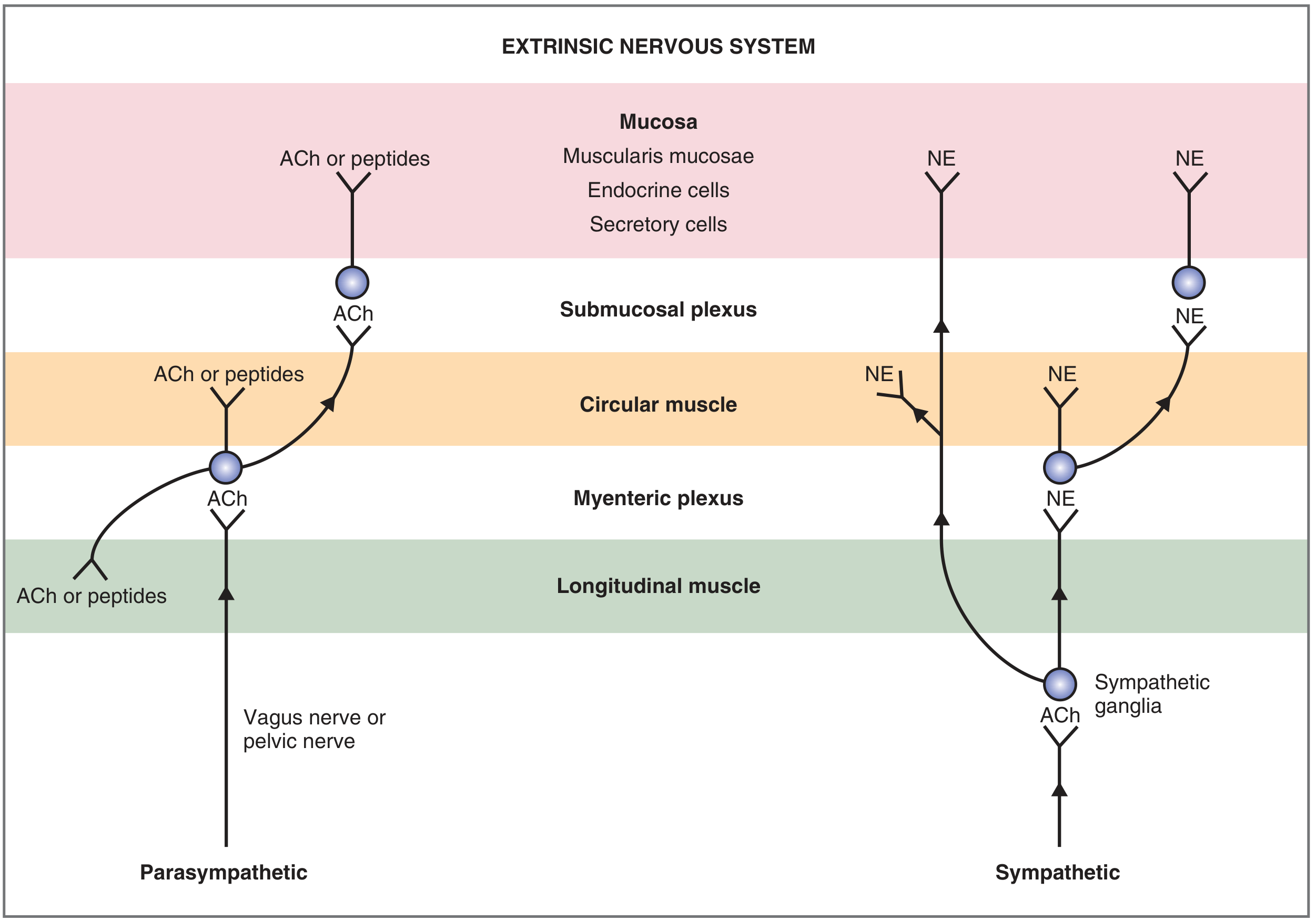
2. Intrinsic Innervation - The Enteric Nervous System (ENS)
The ENS is called the "mini-brain" because it contains all elements of a nervous system - sensory neurons, interneurons, and motor neurons - and can function autonomously even without extrinsic input.
Two Major Plexuses
| Plexus | Location | Main Function |
|---|---|---|
| Myenteric plexus (Auerbach's) | Between longitudinal and circular muscle layers | Controls motility - peristalsis and muscle contractions |
| Submucosal plexus (Meissner's) | Between circular muscle and mucosa | Senses luminal environment, regulates secretion, absorption, and mucosal blood flow |
Neuronal Types in the ENS
- Sensory (afferent) neurons - respond to mechanical, thermal, osmotic, and chemical stimuli in the mucosa.
- Interneurons - integrate sensory information and relay to motor neurons.
- Motor neurons - control smooth muscle contraction/relaxation, secretion, and absorption.
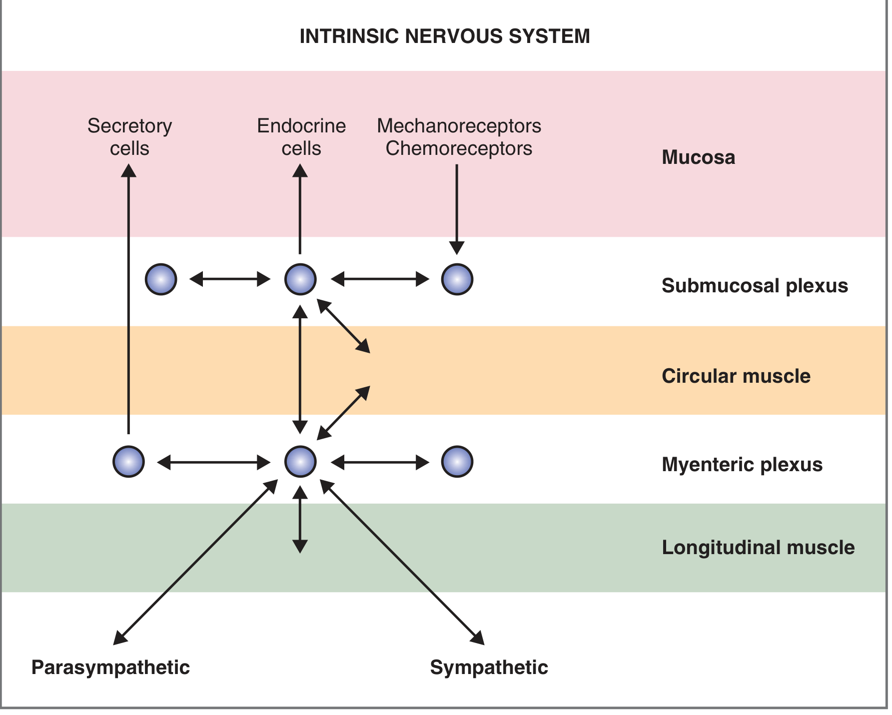
3. Neurotransmitters and Neuromodulators of the ENS
| Substance | Source | Key Actions |
|---|---|---|
| Acetylcholine (ACh) | Cholinergic neurons | Contracts wall smooth muscle, relaxes sphincters, stimulates secretions |
| Norepinephrine (NE) | Adrenergic neurons | Relaxes wall smooth muscle, contracts sphincters |
| VIP (Vasoactive Intestinal Peptide) | Enteric neurons | Relaxes smooth muscle, increases intestinal/pancreatic secretion |
| Nitric Oxide (NO) | Enteric neurons | Relaxes smooth muscle (key inhibitory neurotransmitter) |
| Substance P | Co-secreted with ACh | Contracts smooth muscle, stimulates salivary secretion |
| Enkephalins (opiates) | Enteric neurons | Contracts smooth muscle, decreases intestinal secretion |
| Neuropeptide Y | Enteric neurons | Relaxes smooth muscle, decreases intestinal secretion |
| GRP/Bombesin | Vagal neurons in gastric mucosa | Stimulates gastrin secretion |
(Costanzo Physiology, Table 8.1)
4. Summary - Functional Contrast
| Feature | Parasympathetic | Sympathetic |
|---|---|---|
| Fiber length | Long pre-, short post- | Short pre-, long post- |
| Ganglia location | In gut wall (within plexuses) | Outside gut wall (prevertebral) |
| Neurotransmitter | ACh (pre- and post-) | ACh (pre-), NE (post-) |
| Effect on motility | Increases | Decreases |
| Effect on secretion | Increases | Decreases |
| Effect on sphincters | Relaxes | Contracts |
| Effect on blood vessels | Dilates | Constricts |
Sources: Costanzo Physiology 7th Ed., p. 344-346 | Ganong's Review of Medical Physiology 26th Ed., p. 274 | Goldman-Cecil Medicine, p. 1405 | Sleisenger & Fordtran's GI and Liver Disease
Myenteric and aurbeck plexuses in the git means
The myenteric plexus and Auerbach's plexus are actually the same thing - two names for the same neural network. "Auerbach's plexus" is the eponymous name (named after Leopold Auerbach), while "myenteric plexus" is the anatomical/descriptive name.
Searching sources...
Finding Sources
Finding Sources
Reading File
Reading File
Finding Sources
Reading File
Loading Image
Here is a clear, complete explanation of these plexuses.
Myenteric Plexus = Auerbach's Plexus
They are the same structure. "Auerbach's plexus" is the eponymous name (named after Leopold Auerbach, a 19th-century German anatomist), and "myenteric plexus" is the descriptive anatomical name - both refer to the exact same neural network.
The Two Plexuses of the Enteric Nervous System
The gut wall contains two main nerve networks (plexuses), both part of the Enteric Nervous System (ENS):
| Feature | Myenteric Plexus (Auerbach's) | Submucosal Plexus (Meissner's) |
|---|---|---|
| Location | Between the longitudinal and circular muscle layers (within the muscularis externa) | Between the circular muscle and the mucosa (within the submucosa) |
| Main function | Controls motility - peristalsis, segmentation, muscular contractions | Controls secretion, absorption, mucosal blood flow, and senses the luminal environment |
| What it innervates | Longitudinal and circular smooth muscle | Secretory cells, endocrine cells, mucosal epithelium, submucosal blood vessels |
| Neurons present | Sensory (IPANs), interneurons, excitatory & inhibitory motor neurons | Sensory neurons, secretomotor neurons |
Myenteric (Auerbach's) Plexus - In Detail
- Located between the two muscle layers of the muscularis externa (inner circular + outer longitudinal).
- Has a hierarchical structure: primary plexus (main ganglia network) → secondary plexus (smaller branches) → tertiary plexus (finest branches).
- Contains ganglia spaced at regular intervals and connected by interganglionic fascicles.
- Excitatory motor neurons release ACh and Substance P → contract smooth muscle → propel contents forward (oral to aboral).
- Inhibitory motor neurons release NO, VIP, ATP → relax smooth muscle → allow the segment ahead of a bolus to open (receptive relaxation).
- This creates the "law of the intestine" (Bayliss and Starling): contraction ABOVE a bolus + relaxation BELOW = peristalsis.
Submucosal (Meissner's) Plexus - In Detail
- Located in the submucosa, closer to the mucosal lining.
- Has two layers in humans: one near the mucosa and one near the circular muscle, connected by interganglionic fascicles.
- There is also a third, deeper plexus called the Schabadasch plexus (within the circular muscle - but it contains no ganglia).
- Functions: regulates glandular secretion, water/electrolyte absorption, and blood flow to the mucosa.
Why the Myenteric Plexus is Clinically Important
| Disease | Mechanism |
|---|---|
| Hirschsprung's disease | Absence of myenteric (and submucosal) ganglia in the distal colon → no peristalsis → functional obstruction in newborns |
| Achalasia | Loss of inhibitory neurons in the myenteric plexus of the lower esophagus → failure of LES relaxation |
| Gastroparesis | Damage to myenteric neurons (e.g., in diabetes) → impaired gastric emptying |
| Chagas disease | Trypanosoma cruzi destroys ENS ganglia → megacolon/megaesophagus |
Memory Aid
"Myo" = muscle → Myenteric plexus lies between muscle layers → controls muscle (motility)"Sub-mucosal" → lies below the mucosa → controls mucosal secretion
Sources: Sleisenger & Fordtran's GI and Liver Disease, p. 1954 | Fischer's Mastery of Surgery, p. 2860 | Mulholland & Greenfield's Surgery | Harrison's Principles of Internal Medicine 22E
Function of it .. relation to sympathetic and parasympathetic
The ENS plexuses can work independently but are heavily modulated by sympathetic (inhibitory) and parasympathetic (excitatory) inputs. The parasympathetic "turns up" the ENS, while sympathetic "turns it down."
Searching sources...
Finding Sources
Finding Sources
Reading File
Finding Sources
Reading File
Finding Sources
Reading File
Excellent sources gathered. Here is a thorough, well-organized answer.
Functions of the Myenteric & Submucosal Plexuses + Their Relationship to Sympathetic/Parasympathetic Systems
Functions of Each Plexus
1. Myenteric Plexus (Auerbach's) - "The Motor Plexus"
Primary role: Controls GIT motility
| Function | Mechanism |
|---|---|
| Peristalsis | Coordinates contraction above a bolus + relaxation below (the "law of the intestine") |
| Segmentation | Mixing contractions for digestion and absorption |
| Sphincter control | Relaxes sphincters (LES, pylorus, ileocecal valve, internal anal sphincter) |
| Tone regulation | Sets baseline smooth muscle tone throughout the gut |
- Excitatory motor neurons → release ACh + Substance P → contract longitudinal and circular muscle
- Inhibitory motor neurons → release NO + VIP + ATP → relax smooth muscle (especially important for sphincters)
2. Submucosal Plexus (Meissner's) - "The Secretory/Sensory Plexus"
Primary role: Controls secretion, absorption, and mucosal blood flow
| Function | Mechanism |
|---|---|
| Secretion | Stimulates glandular epithelium to secrete water, electrolytes, and enzymes |
| Absorption | Regulates water and electrolyte absorption |
| Mucosal blood flow | Controls submucosal arterioles (vasodilation during digestion) |
| Sensory monitoring | Detects chemical and mechanical stimuli from the lumen via mechanoreceptors and chemoreceptors |
How Sympathetic & Parasympathetic Connect to the Plexuses
The key concept: Neither the sympathetic nor parasympathetic system talks directly to the smooth muscle as their primary route. Instead, they mostly synapse on the ganglion cells of the myenteric and submucosal plexuses and modulate them. The plexuses are the final common pathway.
Parasympathetic → Plexus Relationship
Vagus nerve (CN X) / Pelvic nerve (S2-S4)
        ↓  [long preganglionic fiber, releases ACh]
Ganglion cells IN the myenteric & submucosal plexuses
        ↓  [short postganglionic fiber, releases ACh or peptides]
Smooth muscle / Secretory / Endocrine cells
Effect on plexus:
- Preganglionic fibers synapse directly onto ENS ganglia → the ENS ganglia are the postganglionic parasympathetic neurons
- Activates and amplifies ENS activity
- Net result: increases motility, increases secretion, relaxes sphincters
"Parasympathetic cholinergic activity generally increases the activity of intestinal smooth muscle." - Ganong's Physiology
Sympathetic → Plexus Relationship
Spinal cord (T5-L3) [short preganglionic, releases ACh]
        ↓
Prevertebral ganglia (celiac, sup/inf mesenteric, hypogastric)
        ↓  [long postganglionic fiber, releases NE]
Synapse on ENS ganglion cells in myenteric/submucosal plexuses
        OR directly onto smooth muscle
Effect on plexus - TWO mechanisms:
| Mechanism | Detail |
|---|---|
| Presynaptic inhibition (main route) | Postganglionic sympathetic fibers end on cholinergic neurons of the ENS. NE activates α2 presynaptic receptors → inhibits ACh release from ENS neurons → suppresses plexus activity |
| Direct muscle inhibition | Some sympathetic fibers bypass the plexus and act directly on smooth muscle cells → relaxation |
| Sphincter contraction | NE acts on α1 receptors of sphincter smooth muscle → contraction |
Net result: decreases motility, decreases secretion, contracts sphincters
"Sympathetic fibers end on postganglionic cholinergic neurons, where norepinephrine inhibits ACh secretion by activating α2 presynaptic receptors." - Ganong's Review of Medical Physiology, p. 471
The Big Picture - How It All Works Together
                    ┌─────────────────────────────┐
                    │      CENTRAL NERVOUS SYSTEM  │
                    └──────────┬──────────────────┘
                               │
              ┌────────────────┴────────────────┐
              ▼                                  ▼
     PARASYMPATHETIC                        SYMPATHETIC
   (Vagus / Pelvic n.)              (Celiac / Mesenteric ganglia)
   Long preganglionic                  Short preganglionic
   ACh → nicotinic                     ACh → nicotinic
              │                                  │
              ▼                                  ▼
    ┌─────────────────────────────────────────────────┐
    │           ENTERIC NERVOUS SYSTEM (ENS)          │
    │   Myenteric plexus ←──→ Submucosal plexus       │
    │  (Auerbach's)              (Meissner's)          │
    │   ~100 million neurons - can act autonomously    │
    └──────────────────────┬──────────────────────────┘
                           │
           ┌───────────────┼──────────────────┐
           ▼               ▼                  ▼
    Smooth muscle    Secretory cells    Endocrine cells
Summary Table
| Feature | Parasympathetic Effect on Plexus | Sympathetic Effect on Plexus |
|---|---|---|
| Where synapse occurs | ON ENS ganglia (long pre → ENS as postganglionic) | ON ENS cholinergic neurons (presynaptic inhibition) OR directly on muscle |
| Neurotransmitter | ACh (at ENS ganglion) | NE (inhibits ENS via α2 receptors) |
| Effect on motility | Increases (via ENS activation) | Decreases (by suppressing ENS) |
| Effect on secretion | Increases | Decreases |
| Effect on sphincters | Relaxes | Contracts (via α1 on sphincter muscle) |
| Effect on blood vessels | Dilates (via VIP/NO from ENS) | Constricts (NE on vessel α1) |
| ENS dependency | ENS is the postganglionic neuron | ENS is the target of inhibition |
Key Point to Remember
The ENS can function entirely on its own (even after cutting all extrinsic nerves). The sympathetic and parasympathetic systems modulate ENS activity - they don't replace it. The ENS is the master controller; the ANS is its volume dial.
Sources: Ganong's Review of Medical Physiology 26th Ed., p. 471 | Costanzo Physiology 7th Ed., p. 344-346 | Goldman-Cecil Medicine, p. 1405
This is a shared conversation. Sign in to Orris to start your own chat.