
Tell me in details about Ca Breast
- Epidemiology and risk factors (including BRCA1/2 and other genes)
- Pathology and classification (DCIS, LCIS, invasive ductal and lobular, special types)
- Molecular subtypes and biomarkers (ER, PR, HER2, Ki-67, Oncotype DX)
- Clinical presentation and examination
- Diagnosis: triple assessment (clinical exam, imaging, biopsy)
- Staging (TNM) and prognostic factors
- Surgical management (breast-conserving surgery, mastectomy, sentinel node biopsy, axillary dissection, reconstruction)
- Adjuvant therapy: radiotherapy, chemotherapy, endocrine therapy, HER2-targeted therapy, CDK4/6 inhibitors
- Metastatic breast cancer
- Screening, prevention and follow-up
- Special situations (male breast cancer, pregnancy, Paget's disease, phyllodes tumour, inflammatory breast cancer)
- Breast cancer is the most common cancer in women worldwide. Most cases are adenocarcinomas arising from the ductal-lobular epithelium, and invasive ductal carcinoma (no special type) is the most common.
- Main risk factors are:
- female sex and increasing age
- prolonged oestrogen exposure (early menarche, late menopause, nulliparity, late first pregnancy, HRT)
- family history and germline mutations (BRCA1/2, TP53, PALB2)
- prior chest radiation, atypical hyperplasia, obesity and alcohol
- Patients usually present with a painless lump. Other signs are nipple retraction or discharge, skin dimpling or peau d'orange, and axillary lymphadenopathy. Many cases are now found on mammographic screening.
- Diagnosis uses triple assessment: clinical exam, imaging (mammography or ultrasound, with MRI in selected patients) and core needle biopsy. Biopsy gives the histology and ER/PR/HER2 status.
- Treatment is multimodal and depends on stage and subtype:
- Surgery (breast-conserving surgery with radiotherapy, or mastectomy) plus axillary staging.
- Systemic therapy by subtype: endocrine therapy for hormone-receptor positive disease, anti-HER2 agents for HER2-positive disease, and chemotherapy for triple-negative or higher-risk disease.
- Prognosis depends mainly on stage at diagnosis, tumour grade, nodal status and molecular subtype. Early-stage disease has a high cure rate.
Which topic do you want in detail?
- Epidemiology and risk factors (including BRCA1/2 and other genes)
- Pathology and classification (DCIS, LCIS, invasive ductal and lobular, special types)
- Molecular subtypes and biomarkers (ER, PR, HER2, Ki-67, Oncotype DX)
- Clinical presentation and examination
- Diagnosis: triple assessment (clinical exam, imaging, biopsy)
- Staging (TNM) and prognostic factors
- Surgical management (breast-conserving surgery, mastectomy, sentinel node biopsy, axillary dissection, reconstruction)
- Adjuvant therapy: radiotherapy, chemotherapy, endocrine therapy, HER2-targeted therapy, CDK4/6 inhibitors
- Metastatic breast cancer
- Screening, prevention and follow-up
- Special situations (male breast cancer, pregnancy, Paget's disease, phyllodes tumour, inflammatory breast cancer)
- Breast cancer is the most common cancer in women worldwide. Most cases are adenocarcinomas arising from the ductal-lobular epithelium, and invasive ductal carcinoma (no special type) is the most common.
- Main risk factors are:
- female sex and increasing age
- prolonged oestrogen exposure (early menarche, late menopause, nulliparity, late first pregnancy, HRT)
- family history and germline mutations (BRCA1/2, TP53, PALB2)
- prior chest radiation, atypical hyperplasia, obesity and alcohol
- Patients usually present with a painless lump. Other signs are nipple retraction or discharge, skin dimpling or peau d'orange, and axillary lymphadenopathy. Many cases are now found on mammographic screening.
- Diagnosis uses triple assessment: clinical exam, imaging (mammography or ultrasound, with MRI in selected patients) and core needle biopsy. Biopsy gives the histology and ER/PR/HER2 status.
- Treatment is multimodal and depends on stage and subtype:
- Surgery (breast-conserving surgery with radiotherapy, or mastectomy) plus axillary staging.
- Systemic therapy by subtype: endocrine therapy for hormone-receptor positive disease, anti-HER2 agents for HER2-positive disease, and chemotherapy for triple-negative or higher-risk disease.
- Prognosis depends mainly on stage at diagnosis, tumour grade, nodal status and molecular subtype. Early-stage disease has a high cure rate.
"breast neoplasms"[MeSH Terms] AND "therapy"[Subheading]
breast cancer treatment guideline update 2026 NCCN ESMO early breast cancer
invasive ductal carcinoma breast mammography spiculated mass
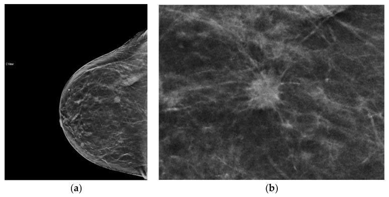
This diagnostic image consists of two panels showcasing synthesized 2D (s2D) mammography in the right craniocaudal (RCC) view. Panel (a) provides a wide field-of-view of the breast, demonstrating heterogeneously dense breast parenchyma with mixed fibrous, glandular, and fatty tissue. A small, high-density mass is visible in the lateral aspect of the breast. Panel (b) is a magnified view of the same mass, highlighting its classic spiculated margins. These spiculations appear as fine, linear radiopaque strands radiating from the central tumor body into the surrounding tissue, creating a starburst or 'crab-like' morphology characteristic of an infiltrative process. The visual findings are pathologically associated with invasive ductal carcinoma (moderately differentiated, ER/PR+, Her2-). This image serves as a clinical example of how tomosynthesis-derived synthesized mammography enhances the conspicuity of architectural distortions and spiculated lesions compared to traditional 2D mammography, particularly in patients with higher breast density.
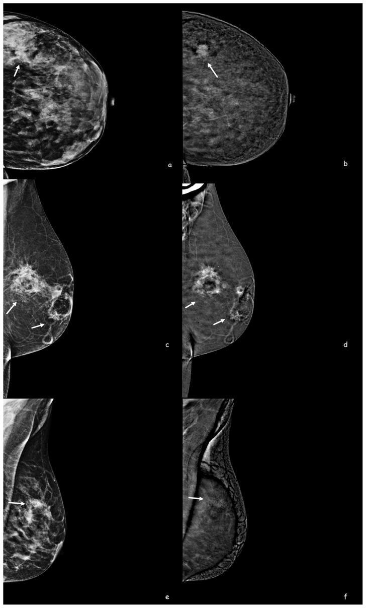
This diagnostic panel displays six images from Contrast-Enhanced Mammography (CEM) illustrating different breast pathologies. Images (a, c, e) are low-energy CEM images, while (b, d, f) are corresponding recombination subtraction images showing iodine uptake. Panel (a-b) demonstrates Invasive Ductal Carcinoma (IDC), presenting as a high-density, irregularly shaped mass in a dense breast with intense contrast enhancement. Panel (c-d) shows fat necrosis, characterized by a spiculated, high-density mass with a central fat-density component and strong contrast enhancement on subtraction. Panel (e-f) illustrates Ductal Carcinoma In Situ (DCIS), appearing as clustered suspicious microcalcifications on the low-energy image; notably, the subtraction image (f) shows no contrast enhancement, exhibiting only calcification artifacts. This comparison highlights the role of CEM in differentiating malignant and benign lesions based on morphology and vascularity, particularly in dense breast tissue where standard mammography may be limited.
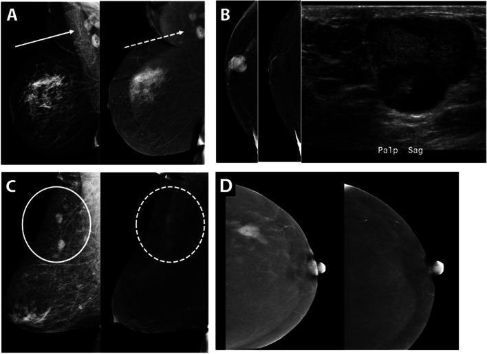
This composite diagnostic image showcases Contrast-Enhanced Mammography (CEM) and ultrasound findings across various breast malignancies. Figure A displays a case of multicentric breast cancer; the low-energy (LE) image (solid arrow) and the recombined image (RI) (dashed arrow) highlight enhancing metastatic right axillary lymph nodes. Figure B focuses on male breast cancer, showing a palpable, enhancing subareolar mass on mammography with a corresponding circumscribed hypoechoic mass with posterior acoustic enhancement on ultrasound, later confirmed as invasive ductal carcinoma. Figure C depicts diffuse B-cell lymphoma manifesting as multiple oval enhancing masses (circled) visible on both LE and RI views. Figure D illustrates the clinical utility of CEM in monitoring treatment response: a pre-treatment RI shows a spiculated, enhancing mass (infiltrating ductal carcinoma), while the post-neoadjuvant chemotherapy RI demonstrates a complete imaging response with total resolution of enhancement and mass, marked by a biopsy clip. The collection serves to teach the diverse morphological and enhancement patterns of primary breast cancers, lymphoma, and axillary metastases, as well as the assessment of therapeutic efficacy.
Carcinoma of the Breast (Ca Breast)
1. Epidemiology
- Breast cancer is the most common cancer in women. About 2.3 million new cases were diagnosed worldwide in 2020, roughly 25% of all cancers in women.
- Incidence ranges from about 27 per 100,000 in Middle Africa and East Asia to 92 per 100,000 in North America.
- In resource-poor countries, for every 2 women diagnosed, 1 dies of the disease.
- Median age at presentation is about 60 years in the UK and USA. It is younger in Asian populations (Bailey & Love's Short Practice of Surgery 28th Ed, p. 951-952).
2. Risk factors
| Category | Factors |
|---|---|
| Hormonal and reproductive | Early menarche, late menopause, nulliparity, first pregnancy after 35 years, HRT for more than 10 years (RR 1.2). Breastfeeding for more than 12 months is protective. |
| Lifestyle | Obesity after menopause (RR 1.29), alcohol (RR 1.05 for light use to 1.46 for more than 4 drinks/day), tobacco (RR 1.14 for 25 or more cigarettes/day) |
| Radiation | Chest irradiation (RR about 6), for example mantle radiotherapy for Hodgkin lymphoma |
| Genetic | BRCA1 (often triple-negative), BRCA2, TP53 (Li-Fraumeni), PTEN (Cowden), PALB2, CHEK2, ATM, CDH1 |
| Other | Age, female sex, first-degree relative with breast cancer, atypical ductal or lobular hyperplasia, LCIS, dense breasts, previous contralateral cancer |
3. Pathology
- About 90% of cases arise from ducts and about 10% from lobules.
- In situ disease has not breached the basement membrane. This includes DCIS and LCIS.
- Invasive disease has breached it. The most common type is invasive carcinoma of no special type (NST, previously "invasive ductal").
- Grade uses the modified Bloom-Richardson (Nottingham) score. It adds up tubule formation, nuclear pleomorphism and mitotic count, each scored 1 to 3. A total of 3 to 5 is grade I, 6 to 7 is grade II, and 8 to 9 is grade III.
- Special types with a better prognosis: mucinous (colloid), tubular, medullary and papillary. Invasive lobular carcinoma is the second most common type. It tends to be multifocal and bilateral and is hard to see on mammography.
- Biomarkers tested on every invasive tumour are ER, PR, HER2 and Ki-67 (Bailey & Love, p. 952).
Molecular subtypes (Bailey & Love, Table 58.4, p. 952)
| Subtype | ER/PR | HER2 | Ki-67 |
|---|---|---|---|
| Luminal A | Positive | Negative | Low |
| Luminal B | Positive | Negative (or positive in some) | High |
| HER2-enriched | Negative | Positive | High |
| Basal-like (triple negative) | Negative | Negative | Usually high |
4. Spread
- Local: the tumour invades breast parenchyma, skin (ulceration, satellite nodules), pectoralis major and the chest wall. Tumour growth factors (FGF, TGF, VEGF) set off a desmoplastic reaction. The collagen contracts and shortens Cooper's ligaments. This causes skin dimpling, puckering and nipple retraction.
- Lymphatic: mainly to axillary nodes (levels I to III), then supraclavicular nodes. Inner-quadrant tumours may also spread to internal mammary nodes.
- Haematogenous: bone is the most common site, followed by lung, liver, brain and pleura.
5. Clinical features
- Symptoms: a painless, hard, irregular lump, usually in the upper outer quadrant. Other symptoms are nipple retraction, bloody nipple discharge, skin changes and axillary swelling.
- Signs: dimpling or tethering, peau d'orange (a sign of locally advanced disease), ulceration, fixity to the pectoral muscle or chest wall, and palpable axillary or supraclavicular nodes.
- Inflammatory carcinoma: diffuse erythema and oedema over more than one-third of the breast. It is aggressive.
- Paget's disease of the nipple: eczematous, scaly nipple and areola, usually with underlying DCIS or invasive cancer.
- Many cases are now detected by screening mammography before any symptom appears.
6. Diagnosis: triple assessment
- Clinical examination of both breasts and all nodal basins.
- Imaging:
- Women under 40 have dense breasts, so use ultrasound first. Women over 40 get mammography with ultrasound.
- Typical mammographic findings are a spiculated mass, clustered pleomorphic microcalcifications and architectural distortion.
- MRI is used for lobular cancer, dense breasts, BRCA carriers, discordant findings and assessing response to neoadjuvant therapy.
- Core needle biopsy, image-guided where needed, with ER, PR, HER2 and Ki-67 testing. FNAC is no longer preferred because it cannot distinguish in situ from invasive disease or test receptors. Axillary ultrasound with node sampling is done if nodes look abnormal.
7. Staging
- T1: 2 cm or less. T2: more than 2 cm to 5 cm. T3: more than 5 cm.
- T4: any size with chest wall involvement (T4a), skin ulceration or satellite nodules (T4b), both (T4c), or inflammatory carcinoma (T4d).
- N1: mobile ipsilateral level I to II nodes. N2: fixed or matted nodes, or internal mammary nodes. N3: infraclavicular, supraclavicular, or 10 or more axillary nodes.
- M1: distant metastasis.
8. Management of early and locally advanced disease
Surgery
- Breast-conserving surgery (BCS) is a wide local excision with clear margins ("no ink on tumour" for invasive disease). It is followed by whole-breast radiotherapy and gives survival equal to mastectomy.
- Mastectomy is used for large tumours relative to breast size, multicentric disease, extensive DCIS, a contraindication to radiotherapy, patient choice, or BRCA carriers choosing risk reduction. Skin-sparing and nipple-sparing techniques with immediate reconstruction (implant or autologous flap) are options.
- Axillary management:
- Sentinel lymph node biopsy (SLNB) is used if the nodes are clinically negative.
- Axillary clearance is done for positive nodes in selected cases. It is often avoided in limited sentinel node disease (ACOSOG Z0011 criteria) or replaced by axillary radiotherapy.
- Targeted axillary dissection is used after neoadjuvant chemotherapy.
- Modified radical mastectomy is still performed in many centres for locally advanced or node-positive disease.
Radiotherapy
- Whole-breast radiotherapy after BCS, often hypofractionated (about 3 weeks) with or without a tumour-bed boost.
- Post-mastectomy radiotherapy for T3 or T4 tumours and 4 or more positive nodes, with consideration for 1 to 3 positive nodes.
- Regional nodal irradiation in selected node-positive patients.
Systemic therapy by subtype
| Subtype | Core treatment |
|---|---|
| Luminal (HR+/HER2-) | Endocrine therapy for 5 to 10 years: tamoxifen (pre-menopausal) or aromatase inhibitors (post-menopausal). Add ovarian function suppression for high-risk premenopausal women. Chemotherapy if genomic score or clinical risk is high. Consider adjuvant CDK4/6 inhibitors (abemaciclib, ribociclib) in high-risk node-positive disease. |
| HER2-positive | Trastuzumab plus pertuzumab with chemotherapy. Neoadjuvant treatment is preferred for larger or node-positive tumours. Residual disease is treated with T-DM1 or trastuzumab deruxtecan according to current guidelines. Add endocrine therapy if HR+. |
| Triple negative | Chemotherapy (anthracycline and taxane, with carboplatin in some). Neoadjuvant pembrolizumab with chemotherapy (KEYNOTE-522). Capecitabine or olaparib (if gBRCA) for residual disease. |
- Neoadjuvant therapy is used to downstage tumours for BCS, assess response (pathological complete response guides post-surgical therapy), and treat locally advanced and inflammatory cancer.
- Bone health: give bisphosphonates or denosumab to women on aromatase inhibitors and to high-risk post-menopausal women.
DCIS and LCIS
- DCIS is treated with BCS plus radiotherapy, or mastectomy. Endocrine therapy is added if ER+.
- LCIS is a risk marker. It is managed by surveillance, with chemoprevention considered.
9. Metastatic (stage IV) disease
- Treatment is palliative and aims to prolong life and preserve quality of life.
- HR+/HER2-: endocrine therapy plus a CDK4/6 inhibitor is the first line. Later options are fulvestrant, PI3K/AKT/mTOR inhibitors, elacestrant for ESR1 mutation, and then chemotherapy.
- HER2-positive: HER2-targeted therapy (trastuzumab, pertuzumab, T-DXd) with chemotherapy.
- Triple negative: chemotherapy, immunotherapy (PD-L1 positive), PARP inhibitors (gBRCA), and sacituzumab govitecan.
- Local measures include bone-modifying agents, palliative radiotherapy, and surgery for fractures or cord compression.
- Cytotoxic therapy is reserved for visceral crisis or endocrine-refractory disease. Visceral crisis means severe organ dysfunction with rapid progression, not just the presence of visceral metastases (NCCN Breast Cancer, Version 4.2026).
10. Screening, prevention and follow-up
- Screening mammography is typically every 1 to 2 years from about age 40 to 50 through 69 to 74, depending on national policy. High-risk women (BRCA carriers, prior chest radiation) get annual MRI plus mammography.
- Prevention includes lifestyle changes (weight, alcohol, exercise), chemoprevention (tamoxifen, raloxifene, anastrozole) for high-risk women, and risk-reducing mastectomy or salpingo-oophorectomy for BRCA carriers.
- Follow-up is clinical review every 3 to 6 months initially, then yearly, with annual mammography. Routine tumour markers and scans are not recommended without symptoms. The NCCN guideline was updated in January 2026 to support the Breast Cancer Index for decisions on extended endocrine therapy (HMP Global).
11. Special situations
- Male breast cancer: about 1% of cases. It is usually ER+ and linked to BRCA2 and Klinefelter syndrome. It is treated like female disease.
- Pregnancy-associated: chemotherapy is possible in the second and third trimesters. Radiotherapy, endocrine therapy and HER2 agents are deferred until after delivery.
- Phyllodes tumour: a fibroepithelial neoplasm graded benign, borderline or malignant. It is treated with wide excision.
- Inflammatory breast cancer: neoadjuvant systemic therapy, then mastectomy with axillary clearance, then radiotherapy.
Sources and currency check
- Core content comes from Bailey & Love's Short Practice of Surgery 28th Ed (Carcinoma of the Breast, p. 951-953).
- The management details come from standard guidelines and my own knowledge, checked against the NCCN 2026 update.
- A PubMed search of recent systematic reviews and meta-analyses found no 2024 to 2025 review that contradicts the above. The results covered fungating tumours, nanoparticles and drug approvals, none of which changes the core management.
- Guidelines change often. Check the current NCCN or ESMO version before making clinical decisions.