
bronchopulmonary segments on hrct and basic hrct lung anatomy from david sutton and hagga
Reading File
Finding Sources
Finding Sources
Finding Sources
Searching Images
labeled bronchopulmonary segments CT anatomy diagram lungs
Searching PubMed
high resolution computed tomography lung anatomy
Searching the Web
bronchopulmonary segments HRCT anatomy radiology reference
Reading File
Reading File
Finding Sources
Finding Sources
Reading Web Page
https://radiopaedia.org/articles/bronchopulmonary-segmental-…
Reading File
Reading File
Loading Image
Loading Image
Finding Sources
Basic HRCT lung anatomy and bronchopulmonary segments
I could not retrieve the original text of David Sutton or Hagga from the available library, so I cannot quote their exact wording or page numbers. The outline below follows standard thoracic HRCT anatomy consistent with those radiology texts, supported by anatomy references.
1. How to orient an axial HRCT image
- Images are conventionally viewed as if looking up from the patient's feet.
- Patient's right is on the left side of the image.
- Anterior is at the top, posterior at the bottom.
- On lung windows:
- Air is black.
- Normal lung is dark grey.
- Vessels, bronchial walls, fissures, and interlobular septa are relatively white.
- HRCT uses thin sections and a high-spatial-frequency reconstruction to show fine lung structures, especially small airways, interstitium, and the secondary pulmonary lobule.
Thoracic CT interpretation is fundamentally based on the secondary pulmonary lobule: a central bronchiole and pulmonary arteriole, surrounding acini, and peripheral interlobular septa containing pulmonary veins and lymphatics. Harrison's Principles of Internal Medicine, 22e, p. 2254.
2. Lobar anatomy on HRCT
Right lung: three lobes
| Lobe | Main location on axial CT | Boundary |
|---|---|---|
| Right upper lobe (RUL) | Mainly upper and anterior lung | Horizontal fissure inferiorly/anteriorly; oblique fissure posteriorly |
| Right middle lobe (RML) | Anterior and medial, adjacent to right heart border | Between horizontal and oblique fissures |
| Right lower lobe (RLL) | Mainly posterior and inferior | Posterior to oblique fissure |
Left lung: two lobes
| Lobe | Main location on axial CT | Boundary |
|---|---|---|
| Left upper lobe (LUL) | Mainly anterior and superior | Anterior to the oblique fissure |
| Lingula | Anteromedial inferior part of LUL, beside the left heart border | Functional counterpart of RML |
| Left lower lobe (LLL) | Mainly posterior and inferior | Posterior to oblique fissure |
Fissures
- The oblique (major) fissure separates the lower lobe from the upper lobe on the left, and from the upper and middle lobes on the right.
- The horizontal (minor) fissure is usually present only on the right and separates RUL from RML. Gray's Anatomy for Students, p. 205.
Practical axial rule:
At the level of the hila, a posterior lesion is usually in a lower lobe. An anterior lesion is usually in an upper lobe, except on the right where a lesion between the fissures can be in the middle lobe.
At the level of the hila, a posterior lesion is usually in a lower lobe. An anterior lesion is usually in an upper lobe, except on the right where a lesion between the fissures can be in the middle lobe.
3. Bronchopulmonary segment: definition
A bronchopulmonary segment is a pyramidal or cone-shaped, functionally independent unit of lung supplied by:
- One segmental (tertiary) bronchus
- Its accompanying branch of the pulmonary artery
The segmental bronchus and pulmonary artery run centrally within the segment. Pulmonary veins usually run between segments, helping to form intersegmental planes. Gray's Anatomy for Students, pp. 209-210. This arrangement is why a segment can often be surgically removed without sacrificing adjacent segments.
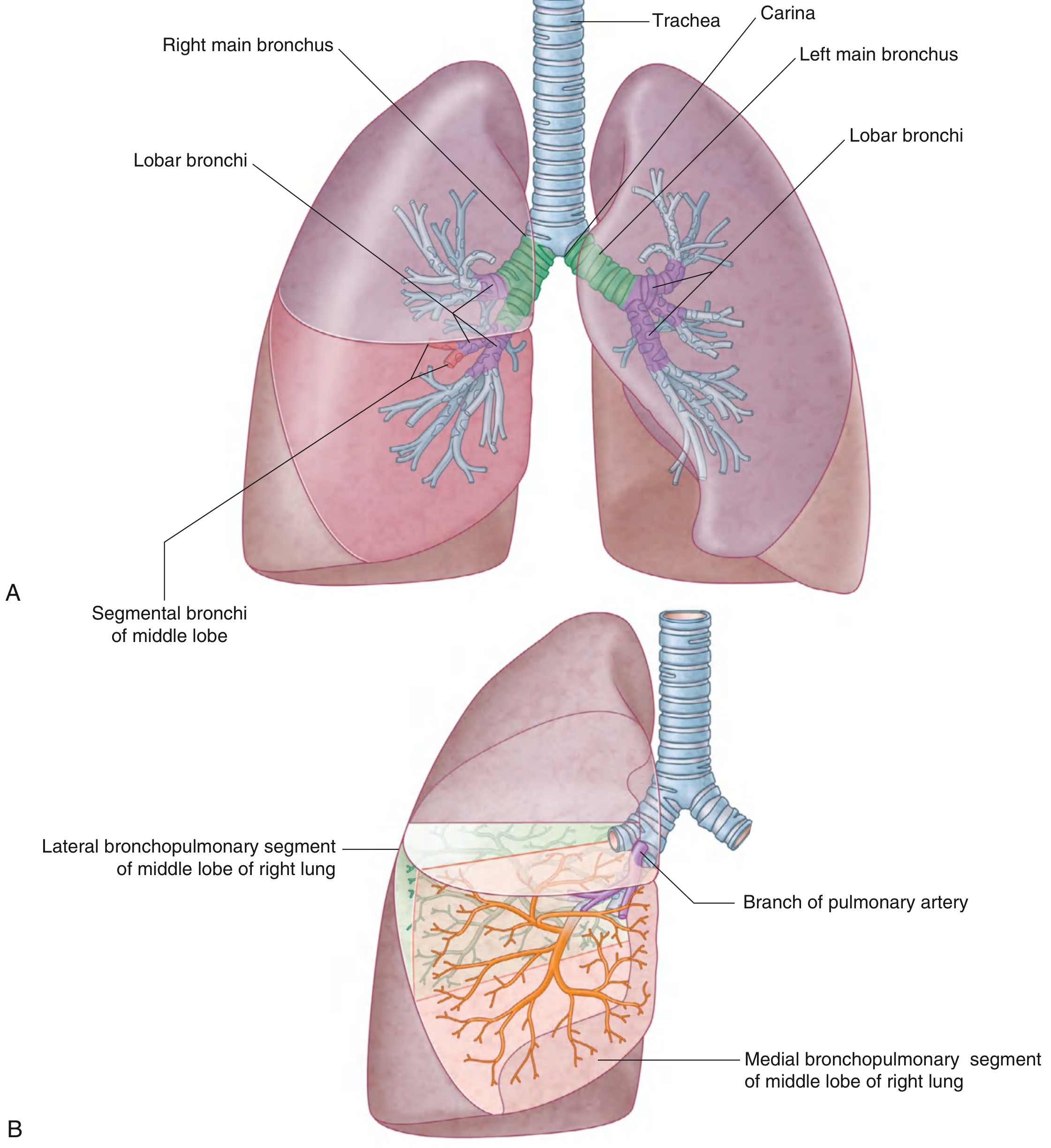
Bronchial tree and segmental organization. Gray's Anatomy for Students, p. 209.
4. Right bronchopulmonary segments
Right upper lobe
| Segment | Name | HRCT location |
|---|---|---|
| S1 | Apical | At apex, uppermost lung |
| S2 | Posterior | Posterior upper lobe |
| S3 | Anterior | Anterior upper lobe |
Right middle lobe
| Segment | Name | HRCT location |
|---|---|---|
| S4 | Lateral | Lateral/anterior RML |
| S5 | Medial | Medial RML, contacts right heart border |
Right lower lobe
| Segment | Name | HRCT location |
|---|---|---|
| S6 | Superior | Posteriorly, at the top of the lower lobe |
| S7 | Medial basal | Medial basal, near mediastinum |
| S8 | Anterior basal | Anterior basal lower lobe |
| S9 | Lateral basal | Lateral lower lobe |
| S10 | Posterior basal | Most posterior basal lung |
Mnemonic: Right lower lobe = Superior + four basal segments: medial, anterior, lateral, posterior.
5. Left bronchopulmonary segments
The left lung is smaller, and certain segments commonly share bronchi. Thus it may be described as having 8 anatomically distinct segments, or 9-10 when the fused components are counted separately.
Left upper lobe
| Segment | Name | HRCT location |
|---|---|---|
| S1+S2 | Apicoposterior | Apex and posterior upper lobe, commonly fused |
| S3 | Anterior | Anterior upper lobe |
| S4 | Superior lingular | Anteromedial, superior lingula |
| S5 | Inferior lingular | Anteromedial, inferior lingula |
Left lower lobe
| Segment | Name | HRCT location |
|---|---|---|
| S6 | Superior | Posterior upper part of LLL |
| S7+S8 | Anteromedial basal | Usually fused on the left |
| S9 | Lateral basal | Lateral basal LLL |
| S10 | Posterior basal | Posterior basal LLL |
The right and left lungs are often described as having 10 segments each, but left apicoposterior (S1+2) and left anteromedial basal (S7+8) segments are frequently fused. Gray's Anatomy for Students, p. 210. A concise current radiology summary gives the same practical classification in this segmental anatomy reference.
6. HRCT landmarks for localizing a lesion
A. Upper lobe versus lower lobe
- Upper lobe disease tends to lie anteriorly at the upper chest.
- Lower lobe disease tends to lie posteriorly, particularly near the bases.
- In supine CT, dependent posterior lower-lobe ground-glass opacity may be positional atelectasis. Prone images can help distinguish this from genuine interstitial abnormality.
B. Middle lobe and lingula
- RML: anterior-medial, contacts the right cardiac border.
- Lingula: anterior-medial left upper lobe, contacts the left cardiac border.
- These are analogous locations. Bronchiectasis or nodularity involving both often suggests a small-airway distribution pattern.
C. Superior segment of lower lobe, S6
- Lies in the posterior upper portion of each lower lobe.
- It may look relatively high on axial images but is still a lower-lobe segment.
- This is a common localization trap.
D. Basal segments
- Seen more clearly on lower cuts.
- Posterior basal segments, S10, lie most posteriorly.
- Anterior basal segments, S8, are more anterior.
- Lateral basal segments, S9, are lateral.
7. Secondary pulmonary lobule: the key HRCT unit
Do not confuse a bronchopulmonary segment with a secondary pulmonary lobule.
| Feature | Bronchopulmonary segment | Secondary pulmonary lobule |
|---|---|---|
| Scale | Large surgical/anatomic unit | Small HRCT unit, about 1-2.5 cm |
| Central structure | Segmental bronchus plus pulmonary artery | Terminal bronchiole plus centrilobular artery |
| Veins | At segment boundaries | In interlobular septa at lobule periphery |
| HRCT relevance | Localizing a lesion for reporting/surgery | Recognizing disease pattern |
HRCT patterns based on lobular anatomy
- Centrilobular nodules: spare the pleural surface and interlobular septa; suggest bronchiolar disease.
- Perilymphatic nodules: along fissures, pleura, bronchovascular bundles, and septa.
- Random nodules: diffuse, including subpleural lung.
- Interlobular septal thickening: at the periphery of secondary lobules.
- Mosaic attenuation and air trapping: often reflect small-airway obstruction, assessed best on expiratory HRCT.
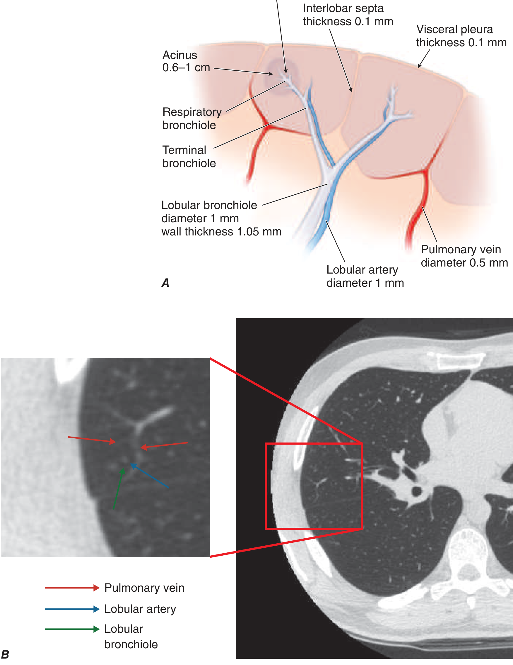
The central airway and artery, with peripheral interstitium, veins, and lymphatics, form the basis for HRCT pattern recognition. Harrison's Principles of Internal Medicine, 22e, p. 2254.
8. Short reporting-style examples
- “Cylindrical bronchiectasis in the right middle lobe medial segment (S5) and lingula.”
- “Subpleural reticulation and traction bronchiectasis predominantly involve the posterior basal lower-lobe segments (S10).”
- “Tree-in-bud nodules are centred in the centrilobular region, indicating small-airway rather than random hematogenous spread.”
- “A focal opacity in the posterior upper part of the lower lobe is in the superior segment (S6), not the upper lobe.”
High-yield recall
- Right: 3 lobes, 10 segments.
- Left: 2 lobes, usually counted as 8-10 because S1+2 and S7+8 commonly fuse.
- Artery and bronchus are intrasegmental.
- Veins are intersegmental.
- Lower lobes are posterior; upper lobes are anterior on axial CT.
- RML and lingula are anterior-medial.
- S6 is posterior but belongs to the lower lobe.
The recent systematic-review literature retrieved does not alter these fundamental anatomic principles.
This is a shared conversation. Sign in to Orris to start your own chat.