
Im 1st year mbbs student in exam imp questions list i saw one question is 5 mark question is give a histology of epithelium give a answer of this question and drawing with label answer give easy to understand English language,when I read this question answer I understand easily, give answer this type when I read this question answer first time so much answer I understand easily in first time and when i write this in exam my sir will give me full out of full marks and easy to read and understand,answer give as 1 st year mbbs [ Anatomy// histology ]and easy to understand and write Thank you
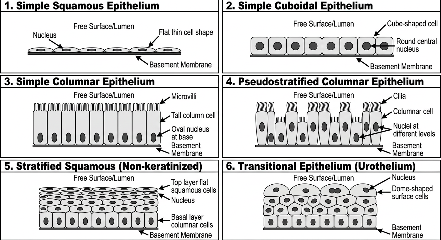
A clean labeled histology diagram showing all major types of epithelium for a 1st year MBBS student, with the following panels clearly drawn and labeled: 1. Simple Squamous Epithelium - flat thin cells, single layer, flat nucleus 2. Simple Cuboidal Epithelium - cube-shaped cells, single layer, round central nucleus 3. Simple Columnar Epithelium - tall column cells, single layer, oval nucleus at base, microvilli on top 4. Pseudostratified Columnar Epithelium - cells appear multilayered but all touch basement membrane, nuclei at different levels, cilia on top 5. Stratified Squamous (Non-keratinized) - multiple layers, top layer flat squamous cells, basal layer columnar 6. Transitional Epithelium (Urothelium) - multiple layers, dome-shaped surface cells (relaxed state) Each panel has a clear title, arrow labels pointing to: basement membrane, free surface/lumen, nucleus, cell shape. Layout: 2 columns, 3 rows. White background, educational medical diagram style, black labels and arrows.
Histology of Epithelium
Definition
Characteristics (Remember: 3 main features)
| Feature | Meaning |
|---|---|
| Closely packed cells | Cells are tightly joined together with very little space between them |
| Polarity | Each cell has a top (apical), side (lateral), and bottom (basal) surface |
| Basement membrane | A thin supporting layer below all epithelial cells; attaches them to connective tissue |
Key point: Epithelium is avascular - it has NO blood vessels. It gets nutrition by diffusion from connective tissue below.
Classification of Epithelium
- Number of layers - Simple or Stratified
- Shape of cells - Squamous, Cuboidal, or Columnar
A. SIMPLE EPITHELIUM (Only ONE cell layer, all cells touch the basement membrane)
1. Simple Squamous Epithelium
- Shape: Cells are flat and thin, like floor tiles
- Nucleus: Flat, in the center
- Location: Blood vessels (endothelium), lungs (alveoli), body cavities (mesothelium)
- Function: Allows passage of substances - filtration and diffusion
2. Simple Cuboidal Epithelium
- Shape: Cells look like small cubes - height = width
- Nucleus: Round, in the center
- Location: Kidney tubules, thyroid gland, small ducts of glands
- Function: Secretion and absorption
3. Simple Columnar Epithelium
- Shape: Cells are tall like columns - height > width
- Nucleus: Oval, near the base of the cell
- Location: Stomach, intestine, gallbladder
- Function: Absorption and secretion; may have microvilli (brush border) on top for better absorption; some cells are goblet cells that secrete mucus
4. Pseudostratified Columnar Epithelium
- Looks like: Multiple layers (because nuclei are at different heights)
- Actually is: A SIMPLE epithelium - ALL cells touch the basement membrane
- Special feature: Usually has cilia on top
- Location: Trachea, bronchi, male reproductive tract (epididymis)
- Function: Mucus secretion and movement (cilia sweep mucus up)
B. STRATIFIED EPITHELIUM (TWO or MORE cell layers)
In stratified epithelium, the shape of the TOP (surface) layer determines the name.
5. Stratified Squamous Epithelium
- Surface cells: Flat and squamous
- Basal layer cells: Columnar or cuboidal (constantly dividing to replace top cells)
- Two types:
- Non-keratinized - top cells are alive, moist; Location: Mouth, esophagus, vagina
- Keratinized - top cells are dead and filled with keratin protein; Location: Skin (epidermis)
- Function: Protection against mechanical friction and abrasion
6. Transitional Epithelium (Urothelium)
- Special type found ONLY in the urinary tract (bladder, ureter, urethra)
- Surface cells: Large, dome-shaped ("umbrella cells") when relaxed; flatten when bladder is full
- Function: Allows stretching (distension) of the urinary tract
- Key feature to remember: Cells CHANGE shape depending on whether bladder is empty or full
Diagram
Actual Histology Photographs (from Textbook)
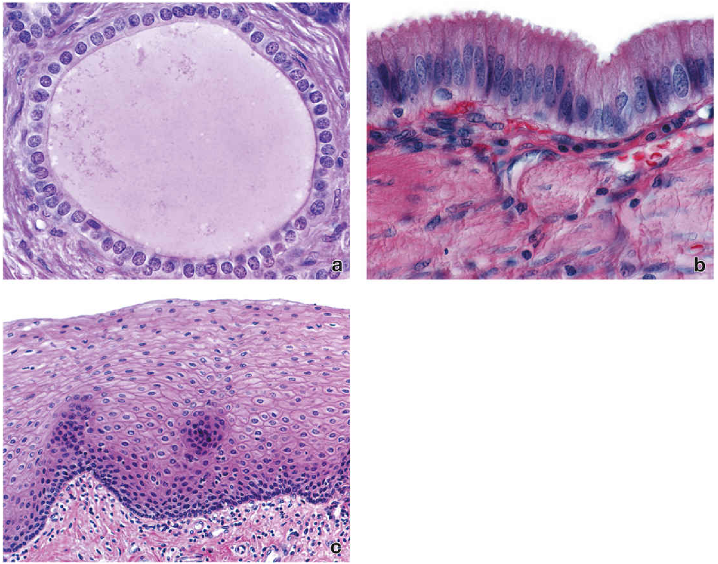
- (a) Pancreatic duct - Simple Cuboidal (one layer, round nuclei)
- (b) Gallbladder - Simple Columnar (one layer, tall cells, oval nuclei at base, microvilli on surface)
- (c) Esophagus - Stratified Squamous (multiple layers, flat cells at surface)
Quick Memory Table (Write this in exam for full marks!)
| Type | Layers | Cell Shape | Nucleus | Location | Function |
|---|---|---|---|---|---|
| Simple Squamous | 1 | Flat | Flat | Alveoli, Blood vessels | Diffusion, filtration |
| Simple Cuboidal | 1 | Cube | Round, central | Kidney tubules, thyroid | Secretion, absorption |
| Simple Columnar | 1 | Tall column | Oval, basal | Intestine, stomach | Absorption, secretion |
| Pseudostratified | 1 (looks like many) | Columnar with cilia | At different levels | Trachea, bronchi | Mucus transport |
| Stratified Squamous | Many | Flat at surface | Oval | Skin, esophagus, mouth | Protection |
| Transitional | Many | Dome-shaped | Round | Urinary bladder, ureter | Stretching |
How to Write in Exam (Tips)
- Start with definition - 1 sentence
- Give 3 characteristics - bullet points
- Classify - Simple vs Stratified, then each type
- For each type: Shape + Nucleus + Location + Function
- Draw a diagram - label: free surface, nucleus, basement membrane, connective tissue
- End with the table - examiners love tables!
Im 1st year mbbs student in exam imp questions list i saw one question is 5 mark question is give a histology of epithelium give a answer of this question and creat a picture [ photo] with label answer give easy to understand English language,when I read this question answer I understand easily, give answer this type when I read this question answer first time so much answer I understand easily in first time and when i write this in exam my sir will give me full out of full marks and easy to read and understand,answer give as 1 st year mbbs [ Anatomy// histology ]and easy to understand and write Thank you
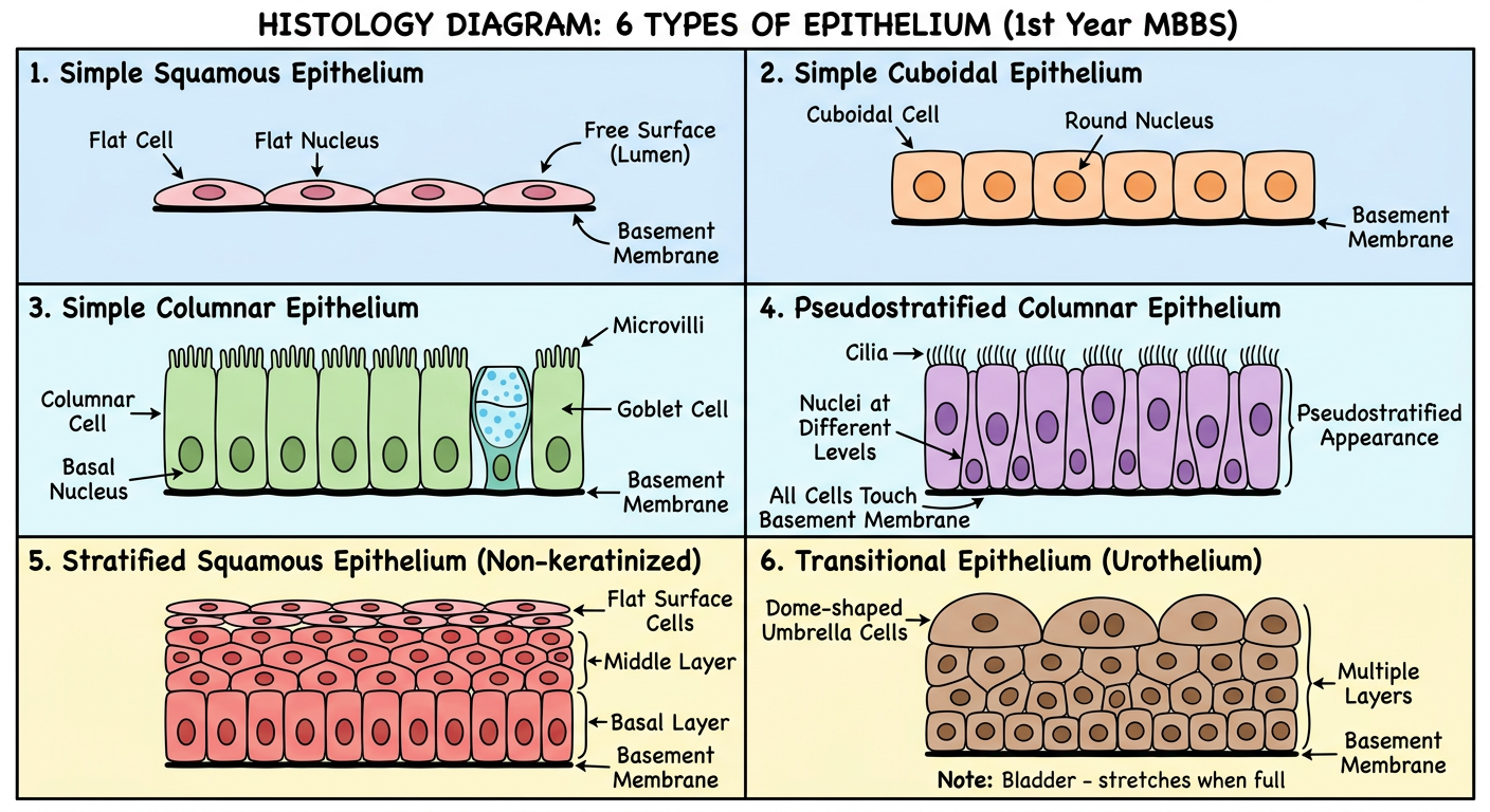
A very clear, clean, educational histology diagram for 1st year MBBS students showing 6 types of epithelium, drawn in a simple cartoon/diagram style (not photorealistic), white background, black outlines, colored cells: Panel 1 - "1. Simple Squamous Epithelium": One single row of very flat, wide cells like floor tiles. Each cell has a flat oval nucleus in the center. A basement membrane line below. Arrow pointing to: flat cell, flat nucleus, basement membrane, free surface (lumen above). Panel 2 - "2. Simple Cuboidal Epithelium": One single row of cube-shaped cells (equal height and width). Each cell has a large round nucleus in the center. Basement membrane below. Arrow labels: cuboidal cell, round nucleus, basement membrane. Panel 3 - "3. Simple Columnar Epithelium": One single row of tall rectangular cells (height much greater than width). Oval nucleus near the base. Small hair-like microvilli on the top (free) surface. One goblet cell (wine-glass shaped, full of mucus). Labels: columnar cell, basal nucleus, microvilli, goblet cell, basement membrane. Panel 4 - "4. Pseudostratified Columnar Epithelium": Cells appear to be in multiple layers because nuclei are at different heights. But ALL cells touch the basement membrane at the bottom. Cilia drawn on top surface. Labels: cilia, nuclei at different levels, all cells touch basement membrane, pseudostratified appearance. Panel 5 - "5. Stratified Squamous Epithelium (Non-keratinized)": Multiple layers of cells. Bottom layer (basal) = tall columnar cells. Middle layers = polygonal cells. Top layer = flat squamous cells. Labels: flat surface cells, middle layer, basal layer, basement membrane. Panel 6 - "6. Transitional Epithelium (Urothelium)": Multiple layers. Top cells are large, dome-shaped umbrella cells. Lower cells are smaller. Labels: dome-shaped umbrella cells, multiple layers, basement membrane. Note: "Bladder - stretches when full". Layout: 2 columns x 3 rows, each panel clearly boxed with its title in bold. Use light blue for simple epithelia panels, light yellow for stratified. Clean educational medical diagram style.
✅ HISTOLOGY OF EPITHELIUM
(5 Marks Answer — 1st Year MBBS | Anatomy / Histology)
📌 DEFINITION
- Covers the outer surface of the body
- Lines the internal hollow organs and body cavities
- Forms glands
It acts as a barrier between the body and the outside world.
📌 GENERAL FEATURES (3 Main Points — Must Write!)
| Feature | Simple Explanation |
|---|---|
| Closely packed cells | Cells sit tightly next to each other, very little space between them |
| Avascular | NO blood vessels inside epithelium; nutrition comes from connective tissue below |
| Basement membrane | A thin layer below ALL epithelial cells — it supports and anchors them |
| Polarity | Each cell has a top side (faces outside/lumen) and a bottom side (faces connective tissue) |
📌 CLASSIFICATION
1. Number of layers → Simple (1 layer) or Stratified (many layers)2. Shape of cells → Squamous (flat), Cuboidal (cube), Columnar (tall)
📌 LABELED DIAGRAM
📌 TYPES OF EPITHELIUM
🔵 A. SIMPLE EPITHELIUM (Only 1 Cell Layer)
1. Simple Squamous Epithelium
- How cells look: Very flat and thin, like floor tiles
- Nucleus: Flat, in the center of the cell
- Where found: Alveoli of lungs, blood vessel walls (called endothelium), body cavities (called mesothelium)
- Function: Allows gases and fluids to pass through easily (diffusion and filtration)
2. Simple Cuboidal Epithelium
- How cells look: Like small cubes — width = height
- Nucleus: Round, in the CENTER of the cell
- Where found: Kidney tubules, thyroid follicles, ducts of small glands
- Function: Secretion (making products) and Absorption (taking in substances)
3. Simple Columnar Epithelium
- How cells look: Tall and thin, like columns — height > width
- Nucleus: Oval, near the BOTTOM (base) of the cell
- Special features:
- Microvilli on top surface → increase absorption area (seen as "brush border")
- Goblet cells mixed in → secrete mucus (look like a wine glass shape)
- Where found: Stomach, small intestine, large intestine, gallbladder
- Function: Absorption and secretion
4. Pseudostratified Columnar Epithelium
- How cells look: Looks like MANY layers BUT it is actually ONLY 1 layer
- Why does it look stratified? Because nuclei are at different heights in the cells
- Key rule: ALL cells touch the basement membrane — that is why it is SIMPLE (not truly stratified)
- Special feature: Has cilia on top to sweep mucus
- Where found: Trachea, bronchi, epididymis (male reproductive tract)
- Function: Mucus secretion + cilia move mucus and trapped dust upward
🟡 B. STRATIFIED EPITHELIUM (2 or More Cell Layers)
In stratified epithelium, the name is given by the shape of the TOP surface cells.
5. Stratified Squamous Epithelium
- How cells look: Many layers stacked on top of each other
- Bottom layer (basal): Columnar/cuboidal cells — actively dividing
- Middle layers: Polygonal (many-sided) cells
- Top layer (surface): Flat, squamous cells
- Two types:
| Type | Top cells | Where found |
|---|---|---|
| Non-keratinized | Alive, moist | Mouth, esophagus, vagina |
| Keratinized | Dead, filled with keratin protein | Skin (epidermis) |
- Function: Protection against friction, wear and tear, and injury
6. Transitional Epithelium (Urothelium)
- Special type found ONLY in the urinary tract
- How cells look:
- Relaxed (empty bladder): Many layers, top cells are large and dome-shaped ("umbrella cells")
- Stretched (full bladder): Cells flatten, fewer apparent layers
- Where found: Urinary bladder, ureter, upper urethra
- Function: Allows the organ to stretch and relax without tearing
📌 QUICK MEMORY TABLE (Write this in exam = Full Marks!)
| Type | Layers | Cell Shape | Nucleus | Location | Function |
|---|---|---|---|---|---|
| Simple Squamous | 1 | Flat | Flat, central | Lungs, blood vessels | Diffusion, filtration |
| Simple Cuboidal | 1 | Cube | Round, central | Kidney tubules, thyroid | Secretion, absorption |
| Simple Columnar | 1 | Tall column | Oval, basal | Intestine, stomach | Absorption, secretion |
| Pseudostratified | 1 (looks many) | Columnar + cilia | Different levels | Trachea, bronchi | Mucus + cilia movement |
| Stratified Squamous | Many | Flat at top | Oval | Skin, esophagus, mouth | Protection |
| Transitional | Many | Dome at top | Round | Bladder, ureter | Stretching |
📌 EXAM WRITING TIP
- Definition (2 lines)
- General features (4 bullet points)
- Classification (simple diagram or heading)
- Each type with: shape + nucleus + location + function
- Draw the labeled diagram
- End with the summary table