
Cold urticaria
cold urticaria skin lesion wheal
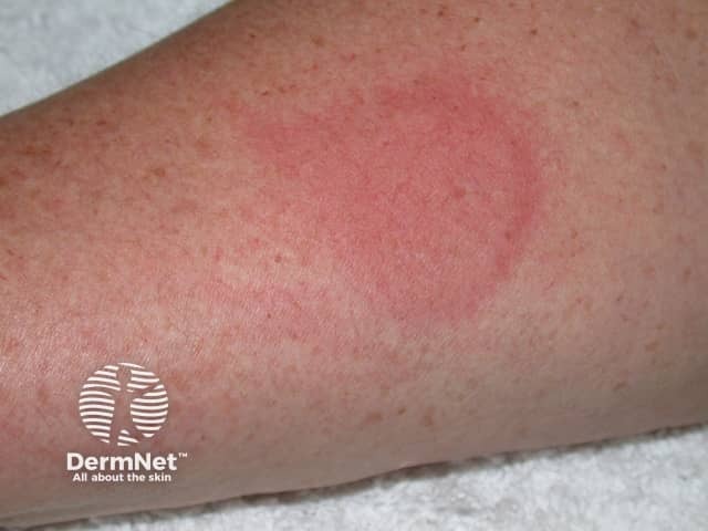
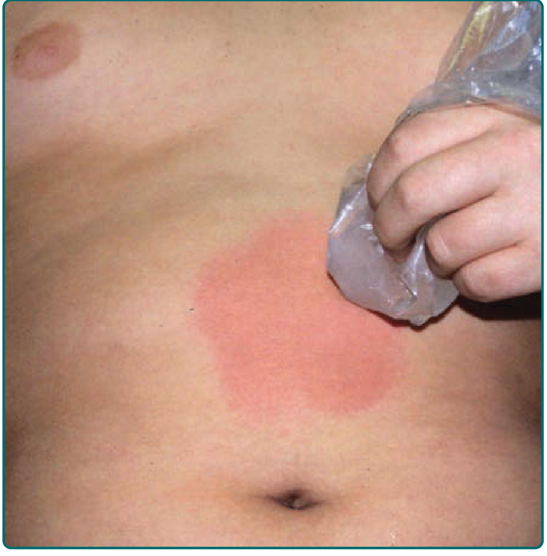
Imaging modality: Clinical photography. Technique: High-resolution color photograph of the skin surface captured after cold provocation with an ice cube. Anatomical location: Forearm skin (cutaneous tissue, integumentary system). Visual features: A well-demarcated, round/oval erythematous wheal with superficial edema and surrounding mild erythema, consistent with an acute urticarial reaction. The lesion is blanchable, pruritic, and mounts within minutes following cold exposure; the center may be slightly lighter; the perilesional area shows diffuse erythema. This is a classic representation of cold urticaria, a physical urticaria caused by exposure to cold temperatures leading to mast cell degranulation and histamine release. The image demonstrates the macroscopic dermatologic hallmark: transient, migratory wheal formation on the skin surface due to superficial dermal edema; no purpura or vasculitis is evident. Clinical significance: diagnostic support for physical urticarias; correlates with ice cube or cold water provocation tests. Differential diagnoses include other forms of urticaria, contact dermatitis, erythema multiforme, urticarial vasculitis, and solar/lattice dermatitis. Management implications include avoidance of cold exposure, antihistamines, and, in refractory cases, omalizumab or H2 blockers. The image is useful for education, dermatology training, and research on cold-induced wheal formation and diagnostic testing. Useful for patient education, and research discussions. Helpful for clinical teaching materials.
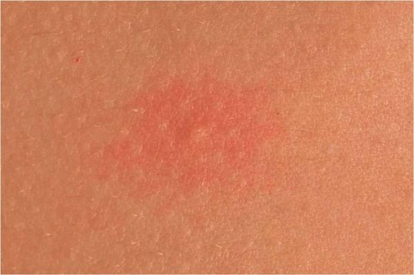
This clinical photograph shows a close-up view of a localized skin lesion on the torso, characteristic of a wheal. The central lesion is a well-circumscribed, erythematous (reddish) patch with a slightly raised, edematous surface compared to the surrounding unaffected skin. The morphology is roughly circular with moderate border definition. The affected area exhibits a subtly uneven texture, contrasting with the smooth, uniform skin tone and fine dermatoglyphic patterns (skin lines) of the adjacent healthy tissue. This image demonstrates the typical presentation of an urticarial lesion (hives), specifically illustrating the transient dermal edema and vascular dilation associated with aquagenic urticaria in an 18-year-old patient. Key educational concepts include morphology of primary skin lesions, recognition of erythematous wheals, and clinical manifestations of physical urticarias.
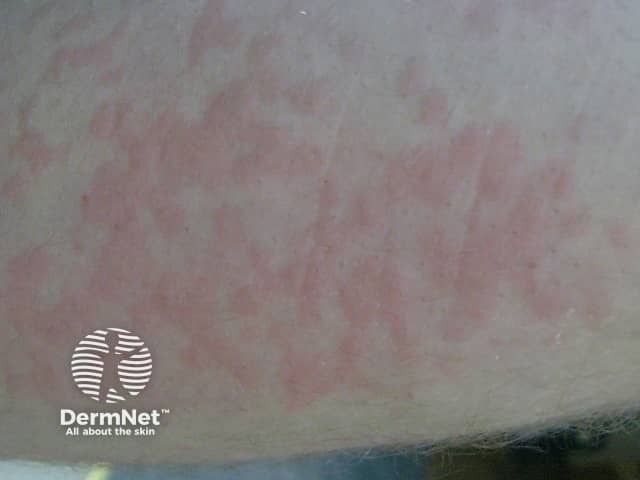
Clinical photography of a cutaneous eruption consistent with cold urticaria. Modality: clinical photograph; technique: standard color photography of exposed skin under diffuse ambient lighting; no staining or contrast. Anatomical location: ventral forearm (exposed integument) showing erythematous, blanchable wheals following cold exposure. Lesions appear as diffuse, pruritic erythematous wheals with slightly raised surfaces; borders are irregular; mild edema within the superficial dermis; lesions may coalesce into larger plaques. Overall, the pattern is mottled erythema with variable intensity, consistent with superficial dermal edema and mast cell–mediated vascular permeability. Pathophysiology: transient dermal edema due to mast cell degranulation triggered by cold stimuli; histology (if biopsy were performed) would show superficial dermal edema with perivascular inflammatory cells and occasional eosinophils. Clinically, onset is rapid after cold contact and resolves within hours after rewarming. Diagnostic significance: supports clinical diagnosis of cold urticaria; helps distinguish from other wheal disorders; ice cube test or temperature-provocation challenges may be used to confirm and stage severity; caution for potential systemic reactions or anaphylaxis with distal cold exposure. Clinical correlation and management: avoid cold exposure, consider non-sedating antihistamines, and document response for educational or research use, including differential diagnoses in teaching cases. This image supports dermatology teaching and case review.
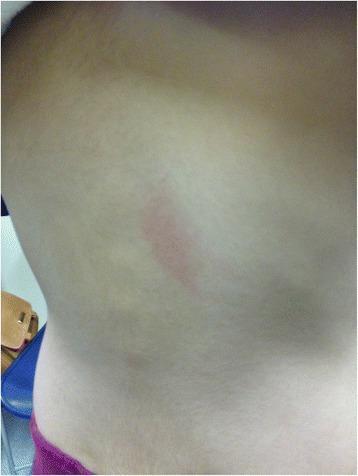
This clinical photograph displays a focal skin lesion on the mid-back of a pediatric patient. The primary finding is a single, vertically-oriented, erythematous plaque or wheal. The lesion is characterized by distinct borders, central redness, and a slightly raised morphology relative to the surrounding healthy skin, which appears otherwise smooth and normochromic. This clinical presentation is characteristic of urticaria (hives), specifically an immediate hypersensitivity reaction. The image demonstrates a localized cutaneous manifestation of a Type I allergic reaction, which in this clinical context occurred 20 minutes following an oral challenge with amoxicillin. This visual serves as an educational example of drug-induced immediate-type allergy in pediatric dermatology and immunology, highlighting the characteristic appearance of a positive oral provocation test result.
"cold urticaria" AND (omalizumab OR treatment)
Cold Urticaria
Classification
Typical Forms
| Type | Key Feature |
|---|---|
| Primary (essential) cold contact urticaria | Most common (~95% of cases); idiopathic; wheals at site of cold contact on rewarming |
| Secondary cold contact urticaria | Associated with cryoproteinemias (cryoglobulins, cryofibrinogen); may have Raynaud or purpura |
Atypical Forms
- Systemic atypical acquired cold urticaria - widespread wheals from core body temperature drop, not local cold contact
- Reflex cold urticaria - generalized cooling induces widespread whealing; ice cube test is negative; risk of anaphylaxis from diving into cold water
- Cold-dependent dermographism
- Cold-induced cholinergic urticaria
- Delayed cold urticaria - erythematous edematous swelling appearing 9-18 hours after cold challenge
Familial (Hereditary) Cold Urticaria
- FCAS1: mutation in NLRP3 (CIAS1), encoding the inflammasome protein cryopyrin
- FCAS2: mutation in NLRP12
- FCAS3: mutation in PLCG2 (phospholipase C-gamma-2)
Clinical Features
- Pruritic wheals and flare develop on cold-exposed areas within minutes of rewarming
- Morphology is flat and widely spread, or may be punctate
- Wheals persist up to 1 hour
- Triggered by: cold weather (rain, wind), cold objects, drinking cold liquids
- In severe cases: swelling of mouth/pharynx after cold liquids
- Palpitations, headache, wheezing, flushing, syncope, abdominal pain
- Loss of consciousness - drowning in cold water has been reported
- Fatal anaphylactic shock can occur with whole-body cold exposure (e.g., cold bath, swimming)
- Cryoglobulinemia (cold urticaria in ~3-4% of these patients)
- Cold agglutinins, cryofibrinogens, cold hemolysins
- Infectious mononucleosis (rare)
- Muckle-Wells syndrome (urticaria + deafness + amyloidosis)
- Helicobacter pylori infection (suggested causative role)
Pathogenesis
- IgE (and rarely IgM) mediated mast cell degranulation
- Histamine is the primary mediator; leukotrienes and platelet-activating factor are also involved
- In familial forms: NLRP3 inflammasome dysregulation drives IL-1β release (not IgE-mediated)
Diagnosis
- An ice cube wrapped in a plastic bag is applied to the forearm skin for 30 seconds to 10 minutes
- Wheals form on rewarming (not during cold contact)
- Water at 7°C (44.6°F) can also be used - less vasoconstriction allows better wheal formation
- Peltier effect-based temperature challenge is an improved method using microprocessor-controlled metals to generate precise skin surface temperatures
⚠️ Do NOT perform the ice cube test in secondary cold urticaria (cryoglobulinemia) - risk of vascular occlusion and tissue ischemia.
⚠️ The ice cube test is negative in familial cold urticaria - place the patient in a 4°C room to reproduce lesions (with caution - anaphylaxis risk).
- Cryoglobulins, cryofibrinogen
- Cold agglutinins, cold hemolysins
- Hepatitis B/C serology, EBV (Monospot)
- Consider lymphoproliferative workup if cryoglobulins detected
Management
Preventive Measures
- Avoid cold wind exposure, cold showers, cold baths, and swimming in cold water
- Avoid drinking cold liquids if oropharyngeal swelling occurs
- Prescribe an epinephrine auto-injector (EpiPen) to all patients given anaphylaxis risk
Pharmacological Treatment
| Line | Agent | Notes |
|---|---|---|
| First-line | Non-sedating (2nd generation) H1 antihistamines | Up to 4× the standard dose; e.g., cetirizine, loratadine, fexofenadine |
| Adjunctive | Sedating (1st generation) H1 antihistamines | Added for breakthrough symptoms |
| Adjunctive | H2 antihistamines (e.g., ranitidine, famotidine) | May add benefit |
| Adjunctive | Montelukast (leukotriene receptor antagonist) | |
| Refractory | Omalizumab (anti-IgE) | RCT of 31 patients stopped early due to benefit at 150 or 300 mg doses; supported by multiple case series |
| Refractory | Cyclosporine, other immunosuppressives | For severe refractory cases |
| Familial (CAPS) | IL-1 receptor antagonists (e.g., anakinra, canakinumab) | Target the NLRP3 inflammasome - antihistamines are not effective |
Desensitization
- Immerse one arm in water at 15°C (59°F) for 5 minutes daily
- Repeated increased cold exposures may be effective in some patients
Prognosis
- In the acquired (primary) form: approximately 50% of cases improve or resolve within 5-6 years
- Disease may be more transient if it follows a viral infection
- Familial forms persist throughout life
- Mean duration in one primary cold contact urticaria series: 6-9 years
- Fitzpatrick's Dermatology, 9th ed., Vol. 1-2 (Chapters 41 and 98)
- Dermatology 2-Volume Set, 5th ed., Chapter 18
- Andrews' Diseases of the Skin, Chapter 7