
Chalizioma
Reading File
A chalazion is a chronic, sterile granulomatous inflammation of a meibomian (tarsal) gland in the eyelid, caused by retained secretions blocking the gland duct. It typically presents as a painless, firm nodule on the upper or lower eyelid.
Searching sources...
Finding Sources
Finding Sources
Searching PubMed
chalazion treatment management
Reading File
Reading File
Reading File
Reading File
Loading Image
Loading Image
Chalazion (Meibomian Cyst)
Definition
A chalazion is a sterile, chronic granulomatous inflammatory lesion (lipogranuloma) of the meibomian gland (or occasionally the gland of Zeis) in the eyelid, caused by obstruction of the gland duct with retained lipid-rich sebaceous secretions. The retained material acts as a foreign body, triggering a granulomatous foreign-body inflammatory reaction.
- Goldman-Cecil Medicine, p. 302
- Kanski's Clinical Ophthalmology 10th ed., p. 54
Pathogenesis & Histology
The blocked meibomian gland secretion forms a collection that stimulates:
- Lipogranulomatous inflammation - extracellular fat deposits
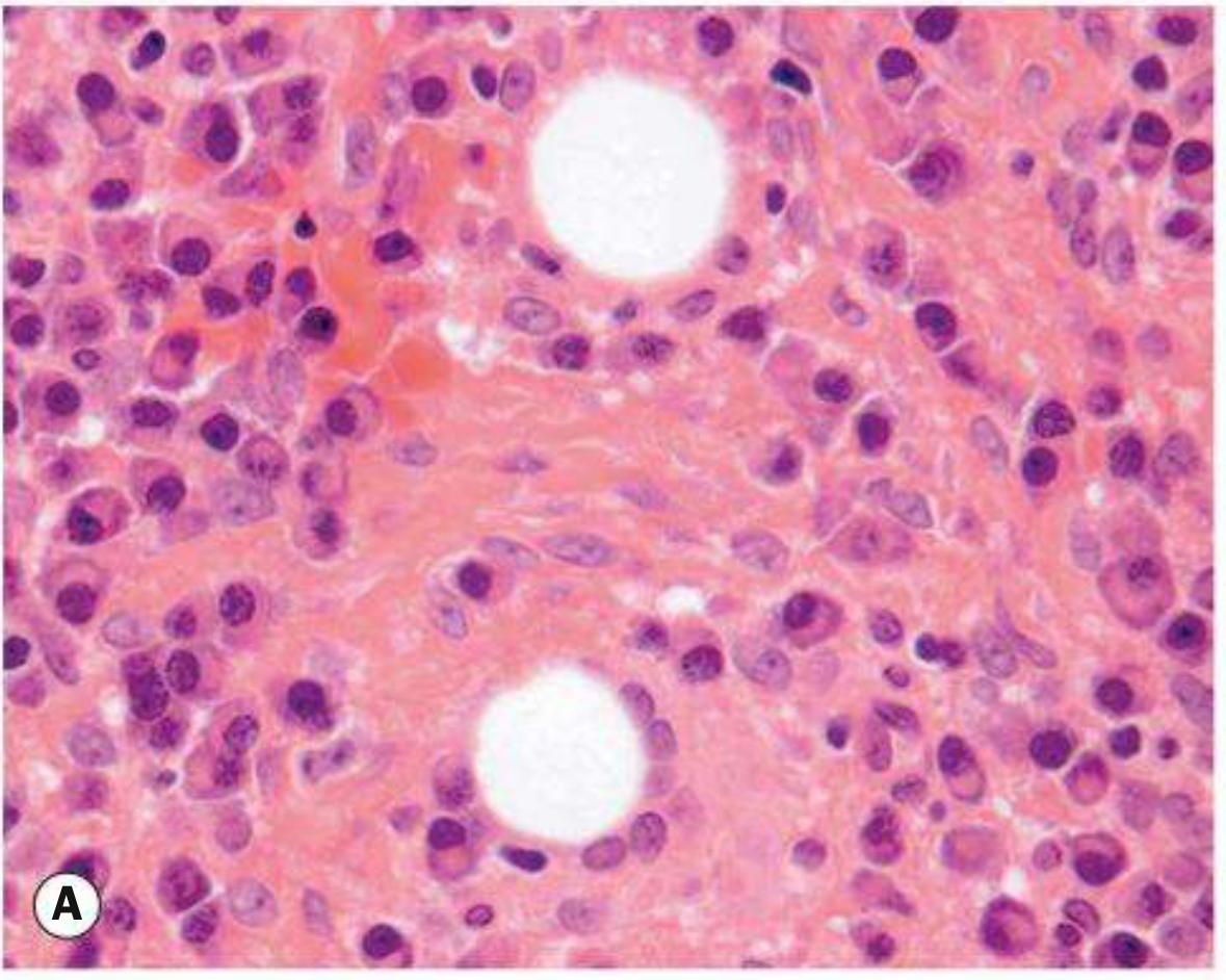
- Surrounded by lipid-laden epithelioid cells and multinucleated giant cells
- Background infiltrate of lymphocytes
Histopathology (Fig. A below): lipogranuloma with large pale epithelioid cells and well-demarcated empty spaces (fat dissolved out during processing)
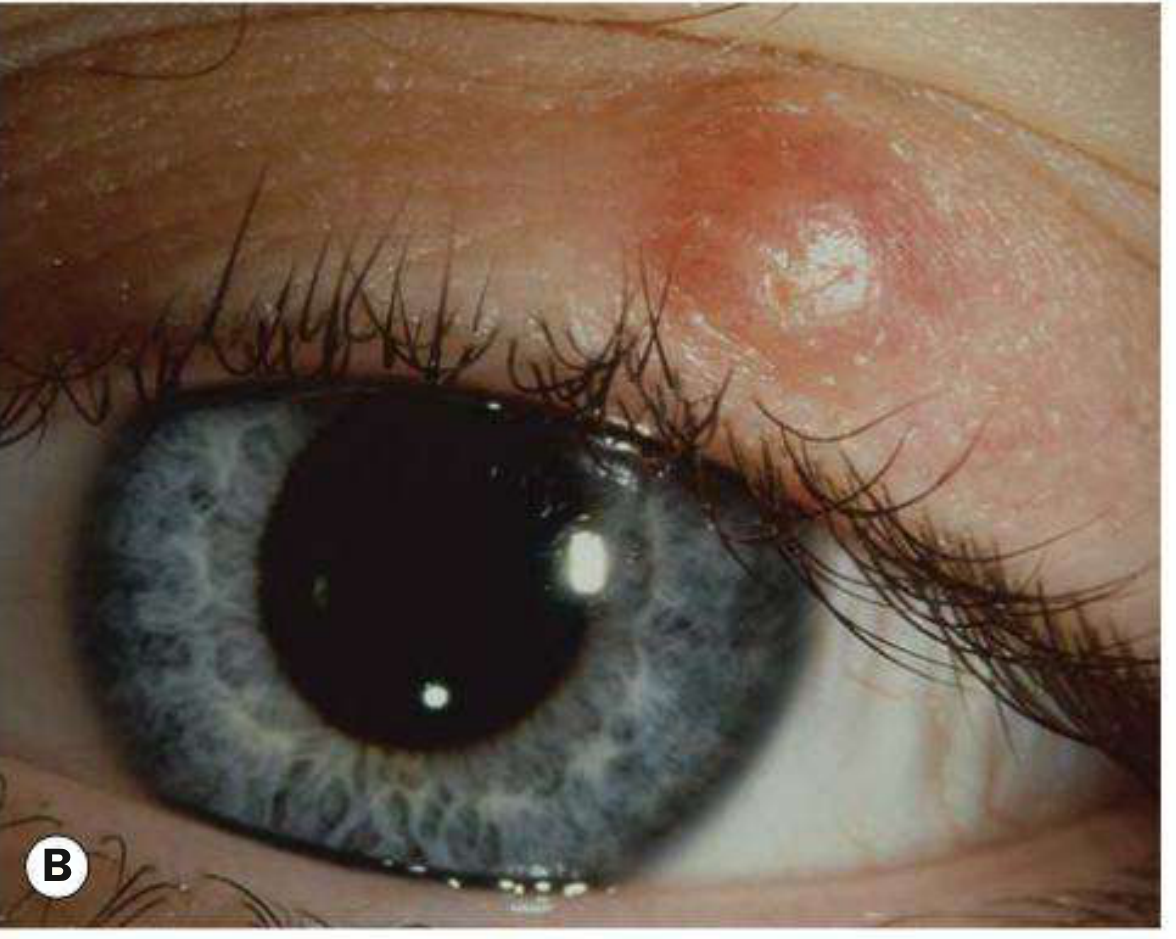
Clinical appearance (Fig. B): painless, firm nodule in the upper or lower eyelid
Associated Conditions
| Condition | Relevance |
|---|---|
| Blepharitis | Commonly co-present |
| Acne rosacea | Associated with multiple/recurrent chalazia |
| Bortezomib (proteasome inhibitor for myeloma) | Predisposes to chalazia within 3 months of initiation |
| Sebaceous carcinoma | Must be excluded in recurrent lesions, especially in older patients |
Symptoms & Signs
Symptoms: Acute or chronic eyelid lump, swelling, tenderness (can be painless or painful)
Critical sign: Visible or palpable, well-defined, subcutaneous nodule in the eyelid
Other signs:
- Blocked meibomian gland orifice
- Eyelid swelling and erythema
- Focal tenderness
- Associated blepharitis or acne rosacea
- Lesion may drain mucopurulent material
Chalazion vs. Hordeolum
| Feature | Chalazion | Hordeolum (Stye) |
|---|---|---|
| Nature | Sterile, chronic granulomatous | Acute bacterial abscess |
| Gland involved | Meibomian (± Zeis) | Meibomian (internal) / Zeis or Moll (external) |
| Pain | Painless or mildly tender | Painful |
| Organism | None (sterile) | Usually Staphylococcus spp. |
| Nodule | Well-defined, firm, non-mobile | Tender, may point at lid margin |
Differential Diagnosis
- Preseptal cellulitis - periorbital erythema, edema, warmth
- Sebaceous carcinoma - recurrent chalazia, eyelid thickening, madarosis, chronic unilateral blepharitis in older patients
- Pyogenic granuloma - benign, red, pedunculated conjunctival lesion
- Forniceal foreign body - especially in contact lens wearers
Key clinical tip: If a chalazion recurs at the same lid position in an older patient, biopsy is mandatory to exclude malignancy (sebaceous carcinoma). - Kanski's Clinical Ophthalmology
Workup
- History: Previous ocular surgery/trauma? Previous chalazia?
- External examination: Palpate eyelid for nodule; look for rosacea
- Slit lamp: Evaluate meibomian glands for inspissation, evert eyelid; assess for madarosis, poliosis, ulceration
Treatment
Step 1 - Conservative (all cases)
- Warm compresses for at least 10 minutes 4x daily with gentle massage over the lesion
- Most lesions resolve over days to weeks
Step 2 - Medical (if needed)
- Topical antibiotic/steroid (e.g., neomycin/polymyxin B/dexamethasone ointment b.i.d. for 1-2 weeks)
- Chronic low-dose doxycycline 20-50 mg p.o. daily to b.i.d. for multiple/recurrent chalazia or ocular rosacea (antibacterial + anti-inflammatory)
Step 3 - Procedural (failure after 3-4 weeks)
Option A: Incision and curettage (I&C)
- Chalazion clamp applied with open side inside the eyelid
- Vertical incision through conjunctival surface
- Curettage of contents
Option B: Intralesional steroid injection
- Triamcinolone 40 mg/mL mixed 1:1 with 2% lidocaine with epinephrine (0.2-1.0 mL total, depending on lesion size)
- Alternatives: betamethasone combinations or dexamethasone sodium phosphate 4 mg/mL
Warning on steroid injection: Can cause permanent depigmentation or skin atrophy at the injection site - especially in dark-skinned individuals. Rarely, vigorous injection can cause retrograde intra-arterial infiltration with central retinal artery occlusion. Use with detailed patient counselling.
All removed specimens - send to pathology
Especially recurrent or atypical chalazia.
Follow-Up
-
Medical therapy: reassess only if lesion persists beyond 3-4 weeks
-
After I&C: reexamine as needed
-
The Wills Eye Manual, pp. 381-383
-
Kanski's Clinical Ophthalmology 10th ed., p. 54
-
Goldman-Cecil Medicine, p. 302
This is a shared conversation. Sign in to Orris to start your own chat.