
Implants in tympanoplasty
Reading File
Tympanoplasty uses grafts and prostheses to reconstruct the tympanic membrane and ossicular chain. The main implant categories are: autografts (temporalis fascia, perichondrium, cartilage), allografts, and synthetic ossicular prostheses (PORPs and TORPs). Let me pull the full details from the medical library.
Searching sources...
Finding Sources
Finding Sources
Finding Sources
Finding Sources
Reading File
Finding Sources
Reading File
Reading File
Reading File
Reading File
Reading File
Searching Images
ossicular prosthesis PORP TORP tympanoplasty
Reading File
Finding Sources
Reading File
Loading Image
I now have comprehensive information from multiple authoritative textbooks. Let me compile the full answer.
Implants in Tympanoplasty
Tympanoplasty uses two broad categories of implants: (1) grafts for tympanic membrane (TM) reconstruction and (2) prostheses for ossicular chain reconstruction (ossiculoplasty). The choice of implant is tailored to the extent of middle ear pathology found at surgery.
Part 1 - Grafts for Tympanic Membrane Reconstruction
Autografts (most common)
| Material | Source | Key Features |
|---|---|---|
| Temporalis fascia | Deep or superficial layer above temporalis muscle | Most widely used; avascular plane = minimal bleeding; easy to handle; excellent graft-take |
| Areolar tissue | Overlying temporalis fascia | Introduced by Moon (1970); excellent take; easier to obtain |
| Perichondrium | Tragus or auricle | Good results; comparable to fascia |
| Cartilage | Tragus, concha cymba, or costal | Preferred for atelectasis, retraction pockets, total perforations, cholesteatoma, and revision cases; more rigid = resists reretraction; "shield" graft technique is standard |
| Fat | Lobule | Used for small perforations (fat-plug myringoplasty) |
| Scar tissue | Surgical scar | Reported with excellent results in select cases |
Temporalis fascia and perichondrium are the go-to materials for straightforward TM reconstruction. Cartilage is increasingly used when standard materials are likely to fail - its rigidity is an advantage in high-risk ears.
"Temporalis fascia and perichondrium are commonly used materials for TM reconstruction. Cartilage may be considered for reinforcement of retraction pockets, atelectatic membranes, and other conditions associated with the increased failure rates of traditional techniques." - Cummings Otolaryngology
Allografts / Xenografts (for revision cases when autografts are unavailable)
- AlloDerm (acellular dermal homograft, LifeCell Corporation) - treated human dermis
- Tutopatch (Tutogen Medical) - xenograft derived from bovine pericardium
Results with these materials are comparable to temporalis fascia, perichondrium, and cartilage. They are a reasonable option in revision cases where autologous tissue is no longer available.
Part 2 - Ossicular Prostheses (Ossiculoplasty)
The goal of ossiculoplasty is to re-establish efficient transfer of vibratory energy from the TM to the stapes footplate, maximizing postoperative hearing gain.
Classification: PORP vs. TORP
| Prosthesis | Full Name | Indication | Lateral End | Medial End |
|---|---|---|---|---|
| PORP | Partial Ossicular Replacement Prosthesis | Stapes superstructure (crura + head) intact | TM / malleus | Stapes head (capitulum) |
| TORP | Total Ossicular Replacement Prosthesis | Stapes superstructure absent (only footplate remains) | TM / malleus | Stapes footplate |
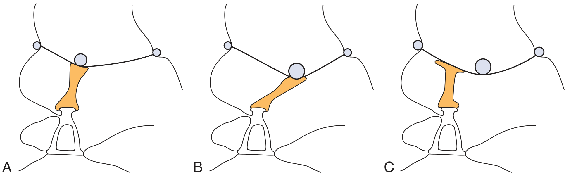
Materials Used for Ossicular Prostheses
1. Autografts
- Incus interposition graft - the patient's own incus is removed, sculpted (an oval acetabulum fashioned in the short process), and repositioned as a bridge. Advantages: immediate availability, biocompatibility, low cost, low extrusion rate. Disadvantages: risk of disease recurrence (cholesteatoma cases), possible fixation to adjacent bone, demineralization/erosion, and time/skill needed to sculpt intraoperatively.
- Autograft cartilage - tragus or auricular/costal cartilage; also used as a "shoe" under TORP base to improve stability on the footplate.
2. Allografts / Alloplastic Materials
Polymers:
- High-density polyethylene sponge - Plastipore / Polycel
- Polytetrafluoroethylene - Teflon
- Silicone rubber
Ceramics:
- Ceravital and Bioglass (glass-ceramics)
- Hydroxyapatite (HA) - calcium phosphate ceramic; achieves true integration with bone without capsule formation; can be porous or dense; disadvantage: brittle, hard to sculpt. Available as the Goldenberg hydroxyapatite prosthesis. Often combined: HA platform + Plastipore shaft (best of both materials)
- Hydroxyapatite bone cement - useful for small defects such as erosion of the lenticular process; hardens within 5 minutes; achieves <10 dB air-bone gap in ~50% of cases
Metals:
- Titanium - now in widespread use. Key properties:
- Extremely low weight (≤4 mg)
- High rigidity - closest in mass to the ossicles it replaces
- Reduces acoustic impedance and sound damping, especially at high frequencies
- Open headplate design: allows surgeon visualization during placement
- Claw-like medial end: secure grip on stapes head
- Not top-heavy (unlike HA) - stays upright
- More user-friendly; shortens learning curve
- Available as Kurz, Spiggle & Theis, Hess-Erga designs (no statistically significant difference between types)
"Allograft prostheses are available in a variety of materials and configurations. Good hearing results, low extrusion rates, and ease of use have led to widespread application of titanium implants." - Cummings Otolaryngology
Hearing Outcomes of Ossiculoplasty
The standard benchmark is air-bone gap (ABG) closure to < 20 dB.
| Prosthesis | ABG < 20 dB | 5-year success |
|---|---|---|
| PORP | 50-85% of cases | ~2/3 |
| TORP | 40-50% of cases | ~1/3 |
| Titanium PORP (Kurz) | ~82% | - |
| Titanium TORP (Kurz) | ~63% | - |
| HA prosthesis (Goldenberg, 233 pts) | 56.8% (ABG < 21 dB) with 5.29% extrusion | - |
TORPs perform less well because the medial strut can be displaced off the footplate center with no bony ledge to secure it. A cartilage "shoe" (4×2 mm oval, hole in center) placed around the TORP base helps stabilize it in the oval window niche.
A meta-analysis of 12 studies (2001-2010) found no statistically significant difference between titanium and non-titanium PORPs/TORPs in hearing outcomes. The severity of middle ear pathology matters more than prosthesis material.
Preventing Extrusion
Extrusion is the main long-term complication of alloplastic prostheses. Key preventive measure: interposing a cartilage disc between the prosthesis head and the TM underside. Studies show this reduces extrusion rates to approximately 4%, with minimal effect on acoustic transmission (confirmed in cadaver studies when cartilage diameter equals prosthesis head diameter).
Staging of Ossiculoplasty
A key decision is whether to do ossiculoplasty at the same sitting as cholesteatoma removal or as a planned second stage:
- Simultaneous reconstruction: reasonable when middle ear anatomy is expected to remain stable
- Staged reconstruction: preferred when mucosa is thickened/infected, bleeding is significant, cholesteatoma recurrence is likely, or Eustachian tube function is poor. A Silastic / Gelfilm / Epidisc sheet is placed at the first stage over the promontory to reduce fibrous adhesions and to establish a stable TM level, making prosthesis length selection easier at the second stage.
Bone Cement (Ionomeric / HA Cement)
For isolated erosion of the lenticular process of the incus, reconstruction with hydroxyapatite bone cement is a simpler alternative:
- Sets within 5 minutes
- ABG < 10 dB in ~50%; ABG < 20 dB in 70-80%
- Mucosa must be stripped and surfaces dried before application, or the cement loosens over time
Summary Table
| Category | Material | Best Use |
|---|---|---|
| TM graft | Temporalis fascia | Standard perforations |
| TM graft | Cartilage (shield/palisade) | Atelectasis, retraction, cholesteatoma, revision |
| TM graft | Perichondrium | Standard; good alternative |
| TM graft | AlloDerm / Tutopatch | Revision when autograft unavailable |
| Ossiculoplasty autograft | Sculpted incus | Incudostapedial gap; cholesteatoma-free |
| PORP - titanium | Kurz, Spiggle & Theis | Stapes superstructure present |
| TORP - titanium | Kurz, Spiggle & Theis | Total stapes superstructure loss |
| PORP/TORP - HA | Goldenberg | Alternative to titanium |
| Bone cement | HA cement | Lenticular process erosion |
| Spacer (1st stage) | Silastic / Gelfilm | Staged ossiculoplasty |
Sources: Cummings Otolaryngology Head and Neck Surgery (Chapter 142); Shambaugh Surgery of the Ear (Chapter 29); Scott-Brown's Otorhinolaryngology Head & Neck Surgery Vol. 2
This is a shared conversation. Sign in to Orris to start your own chat.