
Blood Uric acis test sample should be given on empty stomach
blood uric acid test fasting required empty stomach
Does a Blood Uric Acid Test Require Fasting?
Standard Guidance
- Most labs and healthcare providers say you do not need to be on an empty stomach for a uric acid test
- According to Apollo247 and Metropolis India: "Fasting is typically not required"
- WebMD confirms: "Typically, you don't need to do anything special"
When Some Fasting May Be Advised
- UCSF Health recommends not eating or drinking for 4 hours before the test as a precaution
- If the uric acid test is part of a larger metabolic/lipid panel, fasting may be required for the other tests (e.g., fasting blood sugar, lipid profile)
Things That CAN Affect Results (Regardless of Fasting)
| Factor | Effect |
|---|---|
| High-purine foods (red meat, seafood, organ meats) | Can raise uric acid levels |
| Alcohol consumption | Can raise uric acid levels |
| Certain medications (aspirin, diuretics, vitamin B3) | Can interfere with results |
| Dehydration | Can falsely elevate results |
Bottom Line
- Standalone uric acid test: Fasting is NOT required, but avoiding heavy purine-rich meals and alcohol the night before is good practice
- If part of a panel: Follow your doctor's or lab's specific instructions - fasting may be needed for the other tests in the panel
- Always confirm with your doctor or the specific lab before your test, as protocols can vary
Factors affecting urine output
Factors Affecting Urine Output
Urinary Excretion = Filtration Rate - Reabsorption Rate + Secretion Rate
- Guyton and Hall Textbook of Medical Physiology
1. Glomerular Filtration Rate (GFR)
| Factor | Effect on GFR | Effect on Urine Output |
|---|---|---|
| Blood pressure | Rises with BP (pressure diuresis) | Increases |
| Renal blood flow | Direct relationship | Increases with higher flow |
| Afferent arteriolar dilation | Increases GFR | Increases urine output |
| Efferent arteriolar constriction | Increases GFR | Increases urine output |
| Plasma oncotic pressure | Opposes filtration | High oncotic pressure reduces urine output |
| Bowman's capsule pressure | Opposes filtration | High pressure reduces urine output |
2. Hormonal Factors
a) Antidiuretic Hormone (ADH) / Vasopressin
- Most important hormonal regulator of urine volume
- Secreted by the posterior pituitary in response to raised plasma osmolality or decreased blood volume
- Increases water permeability of collecting ducts via aquaporin-2 channels
- High ADH → concentrated, small volume urine (oliguria)
- Low/absent ADH (e.g., diabetes insipidus) → dilute, large volume urine (polyuria, up to 15-20 L/day)
b) Aldosterone
- Secreted by adrenal cortex in response to angiotensin II, high K+, or low Na+
- Increases Na+ (and water) reabsorption in the collecting duct
- High aldosterone → sodium and water retention → reduced urine output
- Low aldosterone (Addison's disease) → increased urine output, salt wasting
c) Atrial Natriuretic Peptide (ANP)
- Released by atrial stretching (volume overload)
- Promotes sodium and water excretion
- Increases urine output (natriuresis and diuresis)
d) Renin-Angiotensin-Aldosterone System (RAAS)
- Activated by low renal perfusion pressure
- Angiotensin II causes efferent arteriolar constriction and stimulates aldosterone
- Net effect: reduces urine output (sodium and water retention)
3. Tubular Reabsorption
- Glomerulotubular balance - when GFR rises, tubular reabsorption rises proportionally, preventing massive urine losses
- Impaired tubular reabsorption (e.g., in acute tubular necrosis) leads to increased urine output initially, then oliguria
4. Fluid Intake and Hydration Status
- Increased fluid intake → expanded plasma volume → suppressed ADH → increased urine output
- Dehydration → low plasma volume → ADH release → reduced urine output
- Osmotic diuresis: high solute load in filtrate (e.g., glucose in uncontrolled diabetes, mannitol) draws water into tubules → large urine volumes despite volume depletion
5. Blood Pressure and Cardiovascular Status
- Pressure diuresis/natriuresis: rise in arterial blood pressure increases urine output directly
- Heart failure → reduced cardiac output → reduced renal blood flow → low GFR → oliguria
- Shock/hypovolemia → RAAS + ADH activation → oliguria (protective mechanism)
6. Renal Autoregulation
- The kidneys maintain relatively constant GFR over a mean arterial pressure range of 75-160 mmHg via myogenic reflex and tubuloglomerular feedback (TGF via macula densa)
- Outside this range, urine output follows blood pressure changes directly
- Without autoregulation, a 25% rise in BP could increase urine output >30-fold (Guyton and Hall, p. 343)
7. Urinary Tract Obstruction
- Complete obstruction → anuria
- Partial obstruction → can cause normal or even increased urine output (post-obstructive diuresis)
- A fluctuating pattern of oliguria and polyuria strongly suggests obstructive uropathy (Comprehensive Clinical Nephrology, 7th Ed.)
8. Drugs and Medications
| Drug | Effect |
|---|---|
| Diuretics (furosemide, thiazides) | Increase urine output |
| NSAIDs | Reduce GFR and urine output |
| ACE inhibitors/ARBs | May reduce or stabilize urine output |
| Vasopressin analogues (desmopressin) | Reduce urine output |
| Lithium | Causes nephrogenic DI → polyuria |
| Contrast agents | Can cause acute tubular injury → oliguria |
9. Other Factors
- Temperature/sweating: high ambient temperature → fluid loss via sweat → reduced urine output
- Exercise: reduces renal blood flow → decreased urine output
- Diet: high protein diet → increased urea load → osmotic diuresis
- Age: elderly have reduced GFR and concentrating ability
- Fever: increases insensible fluid losses → reduces urine output
- Pregnancy: GFR rises 40-50% → increased urine output tendency
Summary Table
| Factor | Increases Urine Output | Decreases Urine Output |
|---|---|---|
| GFR | High GFR | Low GFR |
| ADH | Low ADH | High ADH |
| Aldosterone | Low aldosterone | High aldosterone |
| ANP | High ANP | Low ANP |
| Blood pressure | Hypertension | Hypotension/shock |
| Fluid intake | High intake | Dehydration |
| Obstruction | Partial/post-obstructive | Complete obstruction |
| Drugs | Diuretics | NSAIDs, vasopressin |
Reasons for renal calculi
renal calculi kidney stones types nephrolithiasis
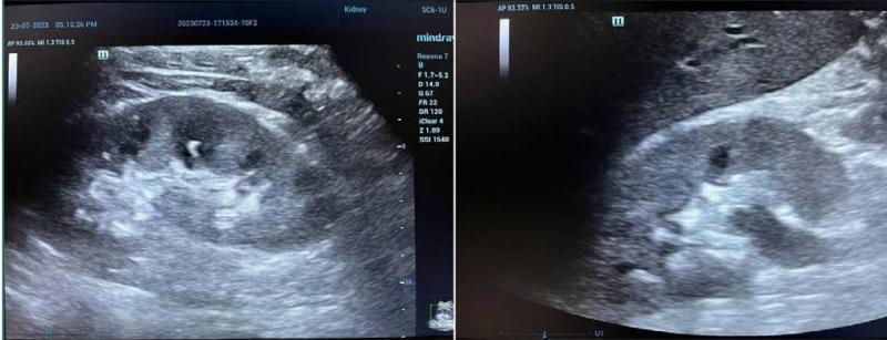
Two grayscale ultrasound images in longitudinal and transverse planes demonstrating renal calculi (nephrolithiasis) within a human kidney. The diagnostic images reveal the renal parenchyma with a clearly defined corticomedullary junction. Inside the renal pelvis/calyceal system, there are distinct hyperechoic (bright white) foci representing renal stones. A key diagnostic feature shown is posterior acoustic shadowing—a dark vertical band extending behind the hyperechoic stones caused by the reflection and absorption of sound waves. The images facilitate the comparison of echogenicity between the relatively hypoechoic renal cortex and the highly echogenic stones. This visual material is used in radiology education to teach the sonographic identification of kidney stones, focusing on morphology, echogenicity, and the presence of acoustic artifacts as definitive diagnostic criteria.
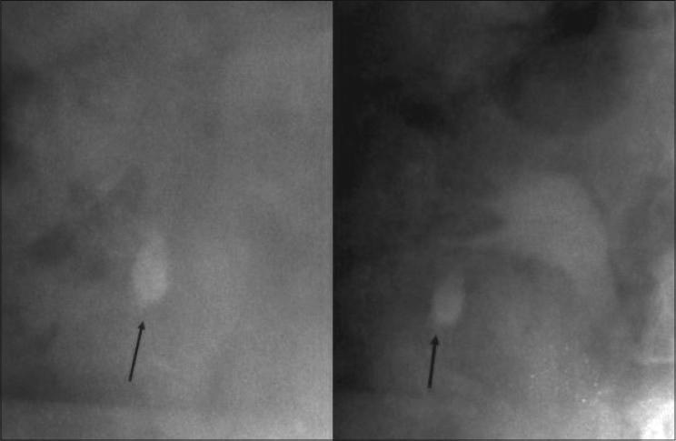
This diagnostic image displays two radiographic views (KUB or fluoroscopy) of the renal region, specifically illustrating nephrolithiasis. In both panels, a prominent, radiopaque, oval-shaped calculus is identified by a black arrow. The stone is situated in the lower pole of the kidney, likely within an inferior calyx. The surrounding renal architecture exhibits varying shades of gray density, with faint outlines of the collecting system and renal parenchyma visible against the darker background of the abdominal cavity. The image serves as a clinical example of complex lower-pole renal calculi, highlighting the challenges of surgical access for percutaneous nephrolithotomy (PCNL) due to the acute angles within the calyces. This material is designed for urological education, focusing on the localization and radiographic identification of renal stones prior to interventional procedures.
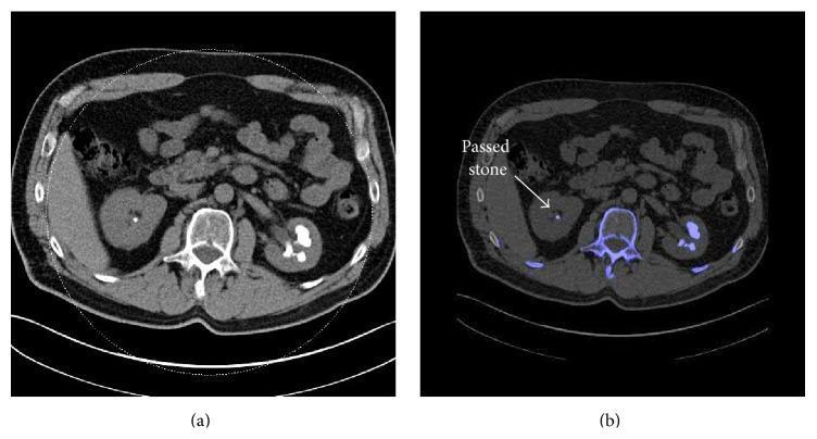
This figure presents a side-by-side comparison of axial abdominal imaging for nephrolithiasis. Image (a) is a conventional non-contrast Computed Tomography (CT) scan showing bilateral renal calculi. The left kidney contains a large, hyperdense staghorn calculus filling the renal pelvis and calyces, while the right kidney shows a smaller, discrete hyperdense stone. Image (b) shows a Dual-Energy CT (DECT) scan with post-processing for stone characterization. In the DECT image, the calculi are color-coded in blue, indicating they are non-uric acid (non-UA) stones (e.g., calcium oxalate, calcium phosphate, or cystine). A white arrow in image (b) highlights a small calculus in the right kidney labeled as a 'passed stone.' The DECT algorithm also applies blue color-coding to other high-density non-uric acid structures, such as the vertebral body. This comparison demonstrates the utility of DECT in distinguishing stone composition based on dual-energy ratios and three-material decomposition, which is critical for determining appropriate clinical management (medical dissolution versus urological intervention).
Reasons for Renal Calculi (Nephrolithiasis / Kidney Stones)
"In all cases, an organic matrix of mucoprotein is present that makes up about 2.5% of the stone by weight."
- Robbins & Kumar Basic Pathology
Types of Renal Calculi and Their Specific Causes
| Stone Type | Frequency | Key Causes |
|---|---|---|
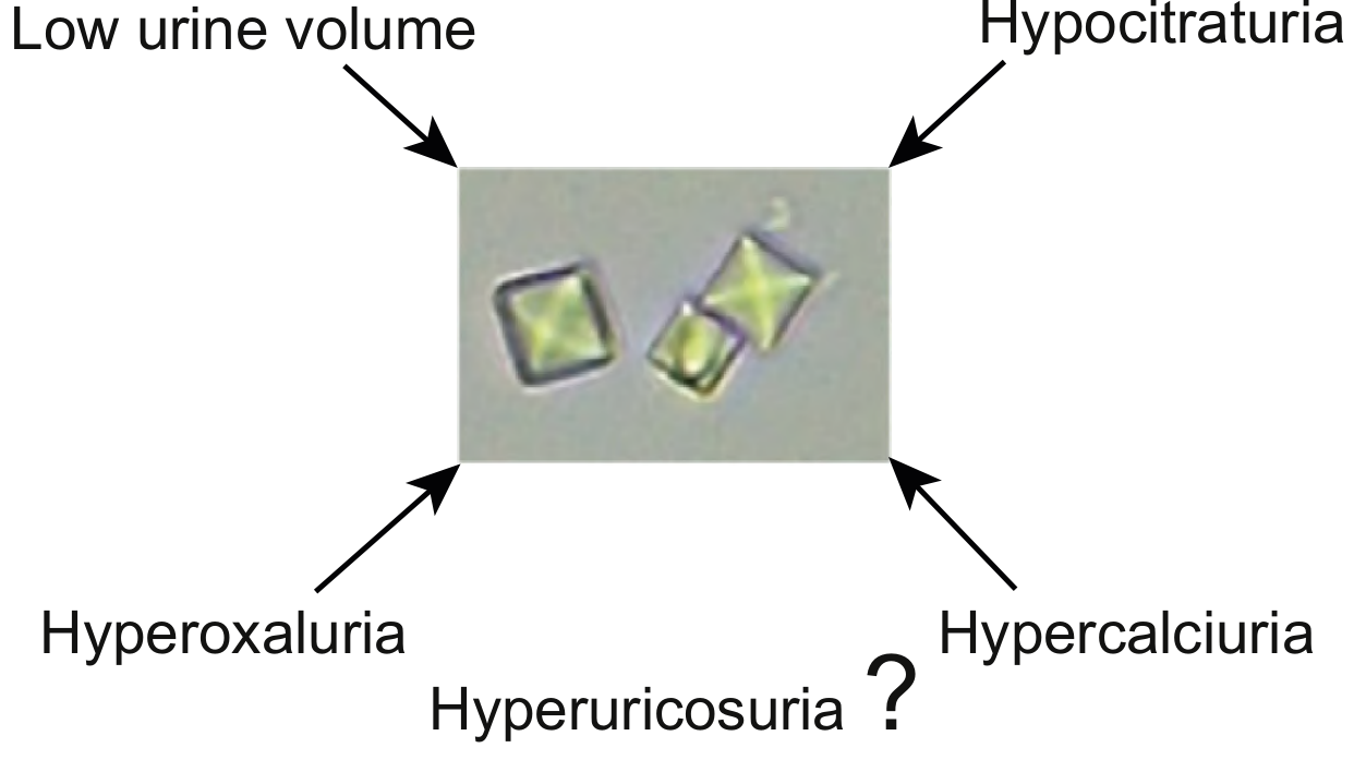
| Calcium oxalate / phosphate | 80% | Hypercalciuria, hyperoxaluria, hypocitraturia, hyperuricosuria |
| Struvite (Mg-NH₃-PO₄) | 10% | Urinary tract infections (urea-splitting bacteria) |
| Uric acid | 6-7% | Hyperuricemia, gout, acidic urine |
| Cystine | 1-2% | Hereditary cystinuria |
1. Urinary Risk Factors (Metabolic Causes)
a) Hypercalciuria (High Urinary Calcium) - Most Common
- Present in 60% of adults with calcium nephrolithiasis
- Three mechanisms (Brenner and Rector's The Kidney):
- Absorptive hypercalciuria - excessive intestinal calcium absorption (most common)
- Renal leak hypercalciuria - defective renal tubular calcium reabsorption
- Resorptive hypercalciuria - excessive bone calcium mobilization (e.g., hyperparathyroidism)
- Also caused by: primary hyperparathyroidism, sarcoidosis, vitamin D overdose, excess calcium supplements
b) Hyperoxaluria (High Urinary Oxalate)
- Sources of excess oxalate:
- Dietary - high intake of spinach, nuts, chocolate, tea
- Enteric (secondary) hyperoxaluria - gut malabsorption (Crohn's disease, short bowel syndrome) leads to excess oxalate absorption
- Primary hyperoxaluria - rare hereditary liver enzyme defect (increased endogenous oxalate production)
c) Hypocitraturia (Low Urinary Citrate) - Key Protective Deficit
- Citrate normally inhibits calcium stone formation by binding calcium in urine
- Low citrate occurs in: renal tubular acidosis, chronic diarrhoea, high-protein diet, hypokalemia
- Deficiency removes an important protective mechanism against crystallization
d) Hyperuricosuria (High Urinary Uric Acid)
- Promotes calcium oxalate stone formation by salting-out calcium oxalate
- Caused by: high purine diet (red meat, seafood, organ meats), gout, rapid cell turnover (cancer therapy, leukaemia)
e) Low Urine Volume
- Found in ~60% of brushite stone formers
- Concentrated urine raises supersaturation of all stone-forming salts
- Key contributor in people living in hot climates or with low fluid intake
f) Abnormal Urinary pH
- Alkaline urine (high pH): promotes calcium phosphate stone formation
- Acidic urine (low pH): promotes uric acid stone formation (uric acid is insoluble in acidic urine)
2. Causes by Specific Stone Type
Calcium Stones (80%)
- Idiopathic hypercalciuria (50% of calcium stones)
- Hypercalcemia from any cause: hyperparathyroidism, sarcoidosis, malignancy, vitamin D toxicity
- Hyperoxaluria
- Hyperuricosuria (promotes salting out of calcium oxalate)
- Hypocitraturia
- No identifiable metabolic cause in 15-20% of cases
Struvite / Infection Stones (10%)
- Almost always due to UTI with urea-splitting organisms:
- Proteus mirabilis (most common)
- Klebsiella, Staphylococcus, Pseudomonas
- These bacteria produce urease, which hydrolyses urea → ammonia + CO₂ → alkaline urine → precipitation of magnesium ammonium phosphate
- Forms staghorn calculi (branching stones filling the renal pelvis)
- (Robbins & Kumar Basic Pathology)
Uric Acid Stones (6-7%)
- Gout (hyperuricemia)
- Tumour lysis syndrome - rapid breakdown of cancer cells (leukaemia, lymphoma, chemotherapy)
- Persistently acidic urine (e.g., diabetes mellitus, metabolic syndrome)
- High-purine diet (50% of uric acid stones have no identifiable cause)
Cystine Stones (1-2%)
- Exclusively seen in cystinuria - a hereditary (autosomal recessive) disorder
- Caused by inactivating mutations in subunits of a dibasic amino acid transporter (SLC3A1 / SLC7A9 genes)
- Leads to failure of tubular reabsorption of cystine, ornithine, lysine, and arginine
- Cystine precipitates in urine → recurrent stones from childhood
3. General Predisposing Factors
| Factor | Mechanism |
|---|---|
| Low fluid intake / dehydration | Concentrated urine, supersaturation of solutes |
| Diet high in purines, oxalate, sodium | Increased lithogenic solute excretion |
| Obesity | Increased urinary calcium, uric acid, and oxalate; insulin resistance lowers urine pH |
| Diabetes mellitus | Acidic urine promotes uric acid stones |
| Family history / genetics | Inherited metabolic defects (cystinuria, primary hyperoxaluria) |
| Recurrent UTI | Struvite stone formation |
| Anatomical abnormalities | Urinary stasis (horseshoe kidney, medullary sponge kidney, UPJ obstruction) promotes crystal accumulation |
| Vitamin A deficiency | Desquamated metaplastic cells act as nidi for stone formation |
| Bowel disease (Crohn's, short bowel) | Enteric hyperoxaluria |
| Immobilization | Bone resorption → hypercalciuria |
| Hot climate / occupation | Increased fluid losses → concentrated urine |
| Drugs | Triamterene, indinavir, topiramate, acetazolamide |
4. Reduced Inhibitors of Stone Formation
- Citrate - binds calcium, prevents crystallization
- Magnesium - forms soluble complexes with oxalate
- Pyrophosphate - inhibits calcium phosphate crystallization
- Nephrocalcin and Tamm-Horsfall protein - urinary proteins that inhibit crystal aggregation
- Trefoil factor 1 (TFF1) - reduced in idiopathic calcium oxalate stone formers (Brenner and Rector's The Kidney)
Imaging: Renal Calculi on CT
Summary: Key Mnemonic
- Hypercalciuria
- Hyperoxaluria
- Hyperuricosuria
- Hypocitraturia
- Low urine volume
- Dysfunctional pH (too acidic or too alkaline)