
Scalp anatomy in detailed explanation and in simple way
scalp anatomy layers SCALP mnemonic detailed explanation
Scalp Anatomy - Detailed & Simple Explanation
What Is the Scalp?
The "SCALP" Mnemonic - 5 Layers
| Letter | Layer | Simple Meaning |
|---|---|---|
| S | Skin | The outermost layer with hair |
| C | Connective tissue (subcutaneous/superficial fascia) | Fatty, fibrous layer under the skin |
| A | Aponeurosis (galea aponeurotica) | Tough sheet of fibrous tissue |
| L | Loose areolar connective tissue | A "slippery" spacer layer |
| P | Pericranium (periosteum) | Membrane covering the skull bone |
Visual Reference
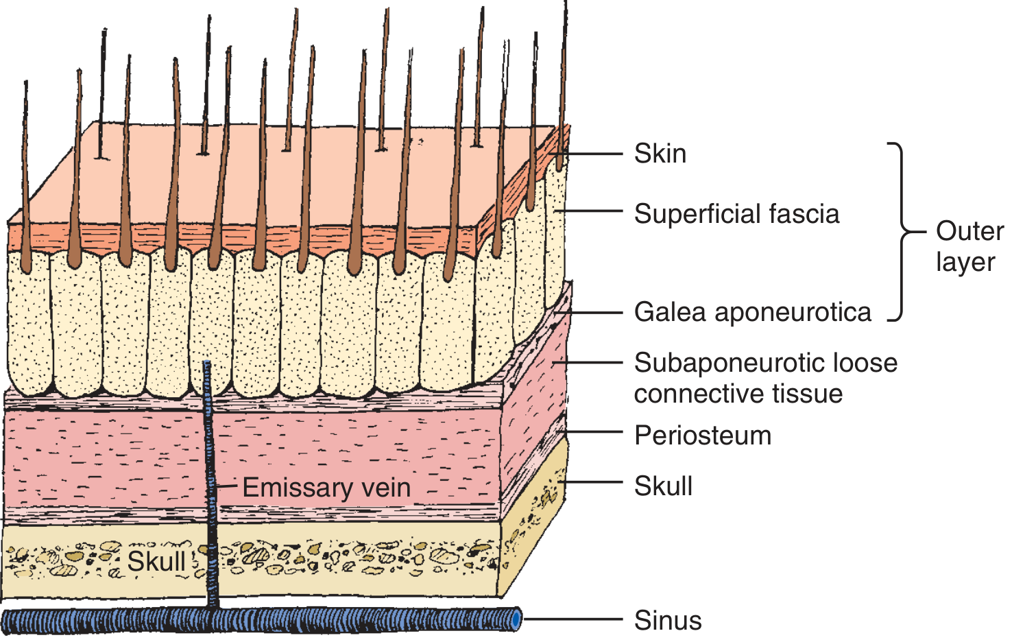
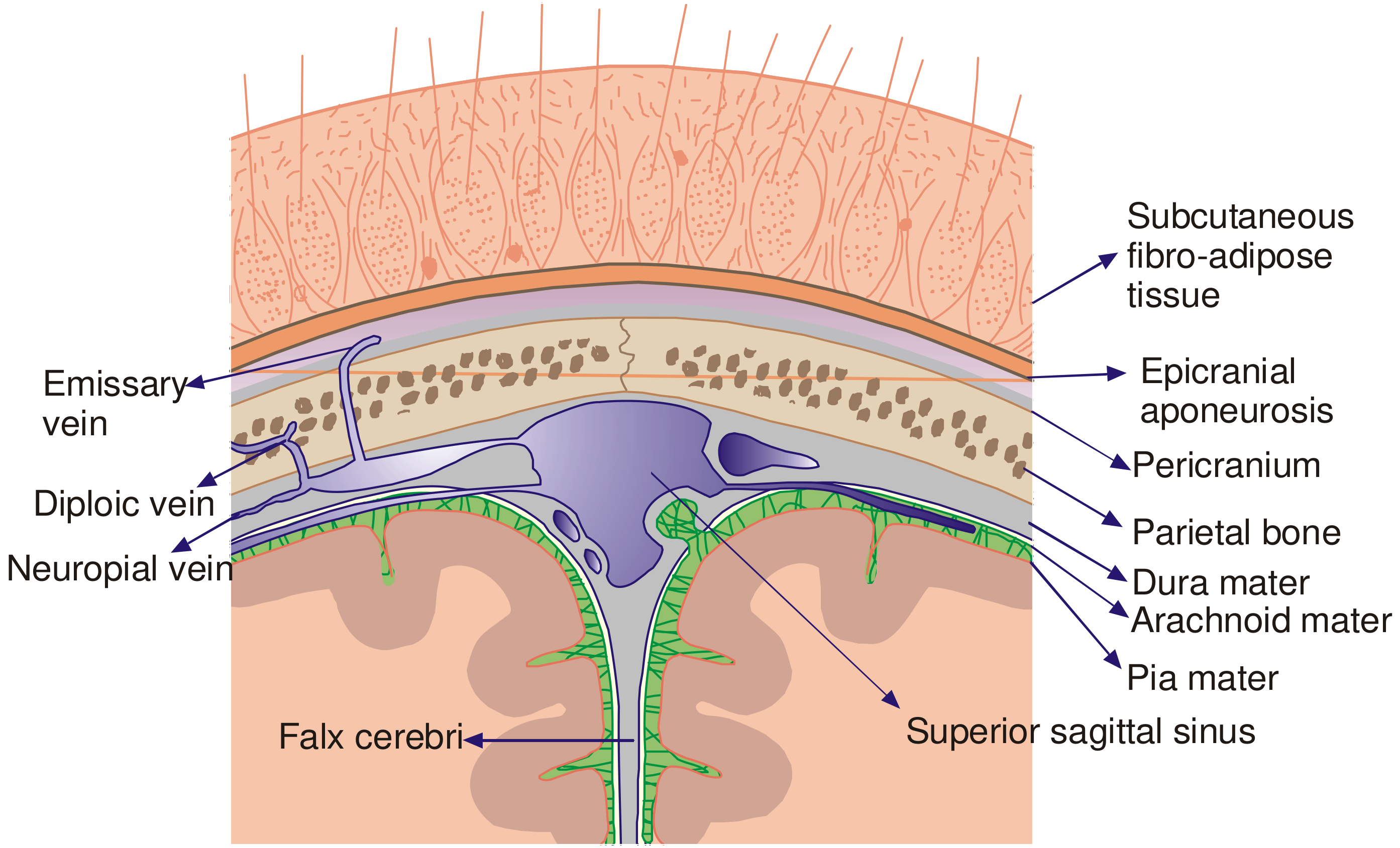
Layer-by-Layer Breakdown
Layer 1 - S: Skin
- The thickest skin in the entire body.
- It is densely hairy and contains numerous hair follicles, sebaceous glands, and sweat glands.
- Because it is thick, it resists tearing but bleeds heavily when cut.
- The skin is tightly bound to the layer below by fibrous septa that run vertically - these septa anchor the skin to the connective tissue and prevent it from sliding easily.
Layer 2 - C: Connective Tissue (Superficial Fascia / Subcutaneous Layer)
- A dense, fibro-fatty layer containing abundant blood vessels, lymphatics, and nerves.
- The blood vessels here are the main source of profuse bleeding from scalp wounds.
- The vessels are held open by the surrounding fibrous septa - this means cut scalp vessels cannot retract or close naturally, so scalp wounds bleed heavily and persistently.
- Contains the terminal branches of the major arteries supplying the scalp.
| Artery | Source | Region Supplied |
|---|---|---|
| Supratrochlear & Supraorbital | Ophthalmic artery (from internal carotid) | Frontal scalp |
| Superficial temporal | External carotid | Temporal (side) scalp |
| Posterior auricular | External carotid | Behind the ear |
| Occipital | External carotid | Back of scalp |
All arteries anastomose (connect) freely with each other, so scalp blood supply is exceptionally rich - this allows scalp wounds to bleed a lot, but also means wounds heal well.
Layer 3 - A: Aponeurosis (Galea Aponeurotica)
- A tough, flat, fibrous sheet (tendon-like sheet) that connects:
- The frontalis muscle (forehead muscle) in front
- The occipitalis muscle at the back of the skull
- Together these muscles + galea form the occipitofrontalis muscle - it is what allows you to raise your eyebrows.
- The skin and superficial fascia are firmly glued to the galea, so these three (S + C + A) act as one unit surgically - they move together and are considered the "outer layer."
- Laterally, the galea blends with the temporal fascia over the temples.
Layer 4 - L: Loose Areolar Connective Tissue (Subaponeurotic Layer)
- A thin, loose, spongy layer between the galea and the pericranium.
- Often called the "danger space" of the scalp - here is why:
- It allows the outer three layers (S + C + A) to slide freely over the skull - this is why the scalp can move when you wiggle your eyebrows.
- It contains emissary veins - valveless veins that connect the scalp veins to the diploic veins inside the skull bone, and further into the intracranial venous sinuses (e.g., superior sagittal sinus).
- Because of these connections: an infection in this layer can spread directly to the meninges, causing meningitis, osteomyelitis of the skull, or even a brain abscess.
- Blood from injury collects here and can spread widely under the galea ("subgaleal hematoma").
Layer 5 - P: Pericranium (Periosteum of the Skull)
- The innermost layer of the scalp - a thin membrane that covers the outer surface of the skull bones.
- Firmly attached to the skull, especially at the suture lines (joints between skull bones).
- It is biologically active - contains osteoblasts and osteoclasts that can repair skull bone.
- It is not firmly attached to the galea, only loosely connected through the areolar tissue above.
- This layer is too thin and delicate to suture reliably - surgeons do not close it in scalp repairs.
Clinically Important Summary: The "3-Layer" Clinical Division
| Clinical Layer | Anatomical Content | Key Feature |
|---|---|---|
| Outer layer | Skin + Subcutaneous tissue + Galea | Firmly adherent; moves as one unit |
| Subaponeurotic layer | Loose areolar tissue | "Danger space" - infection/blood spreads here |
| Pericranium | Periosteum | Attached to skull; cannot be sutured |
Nerves of the Scalp
| Nerve | Origin | Area |
|---|---|---|
| Supratrochlear & Supraorbital | CN V1 (ophthalmic) | Forehead, front scalp |
| Zygomaticotemporal | CN V2 (maxillary) | Temple |
| Auriculotemporal | CN V3 (mandibular) | Temporal/side |
| Lesser & Greater Occipital | C2, C3 cervical | Back of scalp |
| Great Auricular | C2, C3 | Behind the ear |
Why Is the Scalp Clinically Important?
- Massive bleeding: Cut scalp vessels don't retract (held open by fibrous septa) - even small wounds can cause significant blood loss.
- Infection risk: The loose areolar layer (Layer 4) is a direct highway for infection to spread to the skull and brain via emissary veins.
- Scalp avulsions: The loose areolar layer allows the entire outer scalp to be torn off from the skull (degloving injury) - the person can literally be lifted or dragged by the hair and the scalp may tear free.
- Subgaleal hematoma: Blood collects under the galea and can spread across the entire scalp freely, causing a large fluctuant swelling.
- Cephalohematoma vs. subgaleal hematoma in newborns: Cephalohematoma is limited to one bone (periosteum is attached at sutures) - subgaleal hematoma crosses suture lines because it is above the periosteum.
One-Sentence Summary
The scalp has 5 layers (remembered as S-C-A-L-P): Skin (thick, hairy) → Connective tissue (very vascular, bleeds profusely) → Aponeurosis/galea (tough fibrous sheet) → Loose areolar tissue (the "danger space" with emissary veins) → Pericranium (thin membrane on the skull). The outer 3 layers move as one unit; the 4th layer is where infections and blood spread easily to the brain.
Create a visual atlas PDF of scalp anatomy with labeled diagrams
mkdir -p /home/daytona/workspace/scalp-atlas && ls /home/daytona/workspace/scalp-atlas
cd /home/daytona/workspace/scalp-atlas && curl -s -o img_layers.png "https://cdn.orris.care/cdss_images/c0751ecc7bdbf89f87e3b7b1082d5ea2c3517f043050f8d4de09aca95c75494b.png" && curl -s -o img_coronal.png "https://cdn.orris.care/cdss_images/78e61d792696e3900c0b01943b8a41a2ebddc6f56c22aa4e6e7f0011b5e2e87a.png" && ls -lh *.png
~/scalp-atlas/build_atlas.py
"""
Scalp Anatomy Visual Atlas - PDF Generator
Uses reportlab to build a multi-page, richly annotated PDF atlas.
"""

from reportlab.lib.pagesizes import A4
from reportlab.lib.units import cm, mm
from reportlab.lib import colors
from reportlab.lib.styles import getSampleStyleSheet, ParagraphStyle
from reportlab.platypus import (
    SimpleDocTemplate, Paragraph, Spacer, Image, Table, TableStyle,
    PageBreak, HRFlowable, KeepTogether
)
from reportlab.platypus.flowables import Flowable
from reportlab.lib.enums import TA_CENTER, TA_LEFT, TA_JUSTIFY
from reportlab.pdfgen import canvas
from reportlab.lib.colors import HexColor
import os

# ── Paths ─────────────────────────────────────────────────────────────────────
BASE = "/home/daytona/workspace/scalp-atlas"
IMG_LAYERS  = os.path.join(BASE, "img_layers.png")
IMG_CORONAL = os.path.join(BASE, "img_coronal.png")
OUT_PDF     = os.path.join(BASE, "Scalp_Anatomy_Atlas.pdf")

# ── Color palette ─────────────────────────────────────────────────────────────
C_NAVY    = HexColor("#1a2744")
C_TEAL    = HexColor("#0d6e7a")
C_GOLD    = HexColor("#c9860a")
C_LBLUE   = HexColor("#e8f4f8")
C_LGOLD   = HexColor("#fdf3e1")
C_LGREEN  = HexColor("#e8f5e9")
C_LRED    = HexColor("#fce4e4")
C_GREY    = HexColor("#f5f5f5")
C_DGREY   = HexColor("#444444")
C_WHITE   = colors.white
C_BLACK   = colors.black

W, H = A4  # 595.27 x 841.89 pts

# ── Page numbering callback ───────────────────────────────────────────────────
page_num = [0]

def on_page(canvas_obj, doc):
    page_num[0] += 1
    canvas_obj.saveState()
    # Footer bar
    canvas_obj.setFillColor(C_NAVY)
    canvas_obj.rect(0, 0, W, 1.1*cm, fill=1, stroke=0)
    canvas_obj.setFillColor(C_WHITE)
    canvas_obj.setFont("Helvetica", 8)
    canvas_obj.drawCentredString(W/2, 0.38*cm, f"Scalp Anatomy Visual Atlas  •  Page {page_num[0]}")
    canvas_obj.drawString(1.5*cm, 0.38*cm, "© Educational Reference Only")
    canvas_obj.drawRightString(W - 1.5*cm, 0.38*cm, "Sources: Roberts & Hedges | Dikshit FMT | Dermatology 5e")
    # Header bar (skip page 1 – it has its own cover)
    if page_num[0] > 1:
        canvas_obj.setFillColor(C_NAVY)
        canvas_obj.rect(0, H - 1.1*cm, W, 1.1*cm, fill=1, stroke=0)
        canvas_obj.setFillColor(C_WHITE)
        canvas_obj.setFont("Helvetica-Bold", 9)
        canvas_obj.drawString(1.5*cm, H - 0.72*cm, "SCALP ANATOMY VISUAL ATLAS")
        canvas_obj.setFont("Helvetica", 9)
        canvas_obj.drawRightString(W - 1.5*cm, H - 0.72*cm, "Anatomy  |  Clinical Correlates  |  Mnemonics")
    canvas_obj.restoreState()


# ── Style helpers ─────────────────────────────────────────────────────────────
styles = getSampleStyleSheet()

def S(name, **kw):
    base = styles[name]
    return ParagraphStyle(name + "_custom", parent=base, **kw)

TITLE_STYLE   = S("Title", fontSize=28, textColor=C_WHITE, alignment=TA_CENTER,
                  spaceAfter=6, fontName="Helvetica-Bold")
SUBTITLE_STYLE= S("Normal", fontSize=13, textColor=HexColor("#afd8e8"),
                  alignment=TA_CENTER, fontName="Helvetica-Oblique")
H1            = S("Heading1", fontSize=16, textColor=C_NAVY, fontName="Helvetica-Bold",
                  spaceBefore=10, spaceAfter=4)
H2            = S("Heading2", fontSize=13, textColor=C_TEAL, fontName="Helvetica-Bold",
                  spaceBefore=8, spaceAfter=3)
H3            = S("Heading3", fontSize=11, textColor=C_GOLD, fontName="Helvetica-Bold",
                  spaceBefore=6, spaceAfter=2)
BODY          = S("Normal", fontSize=9.5, textColor=C_DGREY, leading=14,
                  alignment=TA_JUSTIFY)
CAPTION       = S("Normal", fontSize=8, textColor=C_TEAL, alignment=TA_CENTER,
                  fontName="Helvetica-Oblique", spaceBefore=3, spaceAfter=6)
SMALL         = S("Normal", fontSize=8, textColor=C_DGREY, leading=12)
MNEM          = S("Normal", fontSize=12, textColor=C_NAVY, fontName="Helvetica-Bold",
                  alignment=TA_CENTER)
DANGER        = S("Normal", fontSize=9.5, textColor=HexColor("#8b0000"),
                  fontName="Helvetica-Bold", leading=14)
BULLET        = S("Normal", fontSize=9.5, textColor=C_DGREY, leading=14,
                  leftIndent=14, bulletIndent=4)


# ── Custom Flowables ─────────────────────────────────────────────────────────

class ColorBox(Flowable):
    """A colored rounded rectangle header band."""
    def __init__(self, text, bg=C_NAVY, fg=C_WHITE, height=1.1*cm, font_size=14):
        Flowable.__init__(self)
        self.text = text
        self.bg = bg
        self.fg = fg
        self.height = height
        self.font_size = font_size
        self.width = W - 3*cm   # set in wrap

    def wrap(self, availWidth, availHeight):
        self.width = availWidth
        return (availWidth, self.height)

    def draw(self):
        c = self.canv
        c.setFillColor(self.bg)
        c.roundRect(0, 0, self.width, self.height, 6, fill=1, stroke=0)
        c.setFillColor(self.fg)
        c.setFont("Helvetica-Bold", self.font_size)
        c.drawCentredString(self.width/2, self.height/2 - self.font_size/3, self.text)


class HRule(Flowable):
    def __init__(self, color=C_TEAL, thickness=1.5):
        Flowable.__init__(self)
        self.color = color
        self.thickness = thickness

    def wrap(self, aw, ah):
        self.width = aw
        return (aw, self.thickness + 2)

    def draw(self):
        self.canv.setStrokeColor(self.color)
        self.canv.setLineWidth(self.thickness)
        self.canv.line(0, 1, self.width, 1)


# ── Table helpers ────────────────────────────────────────────────────────────

def styled_table(data, col_widths, header_bg=C_NAVY, alt_bg=C_GREY):
    ts = TableStyle([
        # Header row
        ("BACKGROUND",  (0,0), (-1,0),  header_bg),
        ("TEXTCOLOR",   (0,0), (-1,0),  C_WHITE),
        ("FONTNAME",    (0,0), (-1,0),  "Helvetica-Bold"),
        ("FONTSIZE",    (0,0), (-1,0),  9),
        ("ALIGN",       (0,0), (-1,0),  "CENTER"),
        ("BOTTOMPADDING",(0,0),(-1,0),  6),
        ("TOPPADDING",  (0,0), (-1,0),  6),
        # Body
        ("FONTNAME",    (0,1), (-1,-1), "Helvetica"),
        ("FONTSIZE",    (0,1), (-1,-1), 8.5),
        ("TOPPADDING",  (0,1), (-1,-1), 5),
        ("BOTTOMPADDING",(0,1),(-1,-1), 5),
        ("LEFTPADDING", (0,0), (-1,-1), 8),
        ("RIGHTPADDING",(0,0), (-1,-1), 8),
        ("GRID",        (0,0), (-1,-1), 0.4, HexColor("#cccccc")),
        ("ROWBACKGROUNDS",(0,1),(-1,-1),[C_WHITE, alt_bg]),
        ("VALIGN",      (0,0), (-1,-1), "MIDDLE"),
    ])
    t = Table(data, colWidths=col_widths, repeatRows=1)
    t.setStyle(ts)
    return t


def highlight_box(text_para_list, bg=C_LBLUE, border=C_TEAL):
    """Wrap content in a colored bordered box via a 1-cell Table."""
    ts = TableStyle([
        ("BACKGROUND",   (0,0), (-1,-1), bg),
        ("LINEAFTER",    (0,0), (0,-1),  3, border),
        ("LINEBEFORE",   (0,0), (0,-1),  3, border),
        ("TOPPADDING",   (0,0), (-1,-1), 8),
        ("BOTTOMPADDING",(0,0), (-1,-1), 8),
        ("LEFTPADDING",  (0,0), (-1,-1), 12),
        ("RIGHTPADDING", (0,0), (-1,-1), 12),
        ("BOX",          (0,0), (-1,-1), 1, border),
    ])
    t = Table([[text_para_list]], colWidths=[W - 3*cm])
    t.setStyle(ts)
    return t


# ── Build story ───────────────────────────────────────────────────────────────

story = []

# ═══════════════════════════════════════════════════════════════════
# PAGE 1 – COVER
# ═══════════════════════════════════════════════════════════════════

class CoverPage(Flowable):
    def wrap(self, aw, ah):
        return (aw, ah)

    def draw(self):
        c = self.canv
        # Full navy background
        c.setFillColor(C_NAVY)
        c.rect(0, 0, W, H, fill=1, stroke=0)

        # Decorative teal band top
        c.setFillColor(C_TEAL)
        c.rect(0, H - 2.5*cm, W, 2.5*cm, fill=1, stroke=0)

        # Decorative gold accent line
        c.setFillColor(C_GOLD)
        c.rect(0, H - 2.7*cm, W, 0.2*cm, fill=1, stroke=0)

        # Gold accent line bottom
        c.setFillColor(C_GOLD)
        c.rect(0, 2.2*cm, W, 0.2*cm, fill=1, stroke=0)

        # Title text
        c.setFillColor(C_WHITE)
        c.setFont("Helvetica-Bold", 32)
        c.drawCentredString(W/2, H - 6*cm, "SCALP ANATOMY")
        c.setFont("Helvetica-Bold", 22)
        c.setFillColor(HexColor("#afd8e8"))
        c.drawCentredString(W/2, H - 7.2*cm, "Visual Atlas")

        # Subtitle
        c.setFillColor(C_GOLD)
        c.setFont("Helvetica-Oblique", 13)
        c.drawCentredString(W/2, H - 8.4*cm,
            "Layers • Vasculature • Nerves • Clinical Correlates")

        # Divider
        c.setStrokeColor(C_TEAL)
        c.setLineWidth(1.5)
        c.line(3*cm, H - 9*cm, W - 3*cm, H - 9*cm)

        # SCALP mnemonic large display
        letters = [("S","Skin"),("C","Connective Tissue"),
                   ("A","Aponeurosis (Galea)"),("L","Loose Areolar Tissue"),("P","Pericranium")]
        y = H - 10.2*cm
        for letter, meaning in letters:
            c.setFillColor(C_GOLD)
            c.setFont("Helvetica-Bold", 22)
            c.drawString(5.5*cm, y, letter)
            c.setFillColor(C_WHITE)
            c.setFont("Helvetica", 13)
            c.drawString(7*cm, y + 0.1*cm, meaning)
            y -= 1.1*cm

        # Bottom info bar
        c.setFillColor(HexColor("#0d2a3a"))
        c.rect(0, 0, W, 2.4*cm, fill=1, stroke=0)
        c.setFillColor(HexColor("#afd8e8"))
        c.setFont("Helvetica", 9)
        c.drawCentredString(W/2, 1.4*cm, "Based on: Roberts & Hedges' Clinical Procedures in Emergency Medicine")
        c.drawCentredString(W/2, 0.9*cm, "P C Dikshit Textbook of Forensic Medicine & Toxicology  |  Dermatology 2-Volume Set 5e")

story.append(CoverPage())
story.append(PageBreak())

# ═══════════════════════════════════════════════════════════════════
# PAGE 2 – OVERVIEW & BOUNDARIES
# ═══════════════════════════════════════════════════════════════════

story += [
    ColorBox("1.  SCALP — OVERVIEW & BOUNDARIES", bg=C_NAVY),
    Spacer(1, 0.4*cm),
    Paragraph(
        "The <b>scalp</b> is the multilayered soft-tissue covering of the skull vault. "
        "It is one of the most vascular structures in the body, with a rich arterial supply "
        "and extensive venous drainage that has direct intracranial connections.",
        BODY),
    Spacer(1, 0.3*cm),
    H2("Anatomical Boundaries"),
]

bounds_data = [
    ["Direction", "Boundary Landmark"],
    ["Anteriorly",  "Supraorbital ridges (brow ridges)"],
    ["Posteriorly", "External occipital protuberance (inion)"],
    ["Laterally",   "Zygomatic arch & external acoustic meatus (ear canal)"],
    ["Superior",    "Vertex of the skull"],
]
story.append(styled_table(bounds_data, [5*cm, 11*cm]))
story.append(Spacer(1, 0.4*cm))

story += [
    H2("The SCALP Mnemonic"),
    Paragraph(
        "Each letter of the word <b>SCALP</b> names one of the five anatomical layers "
        "from superficial to deep:", BODY),
    Spacer(1, 0.25*cm),
]

scalp_data = [
    ["Layer", "Name", "Key Feature"],
    ["S", "Skin",                    "Thickest skin in body; hair follicles, sebaceous & sweat glands"],
    ["C", "Connective tissue (SQ)",  "Dense fibro-fatty; all major vessels & nerves run here"],
    ["A", "Aponeurosis (Galea)",     "Tough fibrous sheet; connects frontalis ↔ occipitalis"],
    ["L", "Loose areolar tissue",    "\"Danger space\" — emissary veins; blood/infection spreads here"],
    ["P", "Pericranium",             "Periosteum of skull; firm at sutures; biologically active"],
]
ts = TableStyle([
    ("BACKGROUND",  (0,0), (-1,0), C_NAVY),
    ("TEXTCOLOR",   (0,0), (-1,0), C_WHITE),
    ("FONTNAME",    (0,0), (-1,0), "Helvetica-Bold"),
    ("FONTSIZE",    (0,0), (-1,0), 9),
    ("ALIGN",       (0,0), (0,-1), "CENTER"),
    ("FONTNAME",    (0,1), (0,-1), "Helvetica-Bold"),
    ("FONTSIZE",    (0,1), (0,-1), 18),
    ("TEXTCOLOR",   (0,1), (0,-1), C_GOLD),
    ("FONTNAME",    (1,1), (-1,-1),"Helvetica"),
    ("FONTSIZE",    (1,1), (-1,-1), 8.5),
    ("TOPPADDING",  (0,0), (-1,-1), 6),
    ("BOTTOMPADDING",(0,0),(-1,-1), 6),
    ("LEFTPADDING", (0,0), (-1,-1), 8),
    ("RIGHTPADDING",(0,0), (-1,-1), 8),
    ("GRID",        (0,0), (-1,-1), 0.4, HexColor("#cccccc")),
    ("ROWBACKGROUNDS",(0,1),(-1,-1),[C_WHITE, C_GREY]),
    ("VALIGN",      (0,0), (-1,-1), "MIDDLE"),
])
t = Table(scalp_data, colWidths=[1.5*cm, 4.5*cm, 10*cm], repeatRows=1)
t.setStyle(ts)
story.append(t)
story.append(PageBreak())

# ═══════════════════════════════════════════════════════════════════
# PAGE 3 – LAYER CROSS-SECTION DIAGRAM
# ═══════════════════════════════════════════════════════════════════

story += [
    ColorBox("2.  LAYERS — CROSS-SECTION DIAGRAMS", bg=C_TEAL),
    Spacer(1, 0.3*cm),
    H2("Figure A: 3-D Exploded Cross-Section of Scalp Layers"),
    Paragraph(
        "The diagram below shows all five layers with their spatial relationships, "
        "the emissary vein passing through the subaponeurotic space, and the "
        "venous sinus beneath the skull.", BODY),
    Spacer(1, 0.3*cm),
    Image(IMG_LAYERS, width=14.5*cm, height=10.5*cm, hAlign="CENTER"),
    Paragraph(
        "Figure A. Cross-section of scalp layers. Note the outer layer (Skin + SQ fascia + Galea) "
        "is surgically considered as one unit. The emissary vein runs through the loose areolar "
        "tissue into the skull and connects to the intracranial venous sinus.\n"
        "Source: Roberts and Hedges' Clinical Procedures in Emergency Medicine", CAPTION),
    Spacer(1, 0.5*cm),
    HRule(),
    Spacer(1, 0.4*cm),
    H2("Figure B: Coronal Section — Scalp to Brain"),
    Paragraph(
        "This deeper view shows the full relationship from scalp skin down to the cerebral cortex, "
        "including the meningeal layers, venous connections, and intracranial structures.", BODY),
    Spacer(1, 0.3*cm),
    Image(IMG_CORONAL, width=14.5*cm, height=10*cm, hAlign="CENTER"),
    Paragraph(
        "Figure B. Coronal section through scalp, skull and brain. Shows emissary vein, "
        "diploic vein, pericranium, parietal bone, dura mater, arachnoid mater, pia mater, "
        "falx cerebri, and superior sagittal sinus.\n"
        "Source: P C Dikshit Textbook of Forensic Medicine and Toxicology", CAPTION),
]
story.append(PageBreak())

# ═══════════════════════════════════════════════════════════════════
# PAGE 4 – LAYER-BY-LAYER DETAIL
# ═══════════════════════════════════════════════════════════════════

story += [
    ColorBox("3.  LAYER-BY-LAYER ANATOMY", bg=C_NAVY),
    Spacer(1, 0.4*cm),
]

layers_detail = [
    {
        "letter": "S",
        "title":  "Layer 1 — SKIN",
        "bg":     C_LGOLD,
        "border": C_GOLD,
        "points": [
            "<b>Thickest skin</b> anywhere in the body.",
            "Contains abundant <b>hair follicles, sebaceous glands</b> (sebum production), and <b>eccrine sweat glands</b>.",
            "Tightly bound to the SQ fascia below by vertical fibrous septa.",
            "The septa <b>prevent vessel retraction</b> when cut — this is why scalp wounds bleed profusely.",
            "Resists tearing; provides protection against minor trauma.",
        ]
    },
    {
        "letter": "C",
        "title":  "Layer 2 — CONNECTIVE TISSUE (Subcutaneous Fascia)",
        "bg":     C_LBLUE,
        "border": C_TEAL,
        "points": [
            "A <b>dense fibro-fatty layer</b> firmly attached to skin above and galea below.",
            "<b>Main source of bleeding</b> in scalp wounds — vessels cannot retract due to surrounding fibrous stroma.",
            "Contains all major scalp <b>arteries, veins, lymphatics, and sensory nerves</b>.",
            "Arteries anastomose freely, giving the scalp exceptional blood supply and wound-healing capacity.",
            "Clinically: direct pressure and rapid wound closure (not vessel ligation alone) controls bleeding.",
        ]
    },
    {
        "letter": "A",
        "title":  "Layer 3 — APONEUROSIS (Galea Aponeurotica)",
        "bg":     C_LGREEN,
        "border": HexColor("#388e3c"),
        "points": [
            "A tough, flat <b>fibromuscular sheet</b> (epicranial aponeurosis).",
            "Connects <b>frontalis muscle</b> (front) to <b>occipitalis muscle</b> (back) = occipitofrontalis muscle.",
            "Fused with skin + SQ fascia above — the three move as one <b>'outer scalp unit'</b>.",
            "Laterally blends with <b>temporalis fascia</b> over the temples.",
            "<b>If a wound gapes open</b>, the galea has been transected — must be sutured separately.",
            "The galea also limits spread of infection and hematoma within the outer layer.",
        ]
    },
    {
        "letter": "L",
        "title":  "Layer 4 — LOOSE AREOLAR TISSUE (Subaponeurotic Space)",
        "bg":     C_LRED,
        "border": HexColor("#c62828"),
        "points": [
            "Also called the <b>'Danger Space'</b> of the scalp.",
            "Allows the outer scalp (S+C+A) to <b>glide freely</b> over the skull — enables eyebrow raising.",
            "Contains <b>emissary veins</b> (valveless) connecting scalp veins → diploic veins → intracranial sinuses.",
            "<b>Blood collects here freely</b> (subgaleal hematoma) — can spread across the entire scalp.",
            "<b>Infection spreads here</b> → emissary veins → skull osteomyelitis, meningitis, brain abscess.",
            "In newborns: subgaleal hematoma (in this layer) crosses suture lines and can be life-threatening.",
        ]
    },
    {
        "letter": "P",
        "title":  "Layer 5 — PERICRANIUM (Periosteum of Skull)",
        "bg":     C_GREY,
        "border": C_DGREY,
        "points": [
            "The <b>periosteum of the outer skull</b> — thin but biologically active.",
            "Firmly attached to skull especially at <b>suture lines</b> (this limits cephalohematoma spread in newborns).",
            "Contains <b>osteoblasts and osteoclasts</b> — participates in bone repair.",
            "Too thin and delicate to hold sutures — not routinely closed in scalp repair.",
            "Separates the scalp from the skull bone; rarely disrupted except in severe injuries.",
        ]
    },
]

for layer in layers_detail:
    paras = [Paragraph(f"<b>{layer['letter']}</b>  {layer['title']}", H2)]
    for pt in layer["points"]:
        paras.append(Paragraph(f"• {pt}", BULLET))
    box = highlight_box(paras, bg=layer["bg"], border=layer["border"])
    story.append(box)
    story.append(Spacer(1, 0.3*cm))

story.append(PageBreak())

# ═══════════════════════════════════════════════════════════════════
# PAGE 5 – BLOOD SUPPLY
# ═══════════════════════════════════════════════════════════════════

story += [
    ColorBox("4.  BLOOD SUPPLY OF THE SCALP", bg=C_TEAL),
    Spacer(1, 0.4*cm),
    Paragraph(
        "The scalp has one of the richest blood supplies in the body. <b>Five paired arteries</b> "
        "supply the scalp — two from the internal carotid system and three from the external carotid "
        "system. They run in the <b>subcutaneous (C) layer</b> and anastomose freely with each other, "
        "creating a complete vascular ring around the scalp.",
        BODY),
    Spacer(1, 0.3*cm),
    H2("Arterial Supply"),
]

artery_data = [
    ["Artery", "Parent Vessel", "System", "Region Supplied"],
    ["Supratrochlear",         "Ophthalmic a. (from ICA)", "Internal carotid", "Medial forehead / front scalp"],
    ["Supraorbital",           "Ophthalmic a. (from ICA)", "Internal carotid", "Lateral forehead / front scalp"],
    ["Superficial temporal",   "External carotid a.",      "External carotid", "Temporal / parietal scalp"],
    ["Posterior auricular",    "External carotid a.",      "External carotid", "Scalp behind ear"],
    ["Occipital",              "External carotid a.",      "External carotid", "Posterior scalp / occiput"],
]
story.append(styled_table(artery_data, [4.5*cm, 4.5*cm, 3.5*cm, 5.5*cm], header_bg=HexColor("#8b0000")))
story.append(Spacer(1, 0.3*cm))

# Key clinical note
note_content = [
    Paragraph("<b>⚠  Why scalp wounds bleed so profusely:</b>", DANGER),
    Paragraph(
        "The fibrous septa of the subcutaneous layer tether arteries to surrounding connective "
        "tissue. When a vessel is cut, it <b>cannot retract or constrict</b> normally. Combined with "
        "the rich anastomotic network, even small lacerations can cause alarming blood loss. "
        "Management: direct compression + rapid suturing (not vessel ligation).",
        SMALL),
]
story.append(highlight_box(note_content, bg=C_LRED, border=HexColor("#c62828")))
story.append(Spacer(1, 0.4*cm))

story += [
    H2("Venous Drainage"),
    Paragraph(
        "Venous drainage parallels the arteries, with additional important connections:", BODY),
    Spacer(1, 0.2*cm),
]
vein_data = [
    ["Vein / Vessel", "Drains To", "Clinical Significance"],
    ["Superficial scalp veins",  "External jugular vein",       "Primary drainage pathway"],
    ["Emissary veins",           "Diploic veins of skull",      "Valveless — can carry infection intracranially"],
    ["Diploic veins",            "Intracranial venous sinuses", "Bridge between scalp and dural sinuses"],
    ["Superior sagittal sinus",  "Confluence of sinuses",       "Risk target if scalp infection untreated"],
]
story.append(styled_table(vein_data, [4.5*cm, 5*cm, 7.5*cm], header_bg=C_NAVY))
story.append(PageBreak())

# ═══════════════════════════════════════════════════════════════════
# PAGE 6 – NERVE SUPPLY
# ═══════════════════════════════════════════════════════════════════

story += [
    ColorBox("5.  NERVE SUPPLY OF THE SCALP", bg=C_NAVY),
    Spacer(1, 0.4*cm),
    Paragraph(
        "The scalp receives sensory innervation from two sources: "
        "<b>branches of the trigeminal nerve (CN V)</b> for the anterior scalp, "
        "and <b>cervical spinal nerves (C2–C3)</b> for the posterior scalp. "
        "Motor supply to the occipitofrontalis muscle comes from the <b>facial nerve (CN VII)</b>.",
        BODY),
    Spacer(1, 0.3*cm),
    H2("Sensory Innervation Map"),
]

nerve_data = [
    ["Nerve", "Origin", "CN / Level", "Area of Scalp"],
    ["Supratrochlear n.",   "Frontal branch of CN V1 (ophthalmic)", "CN V1", "Medial forehead, frontal scalp"],
    ["Supraorbital n.",     "Frontal branch of CN V1",              "CN V1", "Lateral forehead, anterior vertex"],
    ["Zygomaticotemporal n.","Zygomatic branch of CN V2 (maxillary)","CN V2", "Temple region"],
    ["Auriculotemporal n.", "Posterior branch of CN V3 (mandibular)","CN V3", "Lateral scalp, temporal region"],
    ["Lesser occipital n.", "Cervical plexus (C2)",                 "C2",    "Lateral occipital scalp / behind ear"],
    ["Greater occipital n.","Dorsal ramus of C2",                   "C2",    "Posterior scalp (main supply)"],
    ["Third occipital n.",  "Dorsal ramus of C3",                   "C3",    "Inferior posterior scalp"],
    ["Great auricular n.",  "Cervical plexus (C2–C3)",              "C2–C3", "Scalp over parotid, behind auricle"],
]
story.append(styled_table(nerve_data, [3.5*cm, 4.5*cm, 1.8*cm, 6.2*cm], header_bg=C_TEAL))
story.append(Spacer(1, 0.3*cm))

story += [
    H2("Motor Supply"),
    Paragraph(
        "The <b>facial nerve (CN VII)</b> provides motor supply to the frontalis and occipitalis muscles "
        "via its temporal and posterior auricular branches respectively. Damage to the temporal branch "
        "of the facial nerve causes inability to raise the ipsilateral eyebrow.",
        BODY),
    Spacer(1, 0.3*cm),
    H2("Watershed Zone"),
    Paragraph(
        "The scalp has a sensory watershed at approximately the <b>vertex</b> — where trigeminal "
        "territory (anterior) meets occipital nerve territory (posterior). This line corresponds "
        "roughly to a coronal plane through the top of the head.",
        BODY),
    Spacer(1, 0.3*cm),
]

anesthesia_content = [
    Paragraph("<b>🔵  Ring Block for Scalp Anesthesia:</b>", H3),
    Paragraph(
        "To anesthetize the entire scalp, a circumferential 'ring block' is performed by injecting "
        "local anesthetic along the entire hairline, targeting all five nerve pairs listed above. "
        "This is used for scalp surgery, lesion excisions, and laceration repair.",
        SMALL),
]
story.append(highlight_box(anesthesia_content, bg=C_LBLUE, border=C_TEAL))
story.append(PageBreak())

# ═══════════════════════════════════════════════════════════════════
# PAGE 7 – LYMPHATIC DRAINAGE + MUSCLES
# ═══════════════════════════════════════════════════════════════════

story += [
    ColorBox("6.  LYMPHATIC DRAINAGE & MUSCLES OF THE SCALP", bg=C_TEAL),
    Spacer(1, 0.4*cm),
    H2("Lymphatic Drainage"),
    Paragraph(
        "Scalp lymphatics drain to regional lymph node groups depending on the zone of scalp:", BODY),
    Spacer(1, 0.2*cm),
]

lymph_data = [
    ["Scalp Region", "Drains To", "Node Location"],
    ["Frontal / anterior",   "Pre-auricular (parotid) nodes",  "In front of the ear (tragus)"],
    ["Temporal / parietal",  "Pre-auricular & parotid nodes",  "In front of ear / parotid gland"],
    ["Parieto-occipital",    "Post-auricular (mastoid) nodes", "Behind the ear (mastoid process)"],
    ["Occipital",            "Occipital nodes",                "Base of skull posteriorly"],
    ["All regions →",        "Deep cervical chain",            "Along internal jugular vein"],
]
story.append(styled_table(lymph_data, [4.5*cm, 5*cm, 7.5*cm], header_bg=C_NAVY))
story.append(Spacer(1, 0.5*cm))

story += [
    H2("Muscles of the Scalp — Occipitofrontalis"),
    Paragraph(
        "The <b>occipitofrontalis (epicranius)</b> is the primary scalp muscle. It has two bellies "
        "connected by the galea aponeurotica:", BODY),
    Spacer(1, 0.2*cm),
]

muscle_data = [
    ["Belly", "Origin", "Insertion", "Action", "Nerve"],
    ["Frontalis\n(anterior belly)",  "Galea aponeurotica",     "Skin of forehead + eyebrows", "Raises eyebrows;\nwrinkles forehead",   "Temporal branch\nof CN VII"],
    ["Occipitalis\n(posterior belly)","Occipital bone\n(superior nuchal line)","Galea aponeurotica","Pulls scalp posteriorly;\nanchors galea","Posterior auricular\nbranch of CN VII"],
]
ts2 = TableStyle([
    ("BACKGROUND",  (0,0), (-1,0), C_NAVY),
    ("TEXTCOLOR",   (0,0), (-1,0), C_WHITE),
    ("FONTNAME",    (0,0), (-1,0), "Helvetica-Bold"),
    ("FONTSIZE",    (0,0), (-1,0), 9),
    ("FONTNAME",    (0,1), (-1,-1),"Helvetica"),
    ("FONTSIZE",    (0,1), (-1,-1), 8.5),
    ("TOPPADDING",  (0,0), (-1,-1), 6),
    ("BOTTOMPADDING",(0,0),(-1,-1), 6),
    ("LEFTPADDING", (0,0), (-1,-1), 7),
    ("GRID",        (0,0), (-1,-1), 0.4, HexColor("#cccccc")),
    ("ROWBACKGROUNDS",(0,1),(-1,-1),[C_WHITE, C_GREY]),
    ("VALIGN",      (0,0), (-1,-1), "MIDDLE"),
])
t2 = Table(muscle_data, colWidths=[3*cm, 3.5*cm, 3.5*cm, 3.5*cm, 3.5*cm], repeatRows=1)
t2.setStyle(ts2)
story.append(t2)
story.append(Spacer(1, 0.3*cm))
story += [
    H2("Temporoparietal Fascia (TPF)"),
    Paragraph(
        "Lateral to the galea, the scalp fascia transitions into the <b>temporoparietal fascia (TPF)</b>. "
        "The TPF lies just under the subcutaneous tissue of the lateral scalp and is continuous with "
        "the galea. It is a key surgical flap donor site in reconstructive surgery. "
        "It is supplied by the superficial temporal artery.", BODY),
]
story.append(PageBreak())

# ═══════════════════════════════════════════════════════════════════
# PAGE 8 – CLINICAL CORRELATES
# ═══════════════════════════════════════════════════════════════════

story += [
    ColorBox("7.  CLINICAL CORRELATES", bg=C_NAVY),
    Spacer(1, 0.4*cm),
]

clinical = [
    {
        "title": "Scalp Lacerations",
        "bg": C_LRED, "border": HexColor("#c62828"),
        "text": (
            "Scalp lacerations bleed profusely because vessels in the SQ fascia (Layer C) cannot retract. "
            "Wounds that <b>gape open</b> indicate galea transection — the galea must be sutured separately "
            "to restore integrity and prevent infection spreading to Layer L. <b>Stellate lacerations</b> are "
            "common because the inelastic SQ fascia anchors the skin tightly, causing it to split radially "
            "under blunt force."
        )
    },
    {
        "title": "Subgaleal Hematoma (Subaponeurotic Hematoma)",
        "bg": C_LBLUE, "border": C_TEAL,
        "text": (
            "Blood collects in Layer L (loose areolar tissue) between the galea and pericranium. "
            "Because this layer has <b>no fascial barriers</b>, blood can spread over the entire calvarium. "
            "In neonates, subgaleal hematoma is a <b>life-threatening emergency</b> — can hold up to "
            "260 mL blood (the entire neonate blood volume). Crosses suture lines (unlike cephalohematoma)."
        )
    },
    {
        "title": "Cephalohematoma (Neonates)",
        "bg": C_LGOLD, "border": C_GOLD,
        "text": (
            "A subperiosteal hematoma beneath the pericranium (Layer P). Because the pericranium is "
            "attached at <b>suture lines</b>, the hematoma is limited to a single skull bone — it does "
            "<b>not cross</b> suture lines. Contrast with subgaleal hematoma which does cross sutures."
        )
    },
    {
        "title": "Danger Space Infections",
        "bg": C_LRED, "border": HexColor("#c62828"),
        "text": (
            "Layer L contains <b>valveless emissary veins</b> connecting scalp to intracranial sinuses. "
            "Untreated infection in this layer can spread via emissary veins causing: "
            "<b>osteomyelitis</b> of the skull, <b>meningitis</b>, intracranial <b>venous sinus thrombosis</b>, "
            "or <b>brain abscess</b>. Galeal laceration closure is protective."
        )
    },
    {
        "title": "Scalp Avulsion",
        "bg": C_LGOLD, "border": C_GOLD,
        "text": (
            "The looseness of Layer L allows the entire outer scalp (S+C+A) to be avulsed (torn away) from "
            "the skull in one sheet — a degloving injury. This can occur when hair becomes caught in "
            "machinery, or from traction forces during road traffic accidents. The scalp may even be "
            "completely separated from the skull."
        )
    },
    {
        "title": "Scalp Contusion — Wandering Bruise",
        "bg": C_LBLUE, "border": C_TEAL,
        "text": (
            "Blood from a scalp contusion cannot spread downward (blocked by the rigid skull and "
            "pericranium). Instead it spreads laterally in the loose Layer L. A <b>frontal contusion</b> "
            "may migrate downward to appear as a periorbital 'black eye'. A <b>temporal contusion</b> "
            "may later appear behind the ear — misleading examiners about the site of injury."
        )
    },
]

for item in clinical:
    content = [
        Paragraph(f"<b>{item['title']}</b>", H3),
        Paragraph(item["text"], SMALL),
    ]
    story.append(highlight_box(content, bg=item["bg"], border=item["border"]))
    story.append(Spacer(1, 0.25*cm))

story.append(PageBreak())

# ═══════════════════════════════════════════════════════════════════
# PAGE 9 – SUMMARY QUICK-REFERENCE TABLE + MNEMONICS
# ═══════════════════════════════════════════════════════════════════

story += [
    ColorBox("8.  QUICK-REFERENCE SUMMARY", bg=C_TEAL),
    Spacer(1, 0.4*cm),
    H2("Master Summary Table"),
]

summary_data = [
    ["Layer", "Name", "Contents", "Clinical Note"],
    ["S (1)", "Skin",
     "Hair follicles, sebaceous glands, sweat glands, epidermis + dermis",
     "Thickest skin; tightly anchored; resistant to tearing"],
    ["C (2)", "Subcutaneous\nConnective Tissue",
     "Arteries, veins, lymphatics, sensory nerves, fibrous septa",
     "Source of profuse bleeding; vessels cannot retract"],
    ["A (3)", "Aponeurosis\n(Galea)",
     "Fibromuscular sheet; connects frontalis & occipitalis",
     "Wound gaping = galea cut; must be sutured; limits infection"],
    ["L (4)", "Loose Areolar\nTissue",
     "Emissary veins, lymphatics, loose connective tissue",
     "DANGER SPACE — infection/blood spreads intracranially"],
    ["P (5)", "Pericranium",
     "Periosteum; osteoblasts and osteoclasts",
     "Attached at sutures; limits cephalohematoma; too thin to suture"],
]
ts3 = TableStyle([
    ("BACKGROUND",  (0,0), (-1,0), C_NAVY),
    ("TEXTCOLOR",   (0,0), (-1,0), C_WHITE),
    ("FONTNAME",    (0,0), (-1,0), "Helvetica-Bold"),
    ("FONTSIZE",    (0,0), (-1,0), 9),
    ("FONTNAME",    (0,1), (0,-1), "Helvetica-Bold"),
    ("TEXTCOLOR",   (0,1), (0,-1), C_GOLD),
    ("FONTSIZE",    (0,1), (0,-1), 11),
    ("FONTNAME",    (1,1), (-1,-1),"Helvetica"),
    ("FONTSIZE",    (1,1), (-1,-1), 8),
    ("TOPPADDING",  (0,0), (-1,-1), 6),
    ("BOTTOMPADDING",(0,0),(-1,-1), 6),
    ("LEFTPADDING", (0,0), (-1,-1), 7),
    ("GRID",        (0,0), (-1,-1), 0.4, HexColor("#cccccc")),
    ("ROWBACKGROUNDS",(0,1),(-1,-1),[C_WHITE, C_GREY]),
    ("VALIGN",      (0,0), (-1,-1), "MIDDLE"),
    # Color-code danger space
    ("BACKGROUND",  (0,4), (-1,4), C_LRED),
])
t3 = Table(summary_data, colWidths=[1.5*cm, 3.5*cm, 6*cm, 6*cm], repeatRows=1)
t3.setStyle(ts3)
story.append(t3)
story.append(Spacer(1, 0.5*cm))

story += [
    H2("Memory Aids"),
    Spacer(1, 0.2*cm),
]

# Mnemonic card
mnem_data = [
    [Paragraph("<b>S</b>", ParagraphStyle("M", fontName="Helvetica-Bold", fontSize=22, textColor=C_GOLD, alignment=TA_CENTER)),
     Paragraph("<b>Skin</b> — Some", ParagraphStyle("Mb", fontName="Helvetica-Bold", fontSize=11, textColor=C_NAVY))],
    [Paragraph("<b>C</b>", ParagraphStyle("M", fontName="Helvetica-Bold", fontSize=22, textColor=C_GOLD, alignment=TA_CENTER)),
     Paragraph("<b>Connective tissue</b> — Clever", ParagraphStyle("Mb", fontName="Helvetica-Bold", fontSize=11, textColor=C_NAVY))],
    [Paragraph("<b>A</b>", ParagraphStyle("M", fontName="Helvetica-Bold", fontSize=22, textColor=C_GOLD, alignment=TA_CENTER)),
     Paragraph("<b>Aponeurosis</b> — Anatomists", ParagraphStyle("Mb", fontName="Helvetica-Bold", fontSize=11, textColor=C_NAVY))],
    [Paragraph("<b>L</b>", ParagraphStyle("M", fontName="Helvetica-Bold", fontSize=22, textColor=C_GOLD, alignment=TA_CENTER)),
     Paragraph("<b>Loose areolar tissue</b> — Love", ParagraphStyle("Mb", fontName="Helvetica-Bold", fontSize=11, textColor=C_NAVY))],
    [Paragraph("<b>P</b>", ParagraphStyle("M", fontName="Helvetica-Bold", fontSize=22, textColor=C_GOLD, alignment=TA_CENTER)),
     Paragraph("<b>Pericranium</b> — Pizza", ParagraphStyle("Mb", fontName="Helvetica-Bold", fontSize=11, textColor=C_NAVY))],
]
ts_mnem = TableStyle([
    ("BACKGROUND",  (0,0), (-1,-1), C_LGOLD),
    ("TOPPADDING",  (0,0), (-1,-1), 5),
    ("BOTTOMPADDING",(0,0),(-1,-1), 5),
    ("LEFTPADDING", (0,0), (-1,-1), 15),
    ("BOX",         (0,0), (-1,-1), 1.5, C_GOLD),
    ("INNERGRID",   (0,0), (-1,-1), 0.3, HexColor("#e0c060")),
    ("VALIGN",      (0,0), (-1,-1), "MIDDLE"),
])
t_mnem = Table(mnem_data, colWidths=[2.5*cm, 14.5*cm])
t_mnem.setStyle(ts_mnem)

story.append(highlight_box(
    [Paragraph("<b>Mnemonic: SCALP</b>  (the word itself!) — also remembered as:", H3),
     Spacer(1, 0.2*cm),
     t_mnem,
     Spacer(1, 0.2*cm),
     Paragraph('"<i>Some Clever Anatomists Love Pizza</i>"', CAPTION)],
    bg=C_LGOLD, border=C_GOLD
))

story.append(PageBreak())

# ═══════════════════════════════════════════════════════════════════
# PAGE 10 – COMPARISON TABLES (Cephalo vs Subgaleal; Clinical 3-layers)
# ═══════════════════════════════════════════════════════════════════

story += [
    ColorBox("9.  KEY COMPARISONS & CLINICAL LAYERS", bg=C_NAVY),
    Spacer(1, 0.4*cm),
    H2("Cephalohematoma vs Subgaleal Hematoma vs Caput Succedaneum"),
    Paragraph(
        "These three neonatal scalp swellings are commonly confused. The anatomical layer "
        "determines the clinical behavior of each.", BODY),
    Spacer(1, 0.2*cm),
]

compare_data = [
    ["Feature", "Caput Succedaneum", "Subgaleal Hematoma", "Cephalohematoma"],
    ["Layer",         "Above galea\n(SQ tissue, Layer C)", "Below galea\n(Layer L — Loose areolar)", "Below pericranium\n(Layer P — subperiosteal)"],
    ["Crosses sutures?", "YES", "YES (dangerous)", "NO (limited to 1 bone)"],
    ["Onset",         "Present at birth", "Hours after birth", "Hours to days after birth"],
    ["Feel",          "Pitting edema", "Fluctuant, firm",   "Fluctuant, firm"],
    ["Size",          "Limited",       "Can be very large\n(entire calvarium)", "Limited to 1 bone area"],
    ["Blood?",        "No (serum/edema)", "YES — significant hemorrhage", "YES — but limited"],
    ["Dangerous?",    "No",            "YES — can cause shock\nand anemia in neonates", "Usually no"],
    ["Treatment",     "Resolves spontaneously", "Monitor; transfuse if needed", "Resolves in weeks; do not aspirate"],
]
ts4 = TableStyle([
    ("BACKGROUND",  (0,0), (-1,0), C_NAVY),
    ("TEXTCOLOR",   (0,0), (-1,0), C_WHITE),
    ("FONTNAME",    (0,0), (-1,0), "Helvetica-Bold"),
    ("FONTSIZE",    (0,0), (-1,0), 8.5),
    ("FONTNAME",    (0,1), (0,-1), "Helvetica-Bold"),
    ("FONTSIZE",    (0,0), (-1,-1), 8),
    ("TOPPADDING",  (0,0), (-1,-1), 5),
    ("BOTTOMPADDING",(0,0),(-1,-1), 5),
    ("LEFTPADDING", (0,0), (-1,-1), 6),
    ("GRID",        (0,0), (-1,-1), 0.4, HexColor("#cccccc")),
    ("ROWBACKGROUNDS",(0,1),(-1,-1),[C_WHITE, C_GREY]),
    ("VALIGN",      (0,0), (-1,-1), "MIDDLE"),
    ("BACKGROUND",  (2,1), (2,-1), HexColor("#ffeaea")),  # highlight subgaleal column
    ("TEXTCOLOR",   (2,6), (2,7),  HexColor("#8b0000")),
])
t4 = Table(compare_data, colWidths=[3.5*cm, 4.5*cm, 4.5*cm, 5*cm], repeatRows=1)
t4.setStyle(ts4)
story.append(t4)
story.append(Spacer(1, 0.5*cm))

story += [
    H2("Surgical (Clinical) 3-Layer Division of Scalp"),
    Paragraph(
        "Surgeons often simplify the scalp into 3 operational layers:", BODY),
    Spacer(1, 0.2*cm),
]

surgical_data = [
    ["Surgical Layer", "Anatomical Layers Included", "Key Property"],
    ["Outer layer",          "Skin (S) + SQ tissue (C) + Galea (A)", "Firmly fused; moves as one unit; if cut, wound gapes"],
    ["Subaponeurotic layer", "Loose areolar tissue (L)",              "Danger space; easily dissected; spreads blood/infection"],
    ["Pericranium",          "Periosteum (P)",                        "Attached to skull at sutures; too fragile to suture"],
]
story.append(styled_table(surgical_data, [4*cm, 6.5*cm, 7*cm], header_bg=C_TEAL))

story.append(Spacer(1, 0.4*cm))

final_box = [
    Paragraph("<b>One-Sentence Summary</b>", H2),
    Paragraph(
        "The scalp has <b>5 layers (SCALP)</b>: thick <b>S</b>kin anchored to <b>C</b>onnective "
        "tissue (where all vessels run and profuse bleeding originates), a tough <b>A</b>poneurosis "
        "(galea) connecting the forehead and occipital muscles, a <b>L</b>oose areolar 'danger space' "
        "(where blood and infection spread to the brain via emissary veins), and the thin "
        "<b>P</b>ericranium over the skull bone.",
        BODY),
]
story.append(highlight_box(final_box, bg=C_LBLUE, border=C_TEAL))

# ── Build PDF ─────────────────────────────────────────────────────────────────

doc = SimpleDocTemplate(
    OUT_PDF,
    pagesize=A4,
    leftMargin=1.5*cm,
    rightMargin=1.5*cm,
    topMargin=1.8*cm,
    bottomMargin=1.8*cm,
    title="Scalp Anatomy Visual Atlas",
    author="Orris Medical Reference",
    subject="Scalp Anatomy — Layers, Vasculature, Nerves, Clinical Correlates",
)

doc.build(story, onFirstPage=on_page, onLaterPages=on_page)
print(f"PDF saved to: {OUT_PDF}")
cd /home/daytona/workspace/scalp-atlas && python build_atlas.py 2>&1
H1, H2, H3 are ParagraphStyle objects, not functions - they need to be called as Paragraph(text, H2). Let me fix all occurrences:cd /home/daytona/workspace/scalp-atlas && python -c " import re with open('build_atlas.py', 'r') as f: code = f.read() # Replace standalone H1(\"...\"), H2(\"...\"), H3(\"...\") calls with Paragraph(\"...\", Hx) # Pattern: H1(\"text\") -> Paragraph(\"text\", H1) for hx in ['H1', 'H2', 'H3']: # Match: H2(\"...\") or H2('...') pattern = rf'{hx}\((\"[^\"]*\"|\'[^\']*\')\)' repl = rf'Paragraph(\1, {hx})' code = re.sub(pattern, repl, code) with open('build_atlas.py', 'w') as f: f.write(code) print('Done') "
cd /home/daytona/workspace/scalp-atlas && python build_atlas.py 2>&1
ls -lh /home/daytona/workspace/scalp-atlas/Scalp_Anatomy_Atlas.pdf
Scalp Anatomy Atlas
PDF Document · PDF
| Page | Content |
|---|---|
| 1 - Cover | Full navy/teal/gold design with the SCALP mnemonic displayed prominently |
| 2 - Overview & Boundaries | Anatomical limits table + full SCALP layer summary table with gold letter highlights |
| 3 - Cross-Section Diagrams | Both textbook images (3-D exploded layer view + full coronal section to brain), fully captioned |
| 4 - Layer-by-Layer Detail | Color-coded box per layer (gold=Skin, blue=Connective, green=Galea, red=Danger Space, grey=Pericranium) with clinical bullet points |
| 5 - Blood Supply | Complete arterial table (ICA vs ECA sources) + venous drainage with emissary vein pathway + clinical warning box on bleeding |
| 6 - Nerve Supply | Full sensory nerve table (CN V branches + C2-C3), motor supply, watershed zone, ring block technique |
| 7 - Lymphatics & Muscles | Drainage zones table, occipitofrontalis muscle (both bellies), temporoparietal fascia |
| 8 - Clinical Correlates | 6 color-coded clinical boxes: lacerations, subgaleal hematoma, cephalohematoma, danger space infections, avulsion, wandering bruise |
| 9 - Quick Reference | Master summary table + "Some Clever Anatomists Love Pizza" mnemonic card |
| 10 - Key Comparisons | Caput vs Subgaleal hematoma vs Cephalohematoma 3-way comparison table + surgical 3-layer division |