
Notes on bronchopulmonary segments and anaesthetic implications from standard textbooks
Reading File
Now let me search the library for bronchopulmonary segments and anaesthetic implications in parallel.
Finding Sources
Finding Sources
Finding Sources
Finding Sources
Finding Sources
Finding Sources
Good - I can see the library has anesthesiology and anatomy textbooks. Let me search them specifically.
Finding Sources
Finding Sources
Excellent! Found multiple relevant sources. Let me read the key sections in parallel - focusing on anatomy and surgical/anaesthetic implications.
Reading File
Reading File
Reading File
Now let me get the anaesthetic implications from the anesthesiology textbooks and more surgical content.
Finding Sources
Reading File
Reading File
Now let me get the anaesthetic implications from Miller's/Barash and the HPV content:
Reading File
Finding Sources
Now let me fetch the key images from the textbooks to include in the answer:
Loading Image
Reading File
Reading File
I now have rich content from multiple textbooks. Let me compile the comprehensive notes.
Bronchopulmonary Segments: Anatomy and Anaesthetic Implications
(Sources: Gray's Anatomy for Students; Color Atlas of Human Anatomy Vol. 2; Fischer's Mastery of Surgery 8e; Sabiston Textbook of Surgery; Fishman's Pulmonary Diseases and Disorders; Barash Clinical Anesthesia 9e; Miller's Anesthesia 10e)
1. Definition and Structural Basis
A bronchopulmonary segment is the area of lung supplied by a segmental (third-order) bronchus and its accompanying pulmonary artery branch. Key structural features:
- Each segment is shaped like an irregular cone or pyramid, with its apex directed toward the hilum and its base projected peripherally onto the lung surface.
- Each segment is a bronchoarterial unit: the segmental bronchus and accompanying pulmonary artery branch run centrally within it.
- Pulmonary veins run intersegmentally - they pass along the connective tissue at segment margins rather than within segments, demarcating segment boundaries as they converge toward the hilum.
- Two connective tissue systems exist: (1) peribronchial/periarterial connective tissue surrounding the bronchial tree and pulmonary artery, and (2) subpleural connective tissue lining lobe surfaces and forming septa dividing segments and lobules.
- A bronchopulmonary segment is the smallest functionally independent region of lung, and the smallest area of lung that can be isolated and removed without affecting adjacent regions.
(Gray's Anatomy for Students; Color Atlas of Human Anatomy Vol. 2, Internal Organs)
2. Segmental Nomenclature and Count
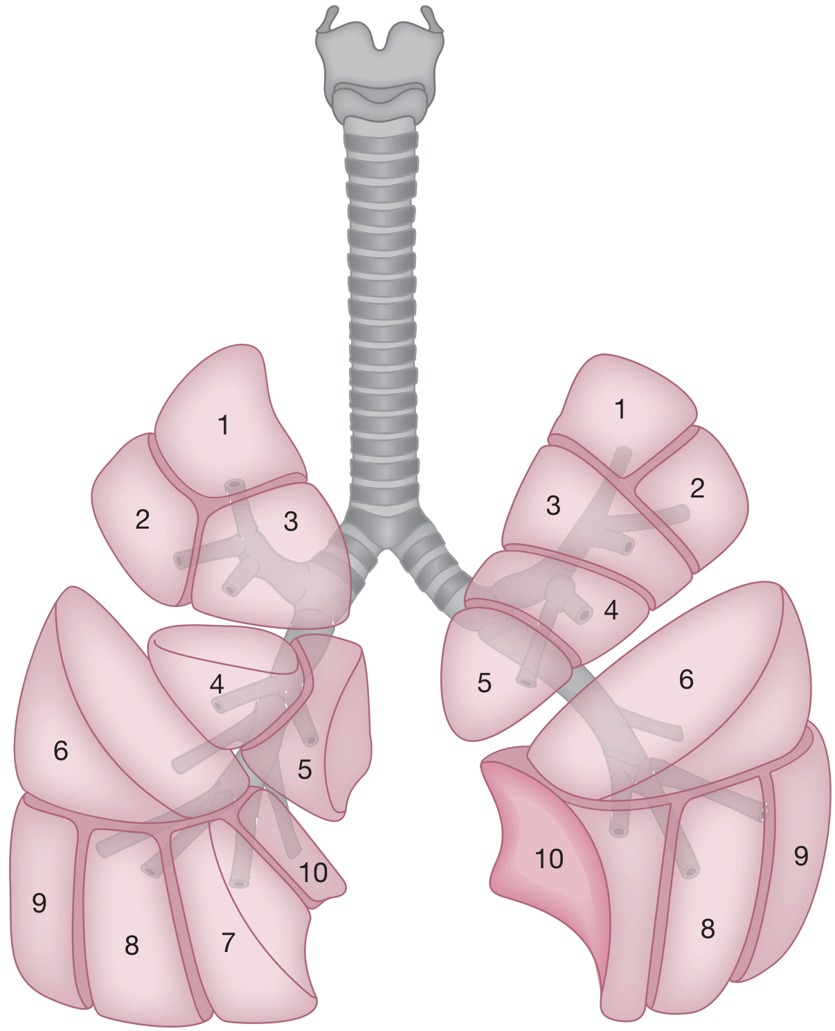
Figure: Bronchopulmonary segments of the human lung (Fishman's Pulmonary Diseases and Disorders)
Right Lung - 10 Segments
| Lobe | Segment | Abbreviation |
|---|---|---|
| Upper (Superior) | Apical | B1 |
| Posterior | B2 (or B3 in some numbering) | |
| Anterior | B3 (or B2) | |
| Middle | Lateral | B4 |
| Medial | B5 | |
| Lower (Inferior) | Superior (Apical) | B6 |
| Medial basal | B7 | |
| Anterior basal | B8 | |
| Lateral basal | B9 | |
| Posterior basal | B10 |
Left Lung - 8 Segments (American nomenclature) or 10 (British)
| Lobe | Segment | Note |
|---|---|---|
| Upper | Apicoposterior | B1+2 (fusion of apical + posterior) |
| Anterior | B3 | |
| Superior lingular | B4 | |
| Inferior lingular | B5 | |
| Lower | Superior (Apical) | B6 |
| Anteromedial basal | B7+8 (B7 absent as separate segment) | |
| Lateral basal | B9 | |
| Posterior basal | B10 |
Key differences: The left lung lacks a separate middle lobe (replaced by the lingula, an embryological homologue). The left upper lobe's apical and posterior segments fuse into a single apicoposterior segment. The medial basal segment (B7) is absent as an independent segment in the left lung.
(Sabiston Textbook of Surgery; Color Atlas of Human Anatomy Vol. 2; Fischer's Mastery of Surgery 8e)
3. Bronchoscopic Anatomy - Endoscopic Clock-Face Positions
This is directly relevant to anaesthesia for airway management, bronchoscopy, and selective bronchial blockade:
Right Lung (as seen endoscopically):
Right Upper Lobe:
- Apical segment (B1) - orifice at 4 o'clock
- Anterior segment (B2) - orifice at 12 o'clock
- Posterior segment (B3) - orifice at 8 o'clock
Right Middle Lobe:
- Lateral segment (B4) - orifice at 3-6 o'clock
- Medial segment (B5) - orifice at 9-12 o'clock
Right Lower Lobe:
- Superior segment (B6) - orifice at 5 o'clock, just distal to the middle lobe bronchus
- Medial basal segment (B7) - orifice at 9 o'clock, typically more proximal than other basal segments
- Anterior basal segment (B8) - orifice at 1 o'clock
- Lateral basal segment (B9) - orifice at 3 o'clock
- Posterior basal segment (B10) - orifice at 6 o'clock
Note: In more than half of patients, a subapical segment exists in the lower lobe with a posterior tertiary bronchus arising anywhere along the inferior lobar bronchus from the superior segment to the final lobar division.
Left Lung (as seen endoscopically):
Left Upper Lobe:
- Upper division - orifice at 8 o'clock (containing apicoposterior B1+3 and anterior B2)
- Lingular division - orifice at 2 o'clock (superior lingular B4 at 10 o'clock; inferior lingular B5 at 2 o'clock)
Left Lower Lobe:
- Superior segment (B6) - orifice at 6 o'clock, just past origin of lower lobe
- Anteromedial basal (B8+7) - orifice at 12 o'clock
- Lateral basal (B9) - orifice at 9 o'clock
- Posterior basal (B10) - orifice at 5 o'clock
(Fischer's Mastery of Surgery 8e)
4. Vascular Supply - Surgical and Anaesthetic Significance
- Pulmonary arteries run centrally within each segment alongside the bronchus (intrasegmental).
- Pulmonary veins run intersegmentally - this is why segmentectomy requires careful vein preservation and why venous bleeding during segmental resection is intersegmental.
- Bronchial arteries supply the airway wall itself from the systemic circulation (superior thoracic aorta or aortic arch): typically two left and one right bronchial artery.
- The right pulmonary artery is longer than the left; it passes horizontally across the mediastinum anterior to the tracheal bifurcation.
- The left pulmonary artery "rides" on the left principal bronchus and crosses over the superior lobar bronchus to the posterior side.
(Sabiston Textbook of Surgery; Gray's Anatomy for Students; Fishman's Pulmonary Diseases and Disorders)
5. Anaesthetic Implications
5a. Bronchoscopy and Airway Management
- Bronchoscopy requires knowledge of bronchopulmonary segments - the clock-face positions above guide the bronchoscopist to specific segmental orifices for lavage, biopsy, or blocker placement.
- Segments are the surgical units of the lung because there is little or no communication between segments at the bronchial level; however, extensive cross-ventilation exists at the parenchymal level (collateral ventilation via pores of Kohn), which has implications for targeted collapse.
5b. One-Lung Ventilation (OLV)
The most direct anaesthetic application of segmental anatomy. OLV is required for:
- Thoracic surgical procedures (lobectomy, pneumonectomy, segmentectomy)
- Oesophagectomy (right thoracotomy/thoracoscopy position)
- Carinal resection and tracheal surgery
Selective segmental oxygen insufflation during OLV: A fiberoptic bronchoscope can be guided under direct vision into a segment remote from the operative site to selectively reinflate it using 5 L/min oxygen flow through the suction port. This improves PaO2 during OLV without interfering with surgical exposure - particularly useful in patients who have had prior contralateral lung resection.
Selective lobar blocking: A bronchial blocker can be placed in a specific lobar bronchus to collapse only the operative lobe while continuing to ventilate the other lobe(s). This requires precise endoscopic knowledge of lobar/segmental anatomy.
(Miller's Anesthesia 10e)
5c. Hypoxic Pulmonary Vasoconstriction (HPV) and Volatile Anaesthetics
- During OLV, the collapsed non-ventilated lung develops hypoxaemia, triggering HPV - vasoconstriction that diverts blood flow away from poorly ventilated lung regions to improve V/Q matching and reduce intrapulmonary shunt (Qs/Qt).
- Volatile anaesthetics (isoflurane, sevoflurane, desflurane) at high concentrations can attenuate HPV in animal models, potentially worsening shunt during OLV. However, clinical effects are less clear, as volatile agents also affect cardiac output, autonomic tone, and humoral factors.
- Non-pharmacological factors that impair HPV include: surgical trauma, temperature, pH, PaCO2, size of the hypoxic segment, and intensity of the hypoxic stimulus.
- In clinical studies of OLV during thoracic surgery, volatile anaesthetics have had minimal effects on PaO2 and Qs/Qt when transitioning from two-lung to one-lung ventilation.
(Barash Clinical Anesthesia 9e)
5d. Postobstructive and Positional Implications
- Dependent lung atelectasis: In the lateral decubitus position for thoracic surgery, gravitational forces preferentially ventilate the non-dependent (operative) lung while perfusion favours the dependent lung - creating V/Q mismatch. Understanding which segments are dependent helps predict where atelectasis and secretion pooling will occur.
- Postobstructive pneumonia: Secondary infection is particularly common in bronchopulmonary segments obstructed by primary or secondary malignancy. The anaesthetist must anticipate purulent secretions, impaired drainage, and risk of soiling the contralateral lung if a double-lumen tube is not correctly positioned.
- Aspiration: Aspiration of gastric contents follows gravitational paths preferentially to the posterior and basal segments (particularly superior segment B6 and posterior basal B10 of the lower lobes in supine patients, and upper lobe posterior segments in erect patients) - relevant to positioning after induction.
- Suctioning during bronchoscopy: Knowledge of segmental anatomy is necessary to direct suction catheters or bronchoscopes to specific segments for lavage, particularly in lobar collapse or mucus plugging.
5e. Surgical Resection Planning and Preoperative Assessment
- Ventilation-perfusion (V/Q) lung scanning measures relative blood flow and ventilation to individual lung regions and is used to predict postoperative FEV1 or DLCO before resection:
Predicted postoperative FEV1 = preoperative FEV1 × (1 - fraction of total perfusion/ventilation to the resected segment(s))
- Segmentectomy (sublobar resection of individual segments) requires precise anatomical knowledge because pulmonary veins running intersegmentally must be carefully identified and preserved or ligated.
- Lymphatic drainage: Lymphatics from most lung segments drain to ipsilateral hilar and mediastinal nodes. However, lymph from the left lower lobe may drain to right mediastinal (paratracheal) lymph nodes - relevant in staging and mediastinoscopy planning.
(Fishman's Pulmonary Diseases and Disorders; Sabiston Textbook of Surgery)
6. Summary Table: Right vs Left Lung Segments
| Feature | Right Lung | Left Lung |
|---|---|---|
| Number of lobes | 3 (upper, middle, lower) | 2 (upper, lower) |
| Number of segments | 10 | 8 (US) / 10 (UK) |
| Middle lobe equivalent | Right middle lobe (B4, B5) | Lingula (B4, B5, superior + inferior) |
| Apical segment | Separate (B1) | Fused with posterior → apicoposterior (B1+2) |
| Medial basal segment | Present (B7) | Absent (fused with anterior basal → B7+8) |
| Main bronchus length | Shorter, more vertical | Longer (passes under aortic arch) |
| Inhaled foreign body risk | Higher (more vertical angle) | Lower |
Key Anaesthetic Take-Home Points
- Bronchoscopy orientation: Know the clock-face positions of all 10 right and 8 left segmental orifices - this guides DLT confirmation, bronchial blocker placement, and targeted lavage.
- Segmental collapse for OLV: Selective lobar/segmental blockers can provide surgical access with less physiological derangement than whole-lung collapse.
- HPV: Volatile agents minimally impair HPV clinically at standard doses during OLV; non-pharmacological factors (surgical trauma, hypocapnia, size of hypoxic zone) are equally important.
- Aspiration positioning: Posterior basal (B10) and superior (B6) segments of lower lobes are most vulnerable in supine patients.
- Preoperative V/Q scanning: Segment-by-segment perfusion/ventilation assessment predicts postoperative lung function when planning resection.
- Left lower lobe lymphatics: Drain contralaterally to right paratracheal nodes - relevant in staging thoracic malignancies.
This is a shared conversation. Sign in to Orris to start your own chat.