
Give me all information important about supine PCNL
supine PCNL percutaneous nephrolithotomy position
supine PCNL percutaneous nephrolithotomy technique advantages complications 2024
PMID: 40086594
PMID: 39692421
supine PCNL percutaneous nephrolithotomy patient positioning setup
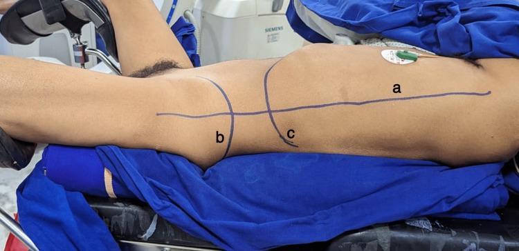
A clinical procedural photograph illustrating the Galdakao-modified supine Valdivia position for percutaneous nephrolithotomy (PCNL). The patient is placed in a modified supine position on a surgical table with blue drapes, utilizing stirrups for leg positioning: the ipsilateral leg is extended straight, while the contralateral leg is flexed and abducted. Bolsters are placed under the hip and chest to elevate the flank, providing surgical access to the renal area. Surface anatomy markings in purple ink identify critical landmarks for the procedure: (a) the posterior axillary line, (b) the iliac crest, and (c) the level of the 12th rib. An ECG electrode is visible on the chest wall. This image serves as an educational guide for urological surgical positioning, demonstrating the necessary surface markers and table configuration required to safely perform supine renal access for stone management.
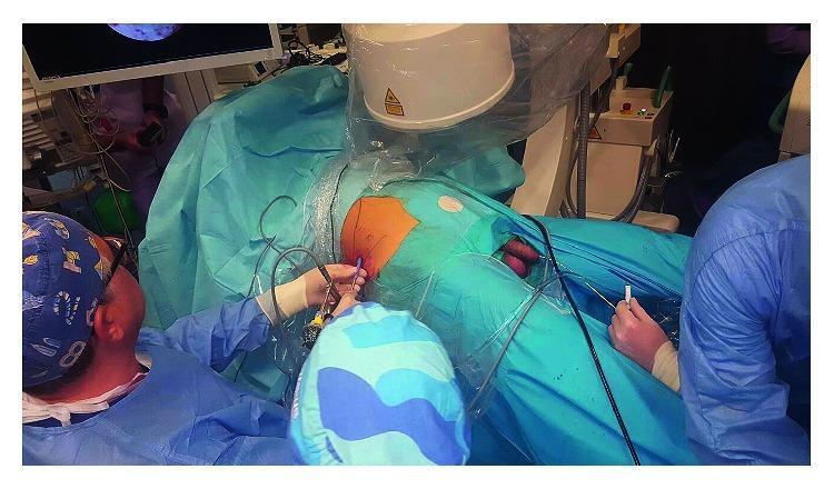
Clinical photograph of a urological surgical procedure, likely Percutaneous Nephrolithotomy (PCNL), demonstrating a modified supine-oblique (Valdivia-Galdakao) patient positioning. The patient is draped in a sterile blue field with the right flank exposed and prepared for percutaneous access. A C-arm fluoroscopy unit is positioned over the patient's torso for intraoperative imaging, with a monitor in the background displaying real-time visual data. The surgical team is arranged to allow simultaneous access: one surgeon operates at the flank, performing a renal puncture or nephroscopy, while an assistant is positioned between the patient's abducted and secured legs to perform retrograde endourological maneuvers (e.g., ureteroscopy or catheter placement). The patient's legs are secured independently in a slightly abducted, non-flexed position to facilitate this dual access to the upper and lower urinary tract. This configuration optimizes workflow for endoscopic combined intrarenal surgery (ECIRS) by providing wide flank exposure and retrograde access to the bladder.
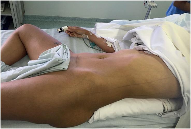
A clinical photograph illustrating the Valdivia position, a specific surgical setup used in urological procedures such as Percutaneous Nephrolithotomy (PCNL). The patient is shown in a modified supine decubitus position on an operating table. To optimize surgical access to the renal area, a large saline bag or firm pillow is placed under the ipsilateral flank, creating a subtle lateral tilt and elevating the kidney. The patient's legs are positioned in a modified lithotomy style: the ipsilateral leg remains extended while the contralateral leg is flexed and abducted. Standard perioperative monitoring is visible, including a pulse oximeter on the index finger and an intravenous line in the forearm. This positioning is designed to allow simultaneous antegrade and retrograde access to the urinary tract while maintaining the hemodynamic advantages of the supine position compared to prone positioning.
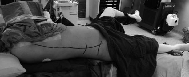
A clinical photograph in grayscale illustrating the 'Giusti position' for supine percutaneous nephrolithotomy (PCNL). The patient is positioned supine with a 15-20 degree rotation toward the contralateral side, supported by padding under the scapula and buttocks to expose the flank. The ipsilateral leg (same side as the stone) is extended and kept straight, while the contralateral leg is abducted and flexed in a cystoscopy or lithotomy position to facilitate simultaneous retrograde endourological access. On the exposed flank, dark surgical skin markings indicate critical anatomical landmarks for renal access, including the posterior axillary line, the lower rib margin, and the iliac crest. This surgical setup is designed for endourological procedures like PCNL, allowing for improved spontaneous fragment passage and ease of simultaneous ureteroscopy or retrograde contrast studies without the need for patient repositioning.
PMID: 39614229
Supine PCNL: Complete Overview
1. Background and History
- Grainger & Allison's Diagnostic Radiology, p. 2170
- Hinman's Atlas of Urologic Surgery, p. 253
2. Indications for PCNL (General)
| Indication | Details |
|---|---|
| Renal pelvic stones | >2 cm |
| Staghorn calculi | Complete or partial |
| Lower pole stones | >1 cm |
| Hard stones | CT density >1000 HU |
| Cystine stones | Relative indication |
| Stones with poor drainage | Calyceal diverticula, horseshoe kidney |
| Failed ESWL or URS | Residual or recurrent stones |
| Special anatomy | Pelvic kidneys (with assistance) |
- Smith and Tanagho's General Urology, p. 143
- Grainger & Allison's Diagnostic Radiology, p. 2170
- Patients with severe cardiopulmonary disease who cannot tolerate prone positioning
- Morbid obesity (abdominal compression in prone reduces venous return and ventilation)
- Concomitant ipsilateral ureteral stones (allows simultaneous retrograde URS)
- Patients with spinal deformities or inability to lie prone
- Cases requiring simultaneous antegrade-retrograde access (ECIRS)
- Solitary kidney (reduces cardiovascular risk)
3. Supine Positional Variants
3a. Classic Valdivia (Total Supine)
- Patient lies completely flat on back
- A small bolster placed under the ipsilateral flank to slightly elevate the kidney
- Ipsilateral leg extended; contralateral leg flexed/abducted (modified lithotomy)
- Allows retrograde access simultaneously
3b. Galdakao-Modified Supine Valdivia (GMSV)
- Most widely used supine variant
- Ipsilateral leg extended flat; contralateral leg in stirrup (flexed and abducted)
- Bolster under ipsilateral hip and chest to create flank elevation
- Key anatomical surface markings: posterior axillary line, iliac crest, 12th rib level
- Allows simultaneous flexible ureteroscopy and PCNL (ECIRS)
3c. Barts Flank-Free Modified Supine (FFMS)
- A newer enhanced variant
- Designed to maximize flank exposure without needing manual support
- Systematic review (PMID 39692421, 2024): surgery duration significantly shorter than prone (MD -15.48 min, p=0.006); no significant difference in stone-free rate, complications, or hospital stay
3d. Giusti Position
- 15-20 degree rotation toward the contralateral side
- Supported by padding under scapula and buttocks
- Skin markings on posterior axillary line, lower rib margin, iliac crest
4. Technical Considerations
Access and Puncture
- In supine PCNL, the posterior calyces are accessed since the kidney rotates slightly anteriorly in the supine position
- Bolsters create slight flank elevation of the operative side to bring the posterior kidney surface into a more accessible plane
- Fluoroscopy or ultrasound can both be used for guidance - same principles as prone approach
- The collecting system tends to collapse slightly compared to prone (gravity-dependent drainage is lost) - this can make puncture marginally more challenging
- The surgical field is smaller than prone and upper pole calyceal puncture is more difficult
- More lateral and anterior puncture carries higher risk of bowel injury - the surgeon must account for bowel position
Simultaneous Retrograde Access (ECIRS)
-
Staghorn calculi requiring both antegrade and retrograde clearance
-
Concomitant ureteral stones
-
Difficult calyces that cannot be reached through the percutaneous tract
-
Comprehensive Clinical Nephrology, p. 2403
No Need for Patient Repositioning
5. Advantages vs. Prone PCNL
| Feature | Supine PCNL | Prone PCNL |
|---|---|---|
| Anesthesia | Safer; better airway access | Higher risk especially in obese/cardiopulmonary compromise |
| Repositioning | None needed | Required after ureteral catheter placement |
| Operative time | Generally shorter | Longer (includes repositioning) |
| Retrograde access | Simultaneous possible (ECIRS) | Also possible but less convenient |
| Surgical field | Smaller | Larger; more working space |
| Upper pole access | More difficult | Easier |
| Multiple tracts | More difficult | Easier (larger back surface) |
| Bowel risk | Lower (retrorenal colon less frequent supine) | Slightly higher retrorenal colon risk |
| Ventilation (obese) | Better | Abdominal compression reduces FRC |
| Hemodynamics | More stable | Prone compression can reduce venous return |
| Radiation | Comparable | Comparable |
- Hinman's Atlas, p. 253
- Miller's Anesthesia, p. 8484
6. Current Evidence: Supine vs. Prone Outcomes
Large Meta-Analysis (PMID 40086594, Lachkar et al., 2025 - 43 studies, 11,774 patients):
- Stone-free rate: No significant difference (RR=0.99, p=0.70)
- Operative time: Significantly shorter in supine (SMD=-0.39, p=0.008)
- Overall complications: Significantly fewer in supine (RR=0.87, p=0.003)
- Major complications: Significantly fewer in supine (RR=0.75, p=0.015)
- Hemoglobin drop, hospital stay, visceral injury, transfusion, sepsis, urinary leakage, pleural effusion: No significant difference
- Conclusion: Supine PCNL is a viable alternative with shorter operative time and fewer complications
Barts FFMS vs. Prone (PMID 39692421, 4 studies, 2024):
- Surgery duration: Significantly shorter for Barts FFMS (MD -15.48 min, p=0.006)
- Stone-free rate, fever, transfusion, nephrostomy use, hospital stay: No significant difference
- Neither position is superior overall - choice should be based on surgeon preference and patient status
Pediatric Meta-Analysis (PMID 39614229, 8 studies, 492 patients, 2024):
- Stone-free rate: No significant difference (OR 0.67, p=0.17)
- Operative time: Significantly shorter in supine (MD 13.75 min, p=0.004)
- Hospital stay: Shorter in supine (MD 0.61 days, p<0.0001)
- Total complication rate: No significant difference
- Conclusion: Supine PCNL equivalent in SFR with shorter operative time and hospital stay in pediatric patients
7. Complications Specific to Supine PCNL
General PCNL Complications (applying to both positions)
| Complication | Rate/Notes |
|---|---|
| Bleeding requiring transfusion | ~5-8%; major hemorrhage 1-3% |
| Sepsis/urosepsis | ~1-5% |
| Urine leakage | Common; usually resolves with drainage |
| Pleural injury (pneumothorax/hydrothorax) | Higher with supracostal/upper pole access |
| Colonic injury | 0.2-0.3%; may actually be lower in supine |
| Pelvic injury/perforation | ~3-4% |
| Hypothermia | From large volumes of irrigation fluid |
| Acute anemia | Bleeding or dilutional |
Supine-Specific Concerns
- Smaller working field - instrument manipulation more limited
- Upper pole access is harder - may require steep angulation; pleural injury risk still present
- Collapsed collecting system in supine - can impair visualization and navigation
- Miller's Anesthesia notes: "Supine positioning...results in a smaller surgical field, collapse of the renal collecting system, and increased difficulty in upper pole calyceal puncture"
- Retrorenal colon risk is lower in supine (2% vs 10% prone)
Monitoring During PCNL (Anesthesia)
- Airway pressures, end-tidal CO2, O2 saturation (for pleural injuries)
- Hemodynamics (for bleeding)
- Temperature (hypothermia from irrigation)
8. Drainage After Supine PCNL
- Standard nephrostomy tube (30Fr tract) - recommended if:
- Second-look procedure planned
- Significant bleeding or collecting system perforation
- Upper pole access with pleural risk
- Multiple tracts created
- High probability of residual stones
- Significant infection
- Tubeless PCNL (ureteral catheter/stent only, no nephrostomy) - gaining support, reduces postoperative morbidity
- Totally tubeless (JJ stent only) - selected patients
- Hinman's Atlas, p. 253
9. Special Populations Favored for Supine PCNL
- Morbid obesity: Less abdominal compression, better ventilation, easier airway access
- Cardiopulmonary compromise: Hemodynamically more stable, airway always accessible
- Spinal deformities / inability to go prone: Cannot tolerate prone positioning
- Solitary kidney: Reduced anesthetic risk
- Horseshoe kidney / pelvic kidney: Supine with US or laparoscopic guidance may be needed
- Simultaneous bilateral stones: Some reports of bilateral supine PCNL in one session
10. Contraindications to PCNL
- Uncorrected coagulopathy
- Active, untreated urinary tract infection (relative - treat infection first)
- Patient anatomy making safe percutaneous access impossible
Summary
- Supine vs prone PCNL meta-analysis 11,774 cases (2025) - Lachkar et al. PMID 40086594
- Barts FFMS vs prone meta-analysis (2024) - Ananda et al. PMID 39692421
- Pediatric supine vs prone meta-analysis (2024) - Porto et al. PMID 39614229