
Classification of vesicobollous lesions
vesicobullous skin lesions classification diagram intraepidermal subepidermal
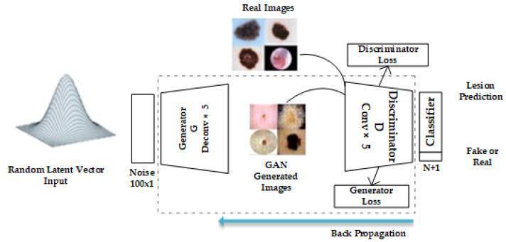
This flow diagram illustrates a Deep Convolutional Generative Adversarial Network (DCGAN) architecture specifically designed for skin lesion classification. The process initiates with a 'Random Latent Vector Input' represented by a Gaussian distribution curve, which is transformed into a 'Noise 100x1' vector. This noise enters a 'Generator (G)' consisting of five deconvolutional layers ('Deconv x 5'). The generator produces synthetic 'GAN Generated Images' depicting various skin lesions. Simultaneously, 'Real Images' (clinical dermatoscopic photographs of benign and malignant lesions) are provided from a training dataset. Both real and generated images serve as inputs for a 'Discriminator (D)' utilizing five convolutional layers ('Conv x 5'). The discriminator computes 'Discriminator Loss' and 'Generator Loss' to facilitate 'Back Propagation' for model optimization. Finally, the network utilizes a 'Classifier' to distinguish between 'Fake or Real' images and provide a 'Lesion Prediction.' This architecture is an advanced diagnostic imaging tool aimed at improving dermatological classification through data augmentation and deep learning.
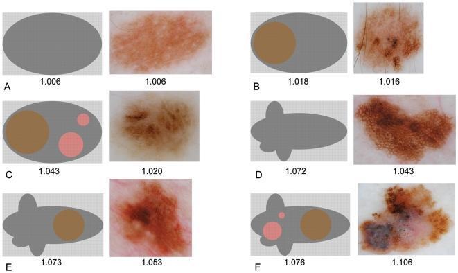
A comparison chart illustrating the classification of pigmented skin lesions through schematic representations and clinical dermoscopic photographs. The figure is organized into six pairs (A-F), demonstrating a progression of morphological irregularity. On the left of each pair, a gray schematic diagram represents the lesion's lacunarity and geometric properties; on the right, a corresponding clinical dermoscopic image shows real-world examples. The progression moves from (A) 'No irregularity'—a smooth oval with homogenous pigmentation—to (B) simple internal asymmetry, (C) hierarchical internal asymmetry with multiple colored internal regions, and (D-F) increasing boundary asymmetry with lobulated borders and complex internal architectural patterns. Key dermoscopic features visible in the clinical images include reticular networks, globules, and varied pigmentation ranging from light tan to dark brown and reddish-purple. The figure serves as a diagnostic educational tool for identifying risk factors in melanocytic lesions, highlighting how mathematical lacunarity values correlate with increasing clinical and structural complexity.
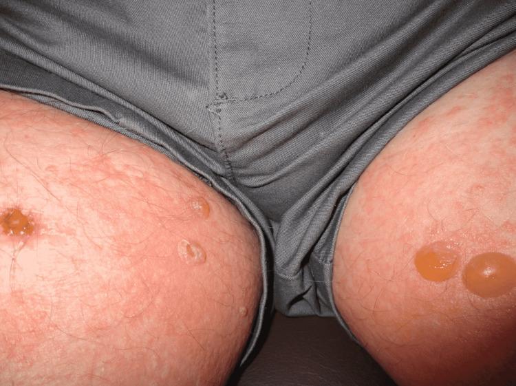
Clinical photograph of the bilateral medial thighs demonstrating multiple tense, serous fluid-filled bullae. The lesions are characteristic of an autoimmune subepidermal blistering disease such as bullous pemphigoid. Visible are several large, dome-shaped blisters with clear to yellowish fluid content. The underlying and surrounding skin shows significant confluent erythema and urticarial plaques, indicating intense localized inflammation. On the left medial thigh, a ruptured bulla with a denuded, darker erosive base is visible, while the right medial thigh shows two prominent, intact tense bullae. The distribution is symmetrical along the inner thighs. This image serves as a classic educational example of bullous dermatitis, highlighting the distinction between tense bullae (subepidermal) and flaccid vesicles (intraepidermal) and their association with erythematous backgrounds in dermatological diagnosis.
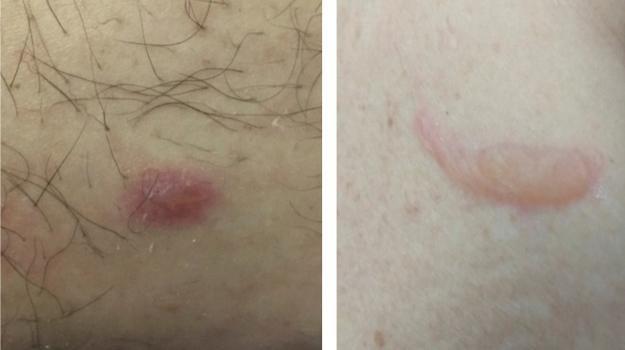
This clinical photograph displays vesico-bullous skin lesions in a patient, categorized as a dermatological manifestation of COVID-19. The image is split into two panels showing different morphological stages. The left panel reveals a small, erythematous, roughly circular vesicle situated on hair-bearing skin. The right panel displays a larger, crescent-shaped bulla. This larger lesion is distinctly elevated and translucent, indicating a fluid-filled subepidermal or intraepidermal space. The surrounding skin in the right panel appears mildly pale but lacks significant peri-lesional inflammation. These lesions represent a varicella-like pattern, which typically appears within the first week of systemic viral symptoms. In a clinical context, such vesico-bullous eruptions are associated with mild to moderate systemic disease and serve as an important diagnostic marker to differentiate COVID-19 related exanthems from other viral or drug-induced reactions.
pemphigus vulgaris flaccid blisters skin erosions
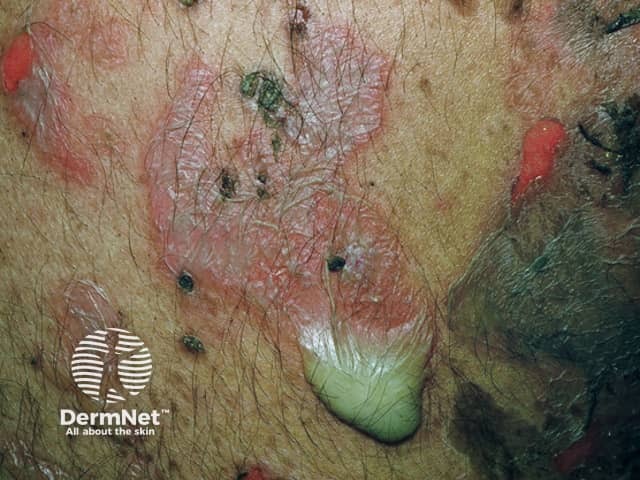
Pemphigus vulgaris, skin clinical photograph. This close‑up image shows erythematous, inflamed skin with ruptured and crusted erosions and several translucent, fragile blisters consistent with flaccid bullae; additional crusting and superficial crusted erosions are evident on the torso. The lesions are distributed across the chest and proximal trunk, with sparing or less involvement of surrounding intact skin. The photographic perspective emphasizes cutaneous involvement with preserved, yet damaged, epidermis and visible fragile vesicles. Clinically, pemphigus vulgaris is an autoimmune blistering disorder characterized by acantholysis and intraepidermal blister formation; the bullae rupture readily, producing painful erosions. The Nikolsky sign may be positive, indicating easy separation of epidermis with gentle pressure. For validation, histology would typically reveal suprabasal acantholysis with a tombstone row of basal keratinocytes; direct immunofluorescence shows intercellular IgG/C3 within the epidermis with desmoglein 3 and desmoglein 1 autoantibodies. Relevance to clinical practice includes prompt recognition for early systemic therapy to reduce progression and mucocutaneous involvement. Differential diagnoses include bullous pemphigoid and dermatitis herpetiformis. This image is relevant for dermatology education, autoimmune blistering disease research, and diagnostic dermato pathology correlation studies, serving as a reference for pattern recognition, disease staging, and treatment planning.
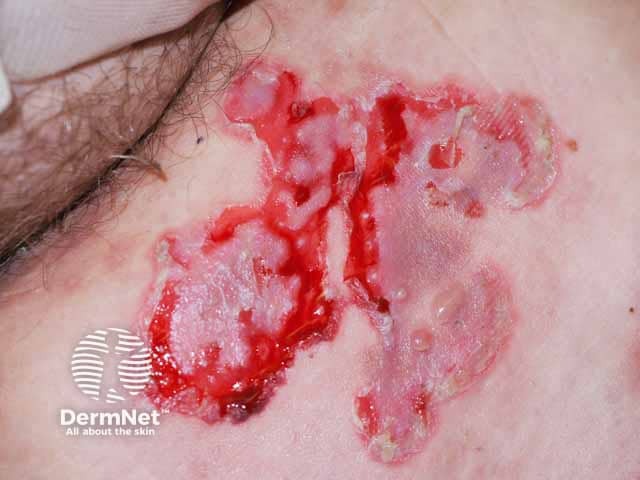
Clinical photography of a pemphigus vulgaris (PV) patient shows diffuse cutaneous erosions and ruptured, flaccid blisters with irregular edges exposing moist, erythematous bases. The close-up, high-magnification view captures superficial losses of epidermal integrity consistent with ruptured pemphigus bullae. Lesions appear tender, crusted at the periphery, and coalesce into larger erosive plaques with surrounding erythema. This presentation is classic for PV, in which intraepidermal acantholysis creates fragile blisters that rupture easily. Mucosal involvement is common in PV but is not depicted in this image. Clinically, a positive Nikolsky sign may accompany the erosions. Histologically, pemphigus vulgaris shows suprabasal acantholysis with tombstone basal cells; immunofluorescence reveals intercellular IgG. The image is relevant for dermatology, medical education, and clinical practice for recognizing active cutaneous PV and guiding management. Common treatment considerations include systemic corticosteroids, steroid-sparing immunosuppressants, and targeted biologics, with careful monitoring for flare progression. For research and training, annotate lesion age, localization, and wound characteristics, and correlate with serology (desmoglein antibodies) and biopsy findings when available. This image exemplifies PV skin involvement and supports differential diagnosis from bullous pemphigoid and other vesiculobullous diseases.
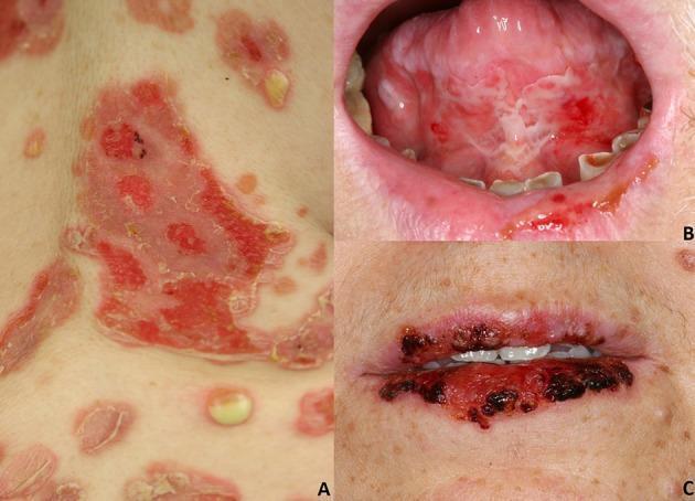
This composite of clinical photographs illustrates the mucocutaneous manifestations of pemphigus variants. Panel A (top left) displays Pemphigus vulgaris on the skin, showing large, irregular, erythematous erosions with peripheral peeling of the epidermis, consistent with ruptured flaccid blisters. A small, intact flaccid vesicle with yellowish fluid is visible in the lower quadrant. Panel B (top right) demonstrates oral involvement in Pemphigus vulgaris, featuring multiple painful-looking erosions and white macerated plaques on the ventral surface of the tongue and labial mucosa. Panel C (bottom right) showcases Paraneoplastic pemphigus, characterized by severe stomatitis with diffuse, hemorrhagic crusting and deep erosions across the entire vermillion border of the upper and lower lips. These images contrast the typical cutaneous and oral erosions of Pemphigus vulgaris with the more severe, hemorrhagic labial involvement often seen in paraneoplastic variants, serving as a diagnostic tool for identifying autoimmune blistering diseases and their clinical severity.
bullous pemphigoid tense blisters erythematous base
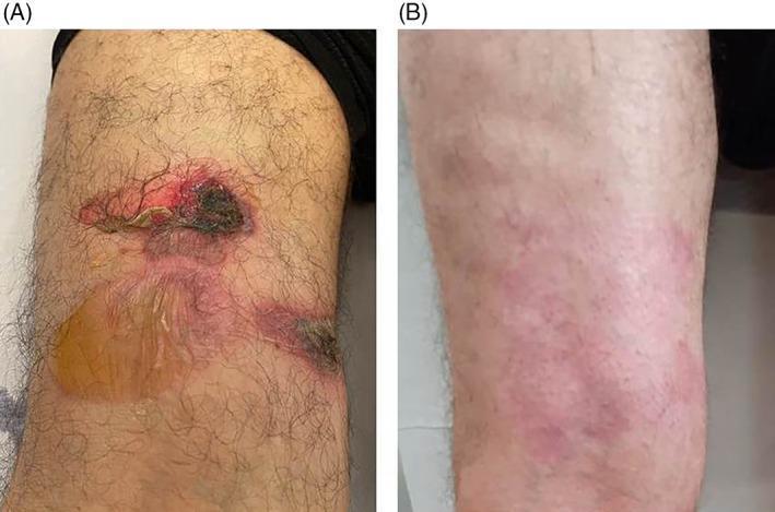
A comparison chart consisting of two clinical photographs (A and B) showing the progression of bullous pemphigoid lesions on a patient's leg. Image A displays a large, tense bulla filled with serous yellowish fluid on an erythematous base. Superior and lateral to this intact blister are several irregular erosions with dark crusting and necrotic-appearing centers, indicative of ruptured bullae. The surrounding skin exhibits significant erythema and inflammation. Image B shows the same anatomical region after three weeks of corticosteroid treatment, demonstrating near-complete resolution. The previously seen blisters and erosions have cleared, leaving behind faint residual macular erythema and post-inflammatory pigmentary changes. This comparison serves as an educational tool for identifying primary autoimmune blistering diseases and monitoring clinical response to systemic therapy.
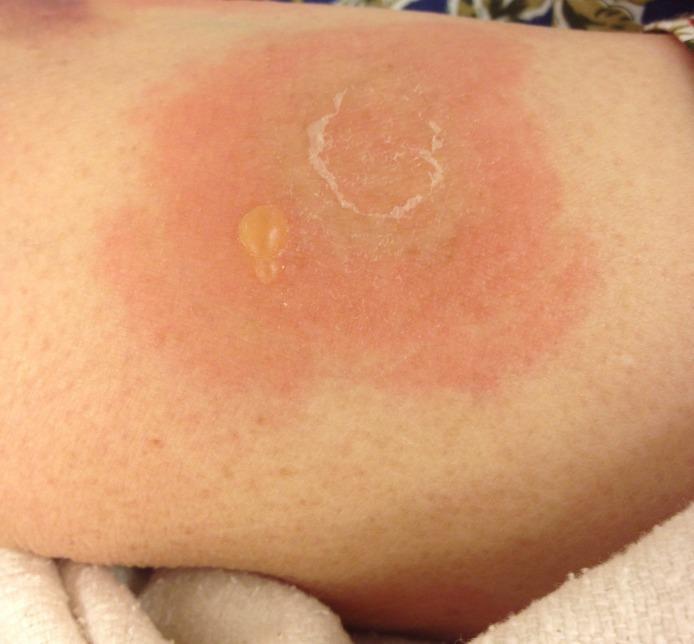
Clinical photograph of the left anterior thigh demonstrating dermatological findings characteristic of an autoimmune blistering disease, specifically bullous pemphigoid. The image shows a large, well-demarcated, circular erythematous and urticarial plaque approximately 5–7 cm in diameter. Centrally located on this inflammatory base is a small, intact, tense bulla containing serous, yellowish fluid. Adjacent to the intact blister is a larger area of epidermal denudation resulting from a ruptured bulla, characterized by a circular erosion with whitish, peripheral remnants of the detached blister roof (collarette of scale). The surrounding skin appears otherwise normal without evidence of scarring or active secondary infection. This visual presentation illustrates the typical clinical morphology of subepidermal blisters arising on an urticarial background, often associated with pruritus and eosinophilia in older adults.
Classification of Vesicobullous Lesions
I. By Anatomical Level of Blister Formation
A. Intraepidermal (Intercellular) Blisters
| Disease | Mechanism / Autoantigen |
|---|---|
| Pemphigus vulgaris | Anti-desmoglein 3 (Dsg3) ± Dsg1; suprabasal acantholysis |
| Pemphigus foliaceus | Anti-Dsg1 only; subcorneal split |
| Paraneoplastic pemphigus | Anti-Dsg3, periplakin, envoplakin; associated with lymphoid neoplasms |
| IgA pemphigus | IgA against desmocollins; subcorneal or intraepidermal split |
B. Subepidermal Blisters - Dermal-Epidermal Junction (Basement Membrane Zone)
| Disease | Autoantigen | Key Feature |
|---|---|---|
| Bullous pemphigoid (BP) | BP180 (collagen XVII) + BP230 | Tense bullae on erythematous/urticarial plaques; elderly |
| Pemphigoid gestationis (Herpes gestationis) | BP180 | Occurs in pregnancy; periumbilical onset |
| Mucous membrane pemphigoid (Cicatricial pemphigoid) | BP180, BP230, laminin-332 | Predominantly mucous membranes; scarring |
| Epidermolysis bullosa acquisita (EBA) | Collagen VII (sub-lamina densa) | Mechanobullous; may mimic DH or BP |
| Linear IgA dermatosis (LAD) / Chronic bullous disease of childhood | BP180/BP230 (97 kDa LAD-1) | IgA linear band at BMZ; "string of pearls" pattern in children |
| Bullous lupus erythematosus | Collagen VII | Autoimmune; often in sun-exposed areas |
| Disease | Mechanism |
|---|---|
| Erythema multiforme | Immune-mediated epidermal necrosis; target lesions |
| Stevens-Johnson syndrome (SJS) | Drug/infection-induced; <10% BSA detached |
| Toxic epidermal necrolysis (TEN) | Drug-induced; >30% BSA; full-thickness epidermal necrosis |
| Porphyria (PCT, VP, EPP) | Phototoxic subepidermal blister in sun-exposed skin |
| Hereditary epidermolysis bullosa (Junctional, Dystrophic) | Genetic defects in structural proteins (laminin-332, collagen VII) |
C. Dermal Papillae Disease (Special Subepidermal Subtype)
| Disease | Feature |
|---|---|
| Dermatitis herpetiformis (DH) | IgA deposits at papillary dermis tips; grouped pruritic vesicles; associated with celiac disease / gluten sensitivity |
II. By Etiology (Harrison's Classification)
I. Primary Mucocutaneous Diseases
- Pemphigus (foliaceus and vulgaris) - intraepidermal
- Bullous pemphigoid - subepidermal
- Gestational pemphigoid - subepidermal
- Cicatricial pemphigoid - subepidermal
- Dermatitis herpetiformis - subepidermal/papillary; associated with gluten enteropathy
- Linear IgA bullous dermatosis - subepidermal
- Epidermolysis bullosa acquisita - subepidermal; associated with inflammatory bowel disease
- Contact dermatitis (allergic and irritant)
- Erythema multiforme
- Stevens-Johnson syndrome
- Toxic epidermal necrolysis
- Bullous fixed drug eruption (including generalized variant)
- Pseudoporphyria (drug- or tanning booth-induced)
- Varicella-zoster virus
- Herpes simplex virus
- Enteroviruses (e.g., hand-foot-and-mouth disease)
- SARS-CoV-2 (varicella-like pattern)
- Staphylococcal scalded skin syndrome (SSSS) - subcorneal split via exfoliatin toxin
- Bullous impetigo
- Bullous tinea
II. Systemic Diseases
- Paraneoplastic pemphigus (+ bronchiolitis obliterans)
- Bullous systemic lupus erythematosus
- Diabetic bullae (bullosis diabeticorum)
- Porphyria (PCT, VP)
- Calcinosis cutis (rare)
Key Diagnostic Differentiating Features
| Feature | Intraepidermal | Subepidermal |
|---|---|---|
| Blister wall | Thin, flaccid | Tense, thick-roofed |
| Rupture | Easily | Resistant |
| Nikolsky sign | Positive | Usually negative |
| Acantholysis | Yes (pemphigus) | No |
| DIF pattern | Intercellular "fishnet" | Linear band at BMZ |
| Main examples | Pemphigus group | Pemphigoid group, DH, EBA |
Clinical Images
- Henry's Clinical Diagnosis and Management by Laboratory Methods, Cutaneous Autoimmune Diseases section - full immunobullous classification table with DIF/IIF patterns and autoantigens
- Harrison's Principles of Internal Medicine 22e (2025), Table 61-12 - etiologic classification of vesicles/bullae
- Fitzpatrick's Dermatology, Vol. 1-2 - subepidermal disease grouping (with and without autoantibodies)