
Internal cranial branches supply to vich branches detail and if possible give flow chart
Reading File
The question is about the internal cranial branches - this is likely about the internal carotid artery intracranial branches. Let me search the medical library for this.
Searching sources...
Finding Sources
Finding Sources
Finding Sources
Finding Sources
Good - found relevant sections. Let me read the detailed content from Gray's Anatomy for Students and the Neuroanatomy book:
Reading File
Reading File
Excellent! Let me read more of this detailed section and also fetch the diagram:
Reading File
Loading Image
Searching Images
internal carotid artery intracranial branches diagram ophthalmic anterior cerebral middle cerebral
Now let me fetch the vascular supply diagram and also get more detailed info on branches of each artery:
Loading Image
Finding Sources
I now have rich information. Let me get a bit more detail on the ophthalmic artery branches and MCA branches, then compose the full answer:
Finding Sources
Finding Sources
I now have comprehensive information. Let me also get the ophthalmic artery branch detail from Gray's:
Reading File
I now have all the information needed for a comprehensive answer. Let me compile it:
Internal Carotid Artery (ICA) - Intracranial Branches
The Internal Carotid Artery (ICA) has no branches in the neck. It enters the skull through the carotid canal, passes through the petrous temporal bone and cavernous sinus, and upon entering the subarachnoid space (supraclinoid portion), gives off its intracranial branches.
Flowchart: ICA Intracranial Branches
INTERNAL CAROTID ARTERY (ICA)
│
├── 1. OPHTHALMIC ARTERY
│ │
│ ├── Central retinal artery → retina
│ ├── Lacrimal artery → lacrimal gland, eyelids
│ ├── Long & short posterior ciliary arteries → eyeball
│ ├── Muscular arteries → extrinsic eye muscles
│ ├── Supra-orbital artery → forehead, scalp
│ ├── Anterior ethmoidal artery → ethmoidal cells, nasal cavity, anterior meningeal branch
│ ├── Posterior ethmoidal artery → ethmoidal cells, nasal cavity
│ ├── Medial palpebral arteries → eyelids
│ ├── Dorsal nasal artery (terminal branch) → nose
│ └── Supratrochlear artery (terminal branch) → forehead
│
├── 2. POSTERIOR COMMUNICATING ARTERY (PComA)
│ └── Connects ICA to Posterior Cerebral Artery (PCA)
│ (forms part of Circle of Willis)
│
├── 3. ANTERIOR CHOROIDAL ARTERY
│ ├── Optic tract
│ ├── Posterior limb of internal capsule
│ ├── Anterior hippocampus
│ ├── Amygdala
│ ├── Globus pallidus (medial segment)
│ └── Enters brain at choroidal fissure
│
└── TERMINAL BIFURCATION
│
├── 4. ANTERIOR CEREBRAL ARTERY (ACA)
│ ├── Connected to contralateral ACA via Anterior Communicating A.
│ ├── Segments: A1 (precommunicating), A2 (postcommunicating), A3 (distal)
│ ├── Callosomarginal artery → follows cingulate sulcus
│ ├── Pericallosal artery → adjacent to corpus callosum
│ └── Supplies: medial frontal lobe, anterior parietal lobe, corpus callosum
│
└── 5. MIDDLE CEREBRAL ARTERY (MCA)
├── M1 segment → gives off lenticulostriate arteries
│ └── Lenticulostriate arteries → striatum, putamen, internal capsule
├── Bifurcates (or trifurcates) in Sylvian fissure into:
│ ├── Superior division → lateral frontal lobe, anterior parietal cortex
│ └── Inferior division → temporal lobe, anterolateral parietal lobe
└── Frontobasal (orbitofrontal) branch → orbital surface of frontal lobe
Detailed Supply of Each Branch
1. Ophthalmic Artery
- First branch of the ICA, given off just after ICA exits the cavernous sinus
- Passes through the optic canal with the optic nerve
- Key clinical point: occlusion of central retinal artery (a branch) → sudden blindness
- Branches listed above supply the entire orbit, eyeball, forehead, scalp, nasal cavity, and ethmoidal sinuses
- Gray's Anatomy for Students
2. Posterior Communicating Artery (PComA)
- Arises from the supraclinoid ICA
- Runs posteriorly to anastomose with the posterior cerebral artery (PCA)
- Forms the lateral limbs of the Circle of Willis
- Clinical: PComA aneurysm → CN III palsy (pupil-involving, painful ptosis)
3. Anterior Choroidal Artery
- Arises from the supraclinoid ICA distal to PComA
- Travels posteriorly over the medial optic tract
- Supplies: optic tract, posterior limb of internal capsule, anterior hippocampus, amygdala, tail of caudate, medial globus pallidus, lateral geniculate body
- Occlusion syndrome: contralateral hemiplegia + hemisensory loss + homonymous hemianopia (the triad)
- Goldman-Cecil Medicine; Adams and Victor's Neurology
4. Anterior Cerebral Artery (ACA)
| Segment | Location | Key Branches | Territory |
|---|---|---|---|
| A1 | Precommunicating | Perforators to anterior hypothalamus | Anterior hypothalamus, septum |
| A2 | Postcommunicating | Orbitofrontal, frontopolar arteries | Medial frontal lobe |
| A3+ | Distal | Callosomarginal, Pericallosal | Medial parietal lobe, corpus callosum |
- Two ACAs anastomose via Anterior Communicating Artery (AComA)
- ACA infarct → contralateral leg weakness (cortical homunculus: leg area is medial)
5. Middle Cerebral Artery (MCA)
- Largest branch of ICA; supplies the largest territory
- Lenticulostriate arteries (from M1) = "arteries of cerebral hemorrhage" - supply internal capsule and basal ganglia; end arteries with no collaterals
- MCA infarct → contralateral face + arm weakness (lateral hemisphere) + aphasia (dominant side) + neglect (non-dominant side)
| Division | Territory |
|---|---|
| Superior | Lateral frontal cortex, motor strip (face/arm), Broca's area |
| Inferior | Superior temporal gyrus, Wernicke's area, parietal cortex |
Summary Table
| Branch | Origin | Key Structures Supplied |
|---|---|---|
| Ophthalmic artery | Just after cavernous sinus | Eye, orbit, forehead, nasal cavity |
| Posterior communicating artery | Supraclinoid ICA | Connects to PCA; Circle of Willis |
| Anterior choroidal artery | Supraclinoid ICA (distal to PComA) | Posterior limb IC, hippocampus, optic tract |
| Anterior cerebral artery | Terminal branch | Medial frontal + parietal lobe, corpus callosum |
| Middle cerebral artery | Terminal branch | Lateral hemisphere, BG, internal capsule |
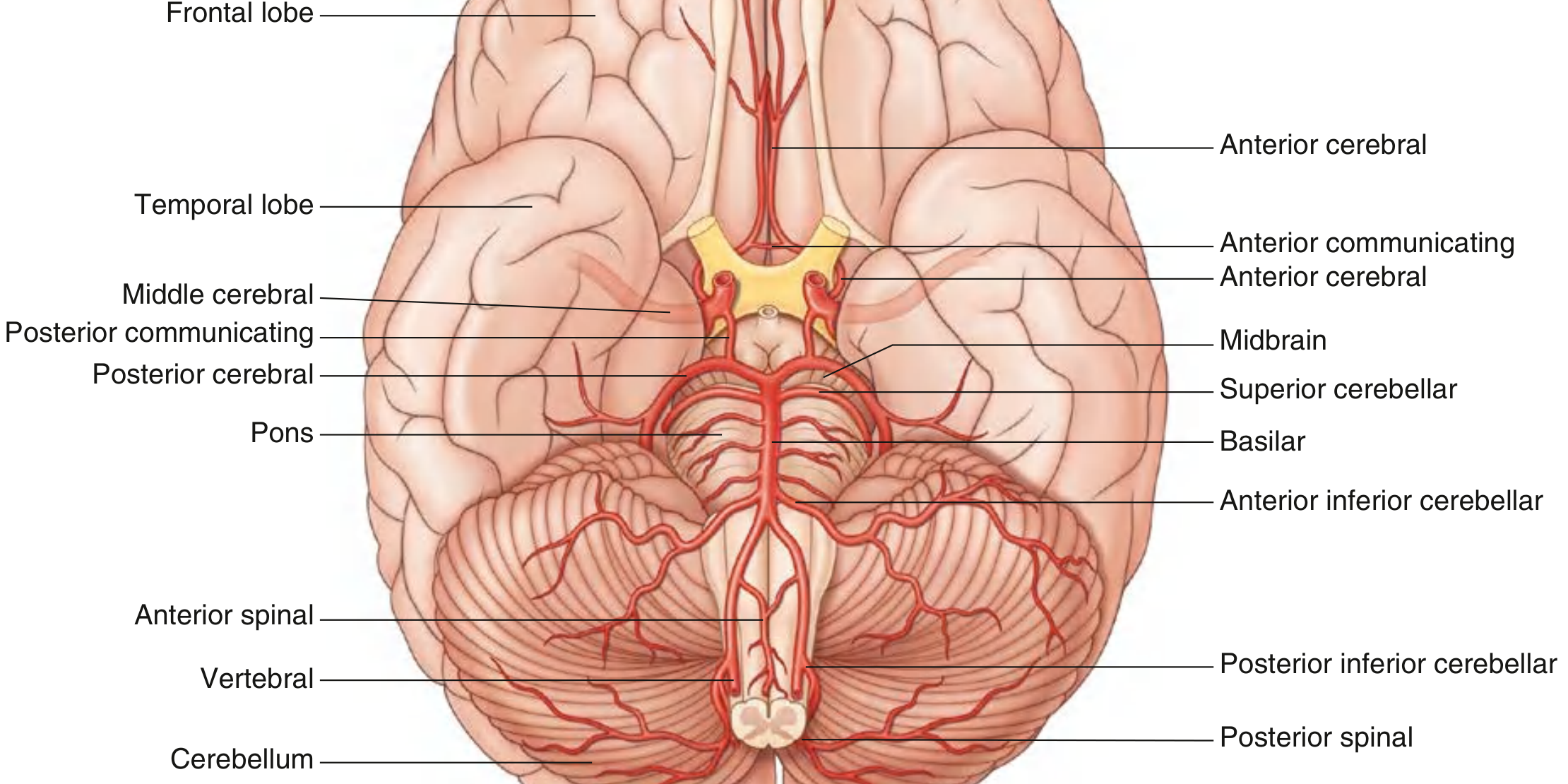
Diagram - Arteries at Base of Brain (from Gray's Anatomy for Students):
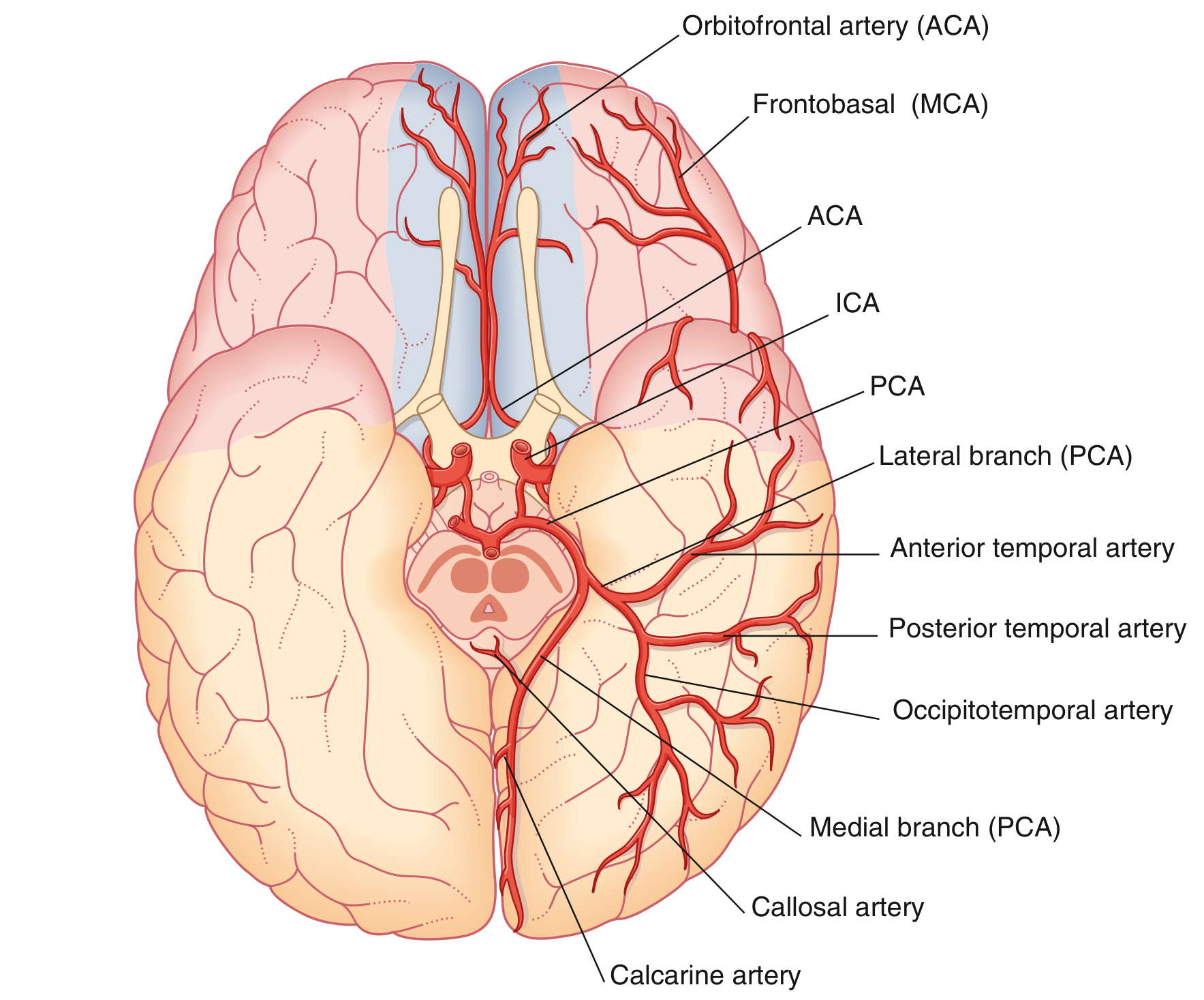
Vascular Supply Territories (ICA vs. Vertebrobasilar):
Sources:
- [Gray's Anatomy for Students](ISBN 9780323934237), pp. 1019, 1307
- [Goldman-Cecil Medicine](ISBN 9780323930345) - Anterior Choroidal and ICA sections
- [Adams and Victor's Principles of Neurology, 12th Ed.](ISBN 9781264264520) - Anterior Choroidal Artery Stroke Syndrome
Vertebral artery
Searching sources...
Finding Sources
Finding Sources
Reading File
Reading File
Finding Sources
Finding Sources
Finding Sources
Loading Image
Reading File
Finding Sources
Searching Images
vertebral artery branches PICA posterior inferior cerebellar artery basilar diagram
I have rich detail from multiple textbooks. Let me now compile the full answer:
Vertebral Artery - Complete Anatomy, Branches & Supply
Origin & Course
The vertebral artery is the first branch of the subclavian artery, arising medial to the anterior scalene muscle at the root of the neck.
Segments (Parts) of the Vertebral Artery:
| Part | Course |
|---|---|
| V1 - Prevertebral | From subclavian artery to foramen transversarium of C6 |
| V2 - Cervical (Foraminal) | Ascends through foramina transversaria of C6 to C2 |
| V3 - Atlantic (Suboccipital) | Curves around posterior arch of C1 (atlas), crosses posterior arch |
| V4 - Intracranial | Enters posterior cranial fossa through foramen magnum; gives branches; joins fellow VA at pontomedullary junction to form basilar artery |
Flowchart: Vertebral Artery Branches
SUBCLAVIAN ARTERY
│
└── VERTEBRAL ARTERY (V1–V3: neck branches — muscular + spinal)
│
└── V4 (INTRACRANIAL) — enters foramen magnum
│
├── 1. MENINGEAL BRANCH (small)
│ └── Supplies dura of posterior cranial fossa
│
├── 2. POSTERIOR INFERIOR CEREBELLAR ARTERY (PICA)
│ ├── Lateral medulla (dorsolateral)
│ ├── Inferior half of cerebellar hemispheres
│ ├── Inferior vermis
│ └── Choroid plexus of 4th ventricle
│
├── 3. POSTERIOR SPINAL ARTERY
│ └── Descends on posterior surface of spinal cord
│ (posterior columns, posterior horns)
│ [Note: more commonly arises from PICA]
│
└── 4. ANTERIOR SPINAL ARTERY (contribution)
└── Both VAs join to form 1 anterior spinal artery
→ descends in anterior median fissure
→ anterior 2/3 of spinal cord
(anterior horns, anterolateral white matter)
│
↓
════════════════════════════════
BASILAR ARTERY (formed by union
of both vertebral arteries at
pontomedullary junction)
════════════════════════════════
│
├── 5. ANTERIOR INFERIOR CEREBELLAR ARTERY (AICA)
│ ├── Lateral caudal pons
│ ├── Inferior surface of cerebellar hemispheres (anterior part)
│ └── Labyrinthine (internal auditory) artery (often from AICA)
│
├── 6. PONTINE BRANCHES (paramedian + circumferential)
│ ├── Paramedian → medial pons (corticospinal fibers, MLF)
│ └── Circumferential → lateral pons
│
├── 7. SUPERIOR CEREBELLAR ARTERY (SCA)
│ ├── Superior cerebellar peduncles
│ ├── Superior vermis + superior cerebellar hemispheres
│ └── Dorsal midbrain (caudal)
│
└── TERMINAL BIFURCATION
│
└── 8. POSTERIOR CEREBRAL ARTERY (PCA) × 2
├── P1 segment → midbrain perforators (thalamic)
├── P2 → posterior thalamus, medial temporal lobe
├── Calcarine artery → primary visual cortex (V1)
├── Posterior temporal artery → inferior temporal lobe
└── Connected to ICA via Posterior Communicating A.
Detailed Supply of Each Branch
1. PICA (Posterior Inferior Cerebellar Artery)
- Largest branch of the intracranial VA
- Supplies the dorsolateral medulla (most clinically important - site of Wallenberg syndrome)
- Supplies inferior half of cerebellum + inferior vermis
- Supplies choroid plexus of 4th ventricle
Wallenberg Syndrome (Lateral Medullary Syndrome) - most often due to ipsilateral VA occlusion (not PICA itself):
- Ipsilateral: Horner syndrome, loss of pain/temp on face, ataxia, dysphagia, hoarseness
- Contralateral: loss of pain/temp on body
- Spared: motor power (corticospinal tract medial = supplied by VA perforators)
2. Posterior Spinal Artery
- Usually arises from PICA (not directly from VA)
- Descends on posterior surface of spinal cord
- Supplies: posterior columns (fine touch, proprioception, vibration) and posterior horns
3. Anterior Spinal Artery
- Single midline artery formed by union of contributions from BOTH VAs
- Descends in anterior median fissure
- Supplies: anterior 2/3 of spinal cord - anterior horns (motor), lateral corticospinal and spinothalamic tracts
- Occlusion → loss of motor + pain/temp below lesion; posterior columns spared (dissociated sensory loss)
4. AICA (Anterior Inferior Cerebellar Artery) - basilar branch
- Supplies lateral caudal pons + anterior inferior cerebellum
- Gives off labyrinthine artery (to inner ear) - occlusion causes vertigo + ipsilateral deafness
- AICA syndrome: ipsilateral facial palsy, deafness, vertigo, Horner + contralateral body pain/temp loss
5. Pontine Branches (basilar)
- Paramedian arteries: corticospinal tract, corticobulbar tract, MLF (medial longitudinal fasciculus)
- Short circumferential: lateral pons structures
6. SCA (Superior Cerebellar Artery) - basilar branch, just before bifurcation
- Superior cerebellar peduncle, superior vermis, superior cerebellum
- Caudal dorsal midbrain
- SCA syndrome: ipsilateral ataxia, contralateral pain/temp loss, CN IV palsy
7. PCA (Posterior Cerebral Artery) - terminal basilar branches
- Perforates: midbrain, thalamus
- Cortical: occipital lobe (visual cortex), inferior + medial temporal lobe
- PCA infarct → contralateral homonymous hemianopia (macular sparing common)
Summary Table
| Branch | Arises From | Key Structures Supplied | Clinical Syndrome if Occluded |
|---|---|---|---|
| Meningeal branch | VA (intracranial) | Posterior fossa dura | - |
| PICA | VA (intracranial) | Dorsolateral medulla, inferior cerebellum | Wallenberg (lateral medullary) syndrome |
| Posterior spinal artery | PICA (usually) | Posterior 1/3 spinal cord | Posterior cord syndrome |
| Anterior spinal artery | Both VAs combine | Anterior 2/3 spinal cord | Anterior cord syndrome |
| AICA | Basilar (caudal) | Lateral caudal pons, inner ear | AICA syndrome (facial palsy + deafness) |
| Pontine branches | Basilar | Pons (paramedian + circumferential) | Locked-in syndrome (bilateral pontine) |
| SCA | Basilar (pre-bifurcation) | Superior cerebellum, dorsal midbrain | SCA syndrome (cerebellar ataxia) |
| PCA | Basilar (terminal) | Occipital lobe, thalamus, medial temporal | Homonymous hemianopia, thalamic syndrome |
Diagram - Arterial Supply to the Brain (Vertebral + Carotid systems):
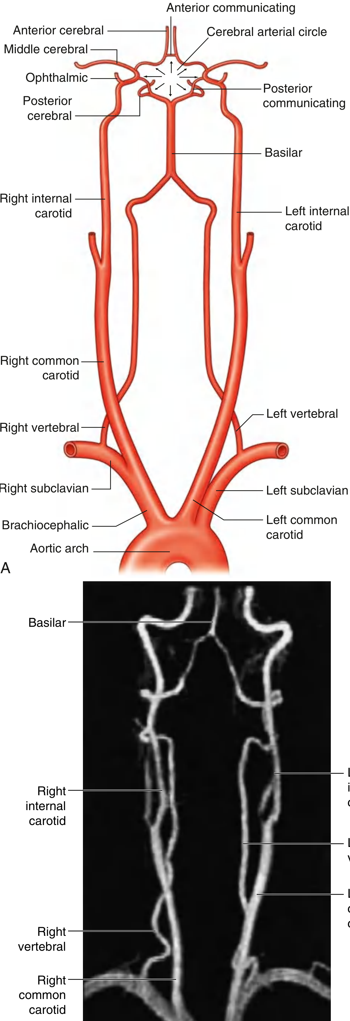
Key Point: The vertebral arteries together with the basilar artery constitute the vertebrobasilar system, which forms the posterior circulation of the brain. This system supplies the brainstem, cerebellum, occipital lobes, thalamus, and medial temporal lobes - structures controlling vital functions, balance, vision, and consciousness.
Sources: Gray's Anatomy for Students (ISBN 9780323934237), p. 1017-1018, 1322; Harrison's Principles of Internal Medicine 22E; Adams and Victor's Principles of Neurology 12th Ed.; Bradley and Daroff's Neurology
Medullary bracg?
The user is asking about medullary branches - the vascular supply to the medulla oblongata (branches of vertebral artery/PICA supplying the medulla). Let me search the library.
Searching sources...
Finding Sources
Finding Sources
Reading File
Finding Sources
Reading File
Finding Sources
Reading File
Finding Sources
Finding Sources
Searching Images
medulla oblongata cross section blood supply vascular territories medial lateral
Finding Sources
Finding Sources
Finding Sources
Loading Image
I now have all the information needed. Here is the complete answer:
Medullary Branches - Blood Supply to the Medulla Oblongata
The medulla is supplied by three groups of arteries, all branches of the vertebral artery and its derivatives, dividing the medulla into three vascular zones.
Flowchart: Medullary Blood Supply
VERTEBRAL ARTERY (intracranial)
│
├─── 1. PARAMEDIAN (ANTEROMEDIAL) BRANCHES
│ │
│ ├── Direct small perforators from Vertebral Artery
│ └── Anterior Spinal Artery branches
│ → Supply: MEDIAL MEDULLA
│ • Pyramid (corticospinal tract)
│ • Medial lemniscus
│ • Hypoglossal nucleus & CN XII fascicles
│
├─── 2. LATERAL (SHORT CIRCUMFERENTIAL) BRANCHES
│ │
│ └── PICA (Posterior Inferior Cerebellar Artery)
│ → Supply: LATERAL MEDULLA (dorsolateral)
│ • Inferior cerebellar peduncle
│ • Vestibular nuclei
│ • Spinal trigeminal nucleus & tract (CN V)
│ • Spinothalamic tract
│ • Descending sympathetic fibers
│ • Nucleus ambiguus (CN IX, X)
│ • Nucleus solitarius (taste)
│ • Dorsal motor nucleus of vagus
│
└─── 3. POSTERIOR BRANCHES
│
└── Posterior Spinal Artery (from PICA)
→ Supply: POSTERIOR MEDULLA
• Gracile nucleus (lower limb proprioception)
• Cuneate nucleus (upper limb proprioception)
• Posterior column nuclei
The Three Vascular Zones of the Medulla
| Zone | Artery | Structures Supplied |
|---|---|---|
| Medial (anteromedial) | Paramedian branches of VA + Anterior Spinal A. | Pyramidal tract (corticospinal), Medial lemniscus, CN XII nucleus + fascicles |
| Lateral (dorsolateral) | PICA (+ VA lateral branches) | Inf. cerebellar peduncle, Vestibular nuclei, Spinal trigeminal n+t, Spinothalamic tract, Descending sympathetics, Nucleus ambiguus, Nucleus solitarius |
| Posterior | Posterior Spinal A. (from PICA) | Gracile & cuneate nuclei, posterior columns |
Clinical Syndromes (Key Exam Topics)
1. Medial Medullary Syndrome (Dejerine Syndrome)
Artery: Paramedian branches of VA or Anterior Spinal Artery
| Structure Damaged | Feature |
|---|---|
| Pyramid (corticospinal) | Contralateral hemiplegia (arm + leg, face spared) |
| Medial lemniscus | Contralateral loss of vibration + proprioception |
| CN XII fascicles / nucleus | Ipsilateral LMN tongue weakness (tongue deviates to side of lesion) |
Memory: Medial = Motor (pyramid) + proprioception (lemniscus) + CN XII The classic triad: contralateral hemiplegia + contralateral position/vibration loss + ipsilateral tongue weakness
2. Lateral Medullary Syndrome (Wallenberg Syndrome)
Artery: Vertebral artery (most common) > PICA (less common)
| Structure Damaged | Feature |
|---|---|
| Inferior cerebellar peduncle | Ipsilateral cerebellar ataxia (limb + gait) |
| Vestibular nuclei | Vertigo, nausea, vomiting, nystagmus |
| Spinal trigeminal nucleus | Ipsilateral face - loss of pain & temperature |
| Spinothalamic tract | Contralateral body - loss of pain & temperature |
| Descending sympathetic fibers | Ipsilateral Horner's syndrome (ptosis, miosis, anhidrosis) |
| Nucleus ambiguus (CN IX, X) | Hoarseness, dysphagia (ipsilateral) |
| Nucleus solitarius | Ipsilateral loss of taste |
Key: Motor power is PRESERVED (pyramids are medial, not affected) Crossed sensory loss = hallmark: ipsilateral face + contralateral body
3. Hemimedullary Syndrome (Babinski-Nageotte Syndrome)
- Combination of medial + lateral medullary infarction
- Caused by occlusion of the ipsilateral VA proximal to PICA and its anterior spinal artery branches
- Features: all findings of both syndromes above combined
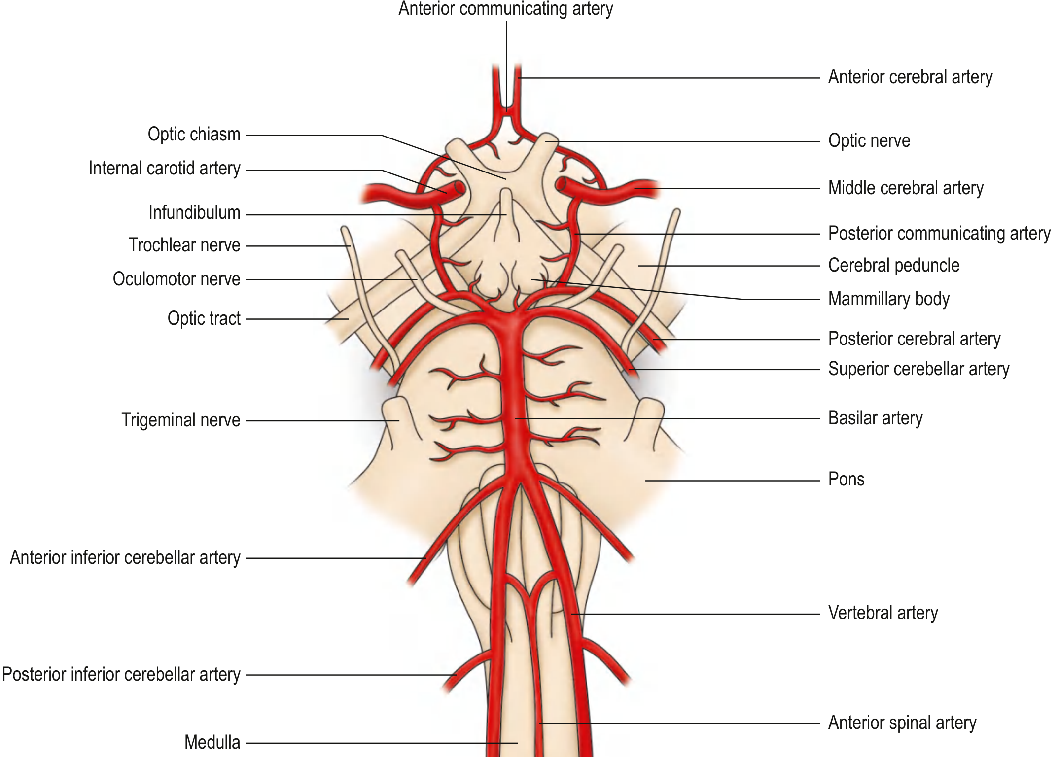
Summary Diagram
Quick Comparison: Medial vs Lateral Medullary
| Feature | Medial (Dejerine) | Lateral (Wallenberg) |
|---|---|---|
| Artery | VA paramedian / Ant. Spinal A. | VA (most common) or PICA |
| Motor | Contralateral hemiplegia | None (motor spared) |
| Sensation | Contralateral proprioception/vibration lost | Crossed: ipsilateral face + contralateral body pain/temp lost |
| CN | Ipsilateral CN XII (tongue) | Ipsilateral CN IX, X (dysphonia, dysphagia) |
| Special | - | Horner's, vertigo, ataxia |
| Prognosis | Moderate | Generally good |
Sources: Neuroanatomy through Clinical Cases, 3rd Ed. - Table 14.7; Localization in Clinical Neurology 8e; Bradley and Daroff's Neurology; Gray's Anatomy for Students
This is a shared conversation. Sign in to Orris to start your own chat.