
Give me easy to understand figure of secondary pulmonary lobule
secondary pulmonary lobule diagram anatomy
pulmonary lobule bronchiole acinus lung structure
A clear, labeled educational diagram of the secondary pulmonary lobule (of Miller). Show a polygonal lobule shape with connective tissue septa at the borders. Inside, label: 1-3 terminal bronchioles entering at the center, branching into respiratory bronchioles, then alveolar ducts and alveolar sacs. Show the centrilobular artery (pulmonary artery branch) running alongside the bronchiole in the center. Show pulmonary veins and lymphatics running in the peripheral septa. Add a small inset showing where the lobule sits within the lung. Use a clean medical illustration style with colored labels: blue for airways, red for arteries, purple for veins, green for lymphatics. Title the diagram "Secondary Pulmonary Lobule (of Miller)".
Secondary Pulmonary Lobule
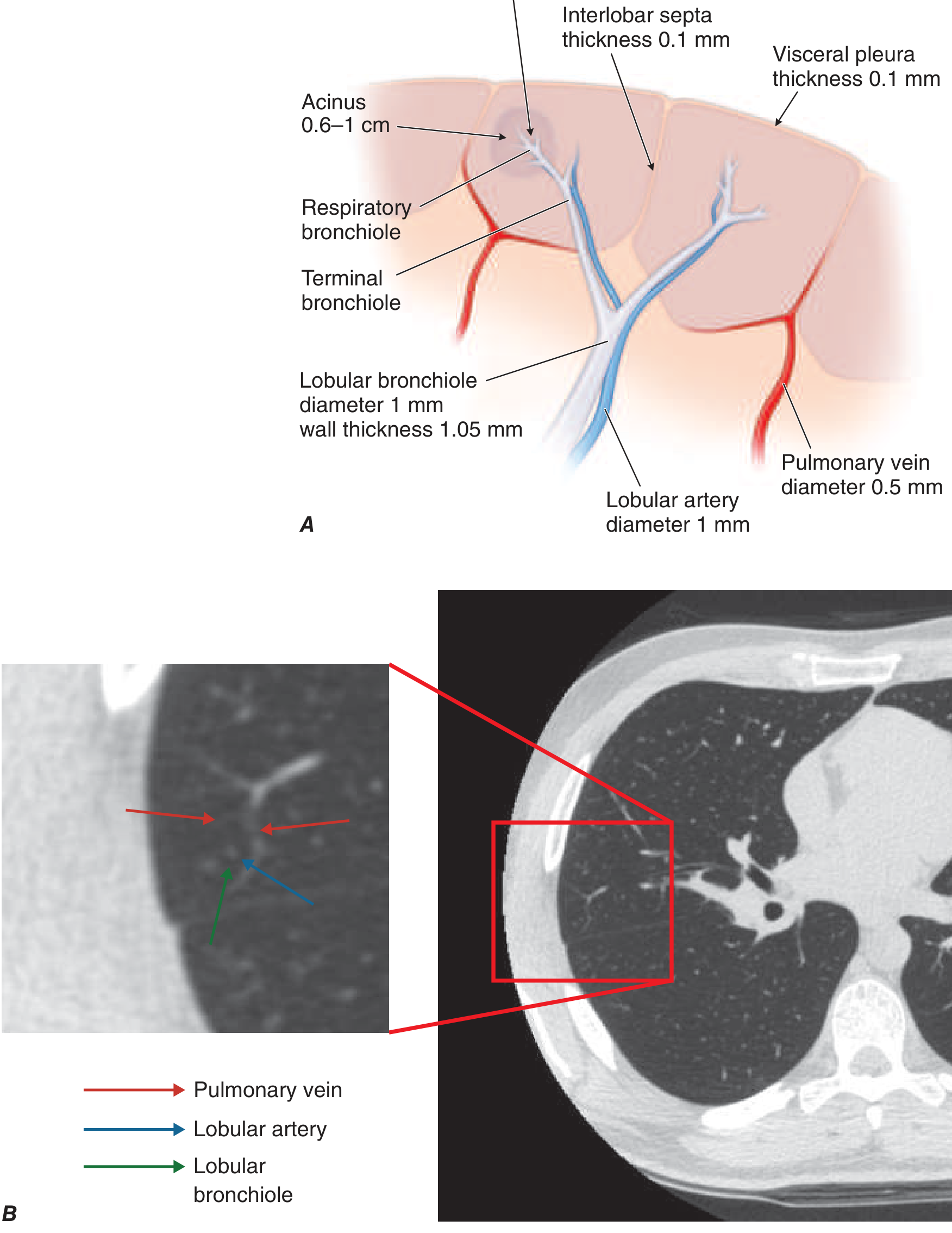
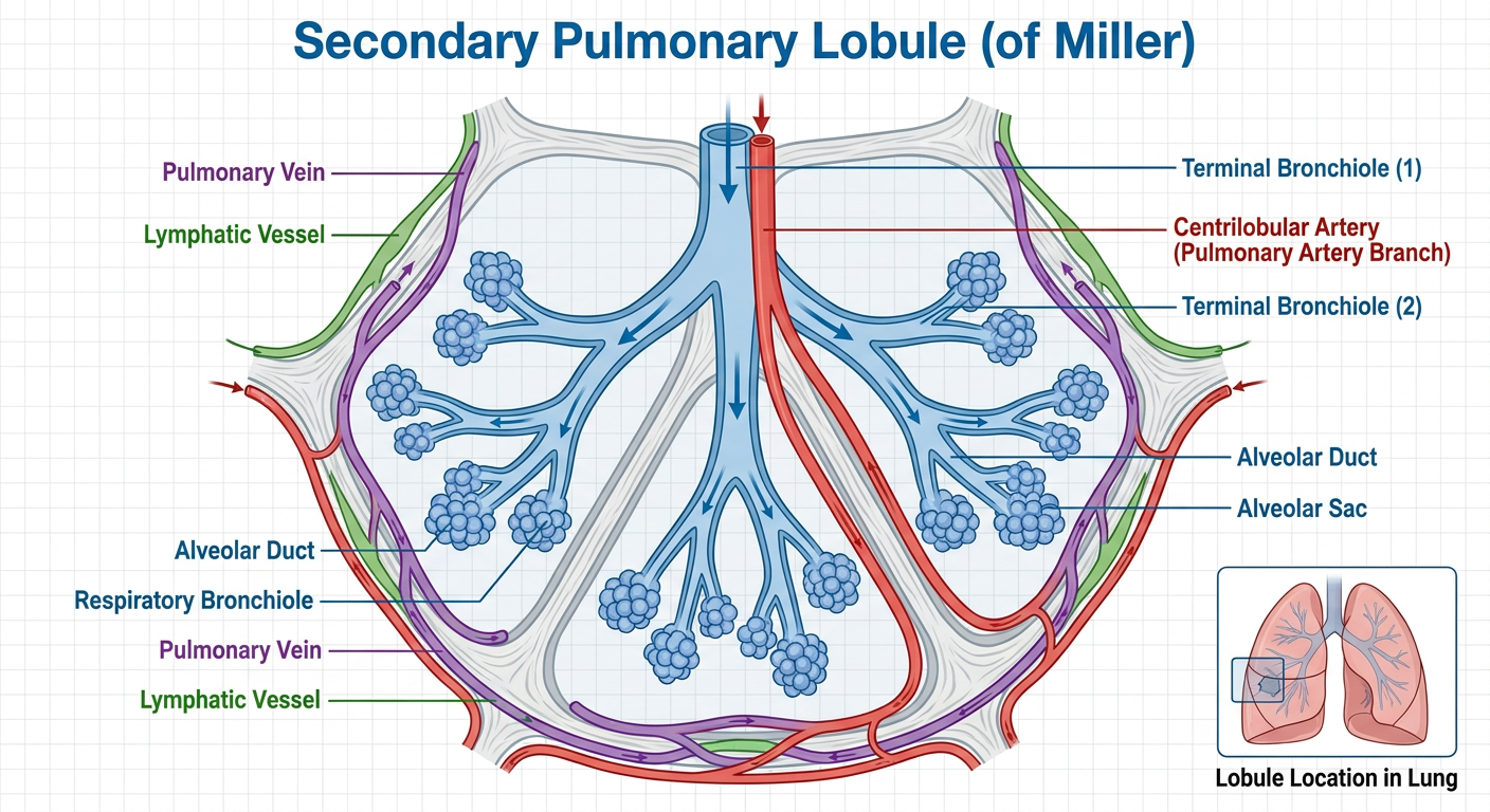
What is the Secondary Pulmonary Lobule?
Key Structures - Easy to Remember
CENTER (Centrilobular) - "The Core"
| Structure | Details |
|---|---|
| Lobular bronchiole | ~1 mm diameter, wall ~0.1 mm |
| Lobular artery (pulmonary artery branch) | ~1 mm diameter, runs alongside bronchiole |
| Terminal bronchiole | branches from lobular bronchiole |
| Respiratory bronchiole | where gas exchange begins |
| Acinus | 0.6-1 cm, the gas-exchange unit |
PERIPHERY (Periseptal) - "The Border"
| Structure | Details |
|---|---|
| Interlobular septa | connective tissue walls, ~0.1 mm thick |
| Pulmonary veins | ~0.5 mm, run in the septa |
| Lymphatics | also run in the septa |
| Visceral pleura | covers the outermost lobules, ~0.1 mm |
Simple Memory Aid
- The central pip = bronchiole + artery (centrilobular)
- The fleshy body = alveolar parenchyma
- The white pith border = interlobular septa with veins + lymphatics
Why It Matters Clinically (on CT/HRCT)
| Pattern | Location in Lobule | Disease Example |
|---|---|---|
| Centrilobular nodules | Center | Bronchiolitis, hypersensitivity pneumonitis |
| Centrilobular emphysema (CLE) | Core destruction | Smoking-related COPD |
| Paraseptal emphysema (PSE) | Peripheral destruction | Apical bullae |
| Panlobular emphysema (PLE) | Entire lobule | Alpha-1 antitrypsin deficiency |
| Interlobular septal thickening | Border/septa | Pulmonary edema, lymphangitic carcinomatosis, ILD |
Smooth vs Nodular vs Irregular septal thickening figure
smooth nodular irregular interlobular septal thickening HRCT comparison
interlobular septal thickening pulmonary edema lymphangitic carcinomatosis HRCT
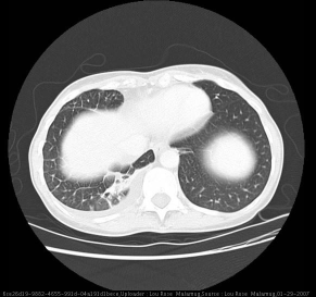
**Imaging Modality:** High-resolution Computed Tomography (HRCT) of the chest.
This diagnostic axial High-Resolution Computed Tomography (HRCT) image of the thorax demonstrates characteristic interstitial lung findings. The primary abnormality is diffuse, smooth interlobular septal thickening, which is most prominent in the anterior segment of the left lung's middle zone, creating a fine reticular pattern. No significant ground-glass opacities (GGO) are associated with this thickening at this level. Additionally, there is evidence of a small, localized pleural effusion along the posterior aspect of the left hemithorax. The mediastinal structures, including the heart and great vessels, are visible, though the primary educational focus remains on the parenchymal interstitial changes. Such patterns are clinically significant for various conditions, including pulmonary edema, lymphangitic carcinomatosis, or specific interstitial lung diseases. The image serves as an educational tool for identifying smooth septal thickening (Kerley lines on CT) and distinguishing secondary lobular anatomy in pulmonary radiology.
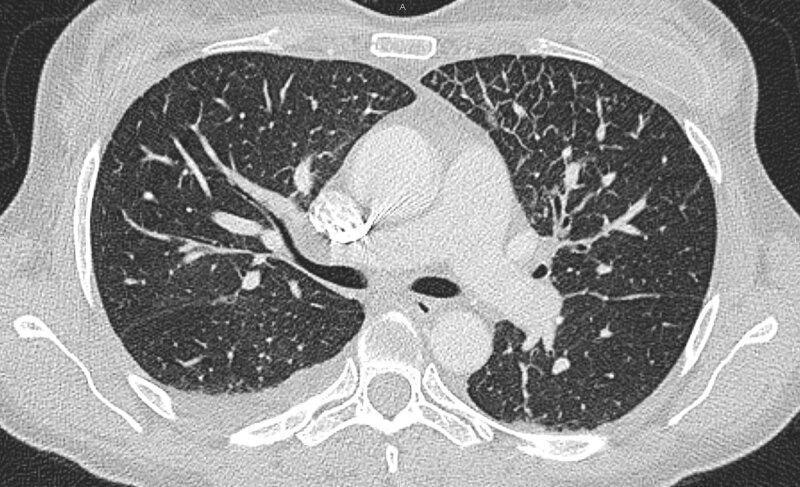
**Imaging Modality:** High-resolution computed tomography (HRCT) of the chest.
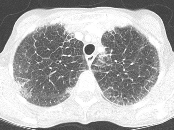
This diagnostic image is an axial high-resolution computed tomography (HRCT) scan of the chest shown in lung window. The scan demonstrates diffuse, bilateral abnormalities within the lung parenchyma, primarily characterized by significant smooth thickening of the interlobular septa. This results in a prominent reticular pattern throughout both lung fields. Additionally, there is evidence of peribronchovascular interstitium thickening, visualized as increased density and haziness surrounding the pulmonary vessels and bronchial structures. Small peripheral areas suggest the beginning of architectural distortion, though the overall appearance is dominated by the interstitial septal lines. These findings are clinically significant in the evaluation of interstitial lung diseases, pulmonary edema, or lymphangitic carcinomatosis. In this specific clinical context, these features represent pulmonary manifestations of amyloidosis. The image serves as an educational example of diffuse interstitial thickening and the resulting alteration of normal lung architecture as seen on diagnostic radiology.
irregular septal thickening usual interstitial pneumonia fibrosis CT
A clean medical educational comparison diagram showing three types of interlobular septal thickening on HRCT chest, displayed side by side as three panels. Each panel shows a schematic cross-section of 4-6 secondary pulmonary lobules (polygonal shapes). Panel 1 labeled "SMOOTH Septal Thickening": uniform, even, straight lines forming the lobule borders - causes listed: pulmonary edema, heart failure, lymphangitic carcinomatosis. Panel 2 labeled "NODULAR Septal Thickening": bumpy/beaded lines with small nodules along the septa - causes listed: sarcoidosis, lymphangitic carcinomatosis, silicosis. Panel 3 labeled "IRREGULAR Septal Thickening": distorted, jagged, uneven lines with architectural distortion of lobule shape - causes listed: IPF/UIP, pulmonary fibrosis, connective tissue disease. Use white background, clear black outlines, with red highlights on the abnormal septa in each pattern. Add a small CT appearance description below each panel.
traction bronchiectasis honeycombing IPF pulmonary fibrosis HRCT irregular reticulation
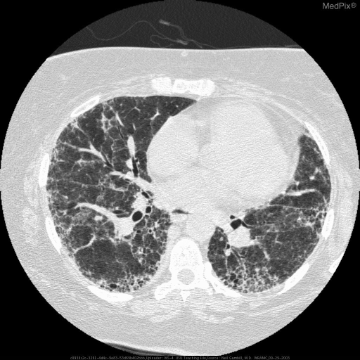
**Imaging Modality:** High-resolution computed tomography (HRCT) of the chest, axial section.
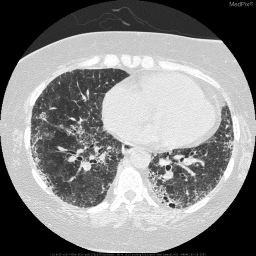
**Imaging Modality:** High-resolution computed tomography (HRCT) of the chest, axial section.
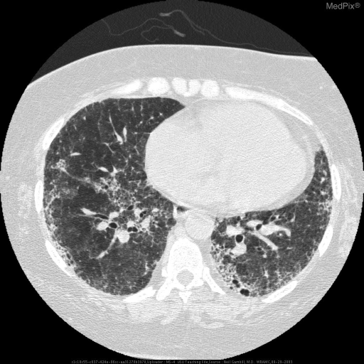
**Imaging Modality:** High-resolution computed tomography (HRCT) of the chest, axial section.
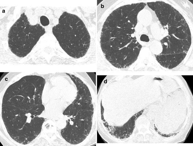
High-resolution computed tomography (HRCT) of the chest consisting of four axial slices (a–d) demonstrating interstitial lung disease. The images show a distribution of pulmonary fibrosis that is characteristically peripheral and subpleural, with a clear basilar predominance (visible in lower slices c and d). Key diagnostic features include fine reticulation (a network of linear opacities), traction bronchiectasis, and traction bronchiolectasis, representing irregular dilation of the airways due to surrounding parenchymal scarring. Notably, there is an absence of cystic honeycombing. This constellation of findings is most consistent with a 'possible Usual Interstitial Pneumonia (UIP)' pattern. Such imaging is critical in the evaluation of chronic fibrosing interstitial pneumonias, including idiopathic pulmonary fibrosis (IPF) and hypersensitivity pneumonitis, where the presence or absence of specific features like honeycombing helps determine the diagnostic certainty of the UIP pattern.
Interlobular Septal Thickening - Smooth vs Nodular vs Irregular
Comparison Diagram
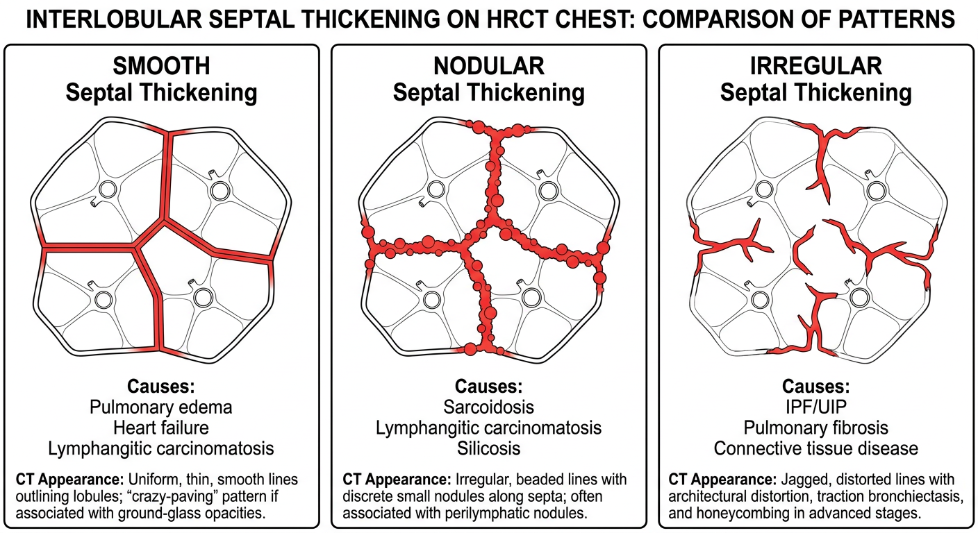
1. SMOOTH Septal Thickening
| Feature | Detail |
|---|---|
| Appearance | Straight, even, uniform lines |
| Mechanism | Fluid / protein / cells filling the septa uniformly |
| Key causes | Pulmonary edema (heart failure), lymphangitic carcinomatosis (early), pulmonary amyloidosis, pulmonary veno-occlusive disease (PVOD) |
| Associated findings | Pleural effusion, Kerley B lines on CXR |
2. NODULAR Septal Thickening
| Feature | Detail |
|---|---|
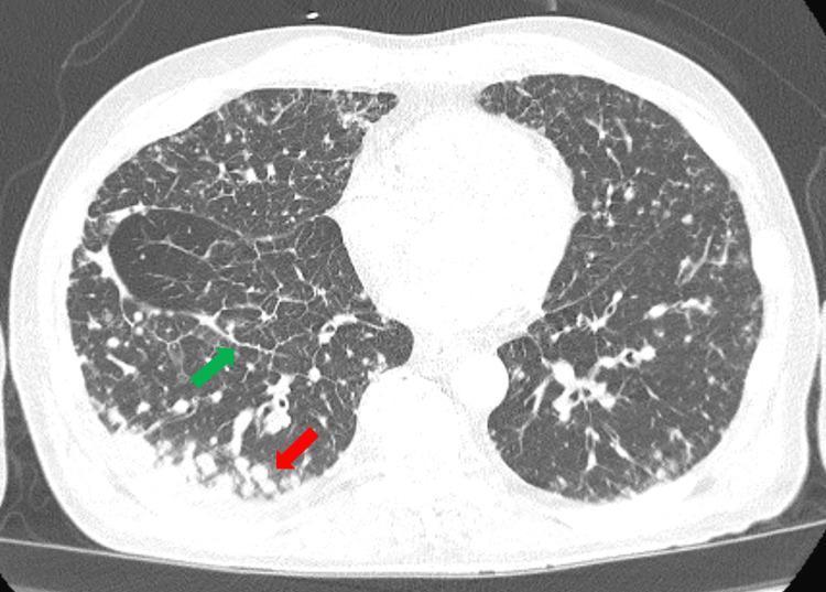
| Appearance | Bumpy, beaded septa - "string of pearls" |
| Mechanism | Granulomas or tumor deposits along lymphatics in septa |
| Key causes | Sarcoidosis, lymphangitic carcinomatosis, silicosis, coal worker's pneumoconiosis |
| Distribution | Perilymphatic - subpleural, along bronchovascular bundles, in fissures |
| Memory trick | "SaLS" = Sarcoid, Lymphangitic carcinomatosis, Silicosis |
3. IRREGULAR Septal Thickening
| Feature | Detail |
|---|---|
| Appearance | Distorted, non-uniform, angular lines; lobule shape destroyed |
| Mechanism | Fibrosis replacing and contracting the interstitium |
| Key causes | IPF/UIP, NSIP, connective tissue disease-ILD, hypersensitivity pneumonitis (fibrotic) |
| Associated findings | Traction bronchiectasis, honeycombing, architectural distortion |
| Distribution | Subpleural, basal > apical |
Quick Comparison Table
| Smooth | Nodular | Irregular | |
|---|---|---|---|
| Septa appearance | Straight, even | Beaded/bumpy | Jagged, distorted |
| Top cause | Pulmonary edema | Sarcoidosis | IPF/UIP |
| Mechanism | Fluid/tumor | Granulomas/deposits | Fibrosis |
| Reversible? | Often yes | Partially | No (fibrosis) |
| Pleural effusion | Common | Uncommon | Absent |
| Traction bronchiectasis | No | No | Yes |
| Distribution clue | Gravity-dependent | Perilymphatic | Subpleural, basal |
Memory Aid: "Smooth = Soaked" (fluid) | "Nodular = Nodding necklace" (beads = granulomas/tumor) | "Irregular = Irreversible" (fibrosis)