
Mydriarics and cycloplegics drugs refer standard books of ophthalmology
mydriatic cycloplegic drugs ophthalmology pupil dilation
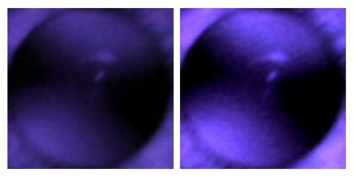
This diagnostic image pair displays infrared (IR) red reflex images of a human pupil, utilized in ophthalmology for assessing refractive errors like myopia and hyperopia. The side-by-side comparison demonstrates the effect of digital image processing on clinical diagnostic data. The left image shows the raw IR capture with lower brightness and diffused contrast. The right image illustrates the same pupil after increasing contrast, which enhances the visibility of crescent-shaped light reflections (photorefractive crescents) within the pupillary aperture. These crescents are critical clinical markers; their morphology and spatial distribution (merged vs. far apart) are used to predict spherical power and refractive state. The images were captured using a non-mydriatic handheld fundus camera, which allows for anterior segment viewing without inducing pupillary constriction. This visual comparison highlights how contrast adjustment can clarify diagnostic features for both manual clinical grading and deep-learning-based AI classification in vision screening.
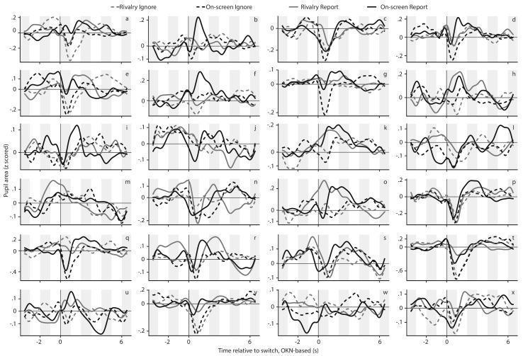
This Comparison Chart displays per-observer pupil response curves under four experimental conditions of binocular rivalry and on-screen replay. The figure is organized into a 6x4 grid of panels (labeled a through x), with each panel representing data from one of 24 unique human observers. The x-axis for every plot denotes the time in seconds relative to a perceptual switch (ranging from -3s to +6s), identified using an optokinetic nystagmus (OKN)-based method. The y-axis represents the z-scored pupil area. Each panel contains four curves corresponding to the study's 2x2 factorial design: Rivalry Ignore (dashed light gray), On-screen Ignore (dashed black), Rivalry Report (solid light gray), and On-screen Report (solid black). These diagnostic-style plots demonstrate the variability and consistency of pupillary constriction (C1) and subsequent dilation (D1) following perceptual switches. The visual material illustrates key concepts in ophthalmology and neuroscience, specifically relating to how the autonomic nervous system and visual cortex modulate pupil size during endogenous and exogenous shifts in perception, with clinical relevance to understanding the neurophysiological markers of cognitive load and sensory transitions.
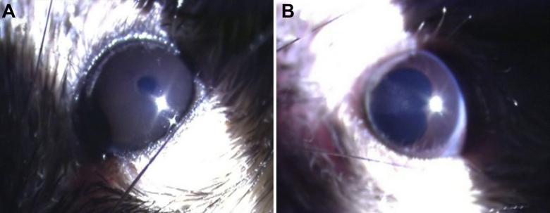
This clinical photograph displays a comparative analysis of the anterior segment of the eye in a medical research model. Image A depicts a healthy eye with a clear anterior chamber and a regular, circular pupil, showing no signs of inflammation. Image B demonstrates pathological changes consistent with Acute Anterior Uveitis (AAU). Key features in image B include an irregular, kidney-shaped pupil following the administration of mydriatic agents. The presence of posterior synechia—adhesions between the iris and the lens—is evident, which prevents uniform pupillary dilation. This side-by-side comparison illustrates the visual hallmarks of an ocular inflammatory response and its effect on pupillary morphology and function. The imagery is essential for studying inflammatory pathways, particularly those involving Toll-like receptor (TLR4) or lipopolysaccharide (LPS)-induced endotoxin uveitis models. Educational concepts include ocular pathology, iris-lens adhesions, and the clinical manifestations of anterior segment inflammation.
Mydriatics and Cycloplegics - Standard Ophthalmology Reference
Definitions
- Goodman & Gilman's Pharmacological Basis of Therapeutics (Table 74-7)
- Katzung's Basic and Clinical Pharmacology, 16th Ed., p.204
Mechanism of Action
Anticholinergic (Muscarinic Antagonist) Cycloplegics/Mydriatics
- Block M3 muscarinic receptors on the sphincter pupillae → pupil dilation (mydriasis)
- Block M3 receptors on the ciliary muscle → paralysis of accommodation (cycloplegia)
- The ciliary muscle receives exclusively parasympathetic innervation; thus, anticholinergics produce complete, reliable cycloplegia.
Sympathomimetic (Pure) Mydriatics
- Act on alpha-1 (α1) adrenergic receptors on the radial/dilator pupillae muscle → pupil dilation only
- No cycloplegic effect - accommodation is preserved
- Example: Phenylephrine
"The dilator smooth muscle is oriented radially and is innervated by the sympathetic nervous system, which causes mydriasis (dilation). At the pupillary margin, the sphincter smooth muscle is organized in a circular band with parasympathetic innervation, which, when stimulated, causes miosis (constriction)."
- Goodman & Gilman's, p.1481
Classification of Drugs
A. Pure Mydriatics (No Cycloplegia)
| Drug | Concentration | Onset | Duration | Mechanism |
|---|---|---|---|---|
| Phenylephrine | 2.5%, 10% | 20 min | 3 hours | α1-adrenergic agonist |
| Hydroxyamphetamine | 1% | 20-30 min | 3-4 hours | Indirect sympathomimetic |
| Adrenaline (epinephrine) | 0.1% | Variable | Short | α1-adrenergic agonist |
"Alpha-adrenocortical stimulant drugs, e.g., phenylephrine, produce a short-lasting mydriasis that is usually sufficient for funduscopic examination. Antimuscarinic drugs should never be used for the sole purpose of mydriasis unless cycloplegia or prolonged action is required."
- Katzung's Basic and Clinical Pharmacology, p.204
B. Cycloplegic-Mydriatics (Anticholinergics) - In Order of Increasing Duration
| Drug | Concentration | Onset (Cycloplegia) | Duration of Cycloplegia | Duration of Mydriasis |
|---|---|---|---|---|
| Tropicamide | 0.5%, 1% | 20-30 min | 15-60 min | 3-6 hours |
| Cyclopentolate | 0.5%, 1%, 2% | 20-45 min | 3-6 hours | 24 hours |
| Homatropine | 2%, 5% | 20-90 min | 12-24 hours | 2-3 days |
| Scopolamine (Hyoscine) | 0.25% | 20-45 min | 3-7 days | 4-7 days |
| Atropine | 0.5%, 1%, 2% | 30-40 min | 5-6 days | 1-2 weeks |
Individual Drug Profiles
1. Atropine
- Most potent and longest-acting anticholinergic cycloplegic
- Concentration: 0.5% (children <12 months), 1% (adults/older children); available as drops and ointment
- Cycloplegia onset: 30-40 min; duration: 5-6 days; mydriasis lasts 1-2 weeks
- Indications: Cycloplegic refraction (especially high hypermetropia or heavily pigmented irides), uveitis/iritis (prevents posterior synechiae), amblyopia penalization therapy, myopia control (low-dose)
- Adverse effects: Photophobia, blurred vision; systemic - flushing, fever, restlessness, dry mouth, tachycardia, urinary retention (especially in children - "mad as a hatter, dry as a bone, hot as a hare, red as a beet")
- Key clinical note (Kanski's): "In a young child the risk of penalization amblyopia should be avoided by always inducing cycloplegia in both eyes at one sitting, particularly if atropine is used." Parents should be instructed to stop drops if signs of systemic toxicity (flushing, fever, restlessness) occur.
2. Scopolamine (Hyoscine)
- Potency and duration: intermediate between homatropine and atropine
- Duration: 3-7 days cycloplegia
- Less commonly used in routine clinical practice
- Also used for motion sickness (transdermal patch)
3. Homatropine
- Synthetic derivative of atropine
- Cycloplegia lasts 12-24 hours; mydriasis lasts 2-3 days
- Particularly valuable for preventing/breaking posterior synechiae in uveitis/iritis
- Used when intermediate duration is needed (Kanski's notes homatropine for CAU management)
4. Cyclopentolate
- The most common agent for cycloplegic refraction in children
- Concentration: 0.5% (infants <6 months), 1% (most children), 2% (darkly pigmented irides)
- Onset: 20-45 min; cycloplegia lasts 3-6 hours, mydriasis lasts 24 hours
- Protocol (Kanski's): "One drop, repeated after 5 minutes, usually results in maximal cycloplegia within 30 minutes, with recovery of accommodation within 2-3 hours."
- Tip: Topical anaesthetic (proxymetacaine) prior to instillation reduces reflex tearing and improves retention.
- Systemic side effects rare but include CNS effects (disorientation, hallucinations) in neonates
5. Tropicamide
- Shortest-acting cycloplegic/mydriatic
- Cycloplegia: 15-60 min; mydriasis: 3-6 hours
- Most commonly used for routine dilated fundus examination in adults
- Incomplete cycloplegia - not ideal for precise cycloplegic refraction in children
- Usually combined with phenylephrine 2.5% for routine dilation
Standard Dilating Regimens (Wills Eye Manual)
Clinical Indications
| Indication | Preferred Agent |
|---|---|
| Routine fundus examination (adults) | Tropicamide 1% + Phenylephrine 2.5% |
| Cycloplegic refraction (children) | Cyclopentolate 1% (preferred), Atropine if needed |
| Cycloplegic refraction (high hypermetropia/pigmented irides) | Atropine 1% |
| Anterior uveitis/iritis - acute | Atropine 1% or Homatropine 5% + Phenylephrine 2.5% |
| Anterior uveitis - chronic/mild | Cyclopentolate 1% at bedtime |
| Breaking/preventing posterior synechiae | Homatropine 5%, Atropine; Mydricaine No. 2 (subconjunctival: atropine + adrenaline + procaine) for resistant PS |
| Amblyopia penalization | Atropine 1% |
| Myopia control | Atropine 0.01% (low-dose) |
"Cycloplegic agents are used in AAU to prevent the formation of posterior synechiae (PS), to break down recently formed synechiae, and to promote comfort by relieving spasm of the pupillary and ciliary muscle. Commonly used anticholinergic agents in order of increasing potency and duration include cyclopentolate (duration 12-24 h), homatropine (3 days) and atropine (7-10 days)."
- Kanski's Clinical Ophthalmology, 10th Ed., p. block3
Contraindications
- Angle-closure glaucoma (narrow anterior chamber angles): Pupil dilation can precipitate acute angle-closure attack by pushing the iris against the trabecular meshwork. Dilating drops are contraindicated in most types of angle-closure glaucoma.
- Shallow anterior chamber: Must be assessed before dilation.
- Systemic anticholinergic sensitivity: Use with caution in elderly (urinary retention, confusion) and children (systemic toxicity).
"Dilating drops are contraindicated in most types of angle-closure glaucoma and in eyes with severely narrow anterior chamber angles. Dilating drops tend to be less effective at the same concentration in darkly pigmented eyes."
- Wills Eye Manual, Appendix A.1
Special Considerations
- Iris pigmentation: Darkly pigmented eyes are less sensitive and require higher concentrations or repeated doses. The cycloplegic effect may be more prolonged in lightly pigmented eyes. (Roberts & Hedges' Clinical Procedures in Emergency)
- Nasolacrimal duct drainage: Digital pressure on the nasolacrimal duct after instillation (or use of ointment form) reduces systemic absorption and toxicity risk, especially in children.
- Mydricaine No. 2 (Kanski's): A subconjunctival injection of adrenaline 0.12 mg + atropine 1 mg + procaine 6 mg in 0.3 mL used to break fresh posterior synechiae when topical drops fail. Cardiovascular monitoring required.
Autonomic Pharmacology Summary

Key Takeaway: Mydriatic vs. Cycloplegic
| Property | Phenylephrine (pure mydriatic) | Tropicamide/Cyclopentolate/Atropine (cycloplegics) |
|---|---|---|
| Pupil dilation | Yes | Yes |
| Cycloplegia | No | Yes |
| Receptor | α1 adrenergic | Muscarinic (M3) |
| Used for refraction | No | Yes |
| Used for fundus exam | Yes (with tropicamide) | Yes |
- Wills Eye Manual (9th Ed.), Appendix A.1 - Dilating Drops
- Kanski's Clinical Ophthalmology: A Systematic Approach, 10th Ed. - Cycloplegia, Uveitis Management
- Katzung's Basic and Clinical Pharmacology, 16th Ed., Table 8-2, p.204
- Goodman & Gilman's Pharmacological Basis of Therapeutics, Table 74-7
- Lippincott Illustrated Reviews: Pharmacology - Atropine, Ophthalmic uses