
Ischemic colitis – aetiology, pathogenesis, clinical features, complications, management
Reading File
Now I'll run parallel searches across the library and PubMed.
Finding Sources
Finding Sources
Searching PubMed
"ischemic colitis"[MeSH Terms] AND management
Excellent - I have rich sources. Now I'll read the main textbook sections in parallel.
Reading File
Reading File
Reading File
Good content. Let me get more from Sabiston (management, complications, risk factors) and Frameworks (clinical features, imaging, treatment):
Reading File
Reading File
Let me get risk factors, classification, and the Sabiston section on clinical features and diagnosis:
Reading File
Reading File
Let me get the remaining Sabiston sections on clinical features, diagnosis and the Current Surgical Therapy section:
Reading File
Reading File
I now have comprehensive information from Sabiston, Frameworks for Internal Medicine, and Yamada's Gastroenterology. Let me fetch the classification/staging image and the flowchart diagram, then compose the full answer.
Loading Image
Loading Image
I now have all the material needed for a comprehensive answer. Let me compile it.
Ischemic Colitis
Ischemic colitis (IC) is the most common form of intestinal ischemia, accounting for more than half of all cases, with an incidence of 7.1-22.9 per 100,000 person-years. It occurs when arterial blood supply to the colon falls below the threshold needed to support cellular metabolic demands. Severity spans a wide spectrum from transient, self-limiting mucosal injury to full-thickness gangrene with perforation.
Sabiston Textbook of Surgery, 21e | Frameworks for Internal Medicine | Yamada's Textbook of Gastroenterology, 7e
Anatomy - The Foundation for Understanding IC
The colon is supplied by two major vessels arising from the aorta:
- Superior mesenteric artery (SMA): ileocolic, right colic, and middle colic branches - supplies ascending colon and proximal two-thirds of transverse colon
- Inferior mesenteric artery (IMA): left colic, sigmoid branches, and superior rectal artery - supplies the remainder
Collateral networks:
- Marginal artery of Drummond - runs parallel to the mesenteric margin from cecocolic to rectosigmoid junction; receives flow from both SMA and IMA terminal branches. Absent at the splenic flexure in up to 18% of people.
- Arc of Riolan (meandering mesenteric artery) - an inconstant vessel connecting the SMA/middle colic to the IMA/left colic near the mesenteric root. When present and prominent, it often signals occlusion of one of the major vessels.
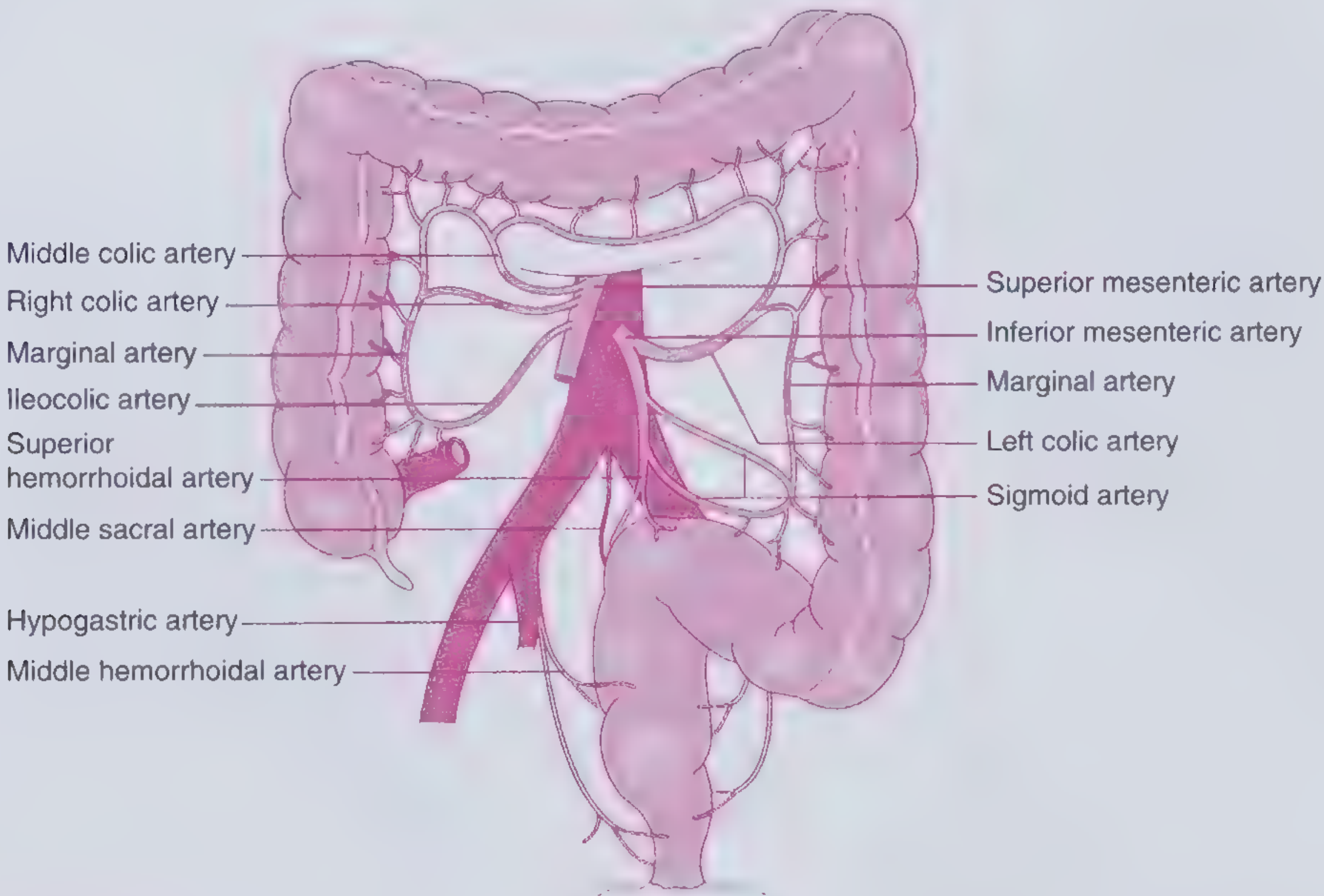
Watershed areas - regions where collateral circulation is inconsistent and therefore most vulnerable:
| Watershed Area | Anatomical Name | Basis |
|---|---|---|
| Splenic flexure | Griffiths point | Distal SMA territory meets distal IMA territory; marginal artery absent here in up to 50% |
| Rectosigmoid junction | Sudeck's point | End of IMA territory; sparse collaterals from internal iliac |
| Right colon / ileocecal region | - | Less well-developed collateral plexus |
Aetiology
IC is broadly classified into nonocclusive (vast majority) and occlusive causes.
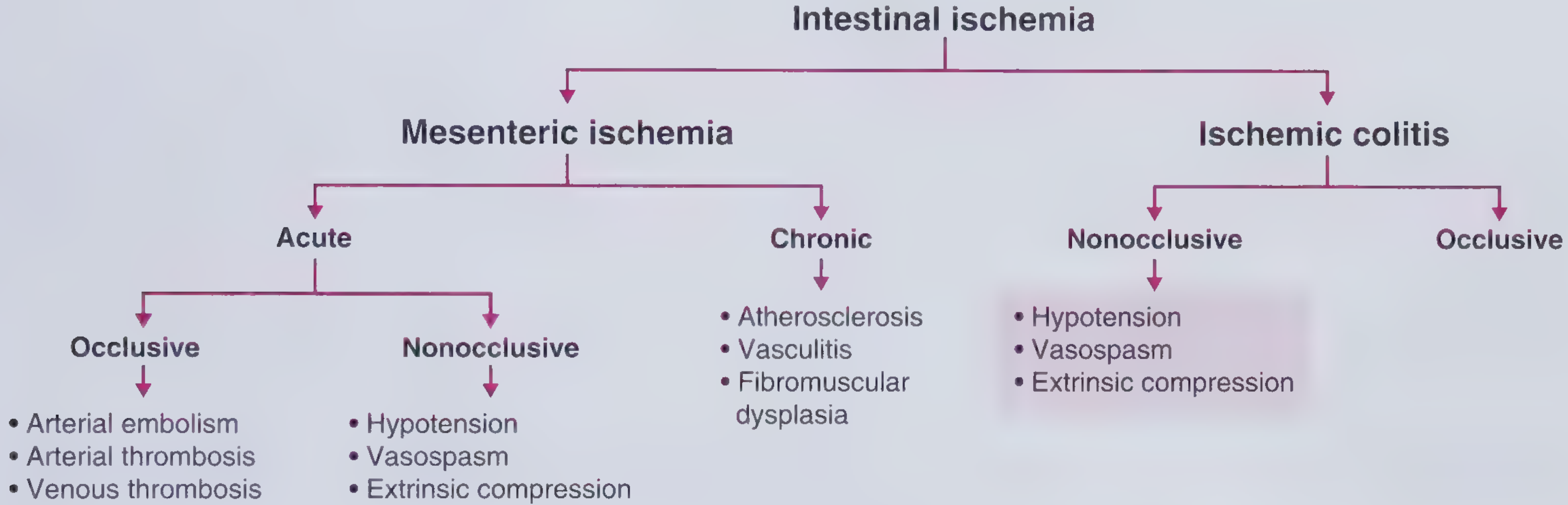
Nonocclusive Causes (most common)
These affect predominantly the watershed regions.
1. Hypoperfusion / low-flow states:
- Septic shock, hemorrhagic shock, cardiogenic shock
- Congestive heart failure
- Dehydration
- Post-major surgery (especially aortic surgery, AAA repair with IMA ligation)
2. Vasospasm:
- Cocaine and methamphetamine use (particularly in young patients without vascular risk factors)
- Vasopressor drugs (e.g., norepinephrine)
- Serotonergic drugs: sumatriptan, alosetron
- Decongestants: pseudoephedrine
3. Extrinsic compression:
- Diverticulitis, volvulus, tumor, adhesions, intestinal prolapse causing raised intraluminal pressure
4. Long-distance running/strenuous exercise - splanchnic vasoconstriction to redirect blood to muscles; approximately a quarter of marathon runners have occult rectal bleeding afterward.
Occlusive Causes (less common)
Affect the territory of the occluded vessel rather than watershed areas.
- Arterial embolism (e.g., from atrial fibrillation) - IMA rarely involved due to its small caliber compared with SMA
- Arterial thrombosis - on a background of atherosclerosis
- Venous thrombosis - in hypercoagulable states (nephrotic syndrome, antiphospholipid syndrome, OCP use, malignancy)
- Iatrogenic - aortoiliac instrumentation, endovascular AAA repair, post-colonoscopy
Drug-Related Causes
| Category | Examples |
|---|---|
| Vasoactive/vasospastic | Cocaine, methamphetamine, sumatriptan, pseudoephedrine, vasopressors |
| Constipation-inducing | Opioids, other constipation-inducing agents |
| Hormonal | Oral contraceptives, female hormones |
| Immunomodulatory | Anti-TNF-α agents, interferon-α/β |
| Chemotherapy | Taxanes |
| Prokinetics/bowel prep agents | Kayexalate (in sorbitol suspension - osmotic ischemia) |
Infectious Causes
- E. coli O157:H7 (enterohemorrhagic, Shiga toxin-producing): the prototype - direct cytotoxic effect on endothelium causes mucosal ischemia ± hemolytic uremic syndrome; fibrin thrombi seen on biopsy
- Clostridioides difficile: can produce ischemic-pattern injury
Risk Factors
| Category | Specific Conditions |
|---|---|
| Demographic | Age >60, female sex (female preponderance) |
| Vascular | Hypertension, diabetes mellitus, dyslipidemia, atherosclerosis, ischemic heart disease, cerebrovascular disease, PVD |
| Cardiac | Atrial fibrillation, heart failure |
| GI | Constipation, IBS, prior abdominal surgery |
| Haematologic | Hypercoagulable states, sickle cell disease |
| Respiratory | COPD |
| Autoimmune | SLE, rheumatoid arthritis |
Pathogenesis
The colon receives less blood flow per gram of tissue than the small intestine, and its microvasculature is buried within a thicker wall - making it inherently more susceptible to hypoperfusion.
When blood flow falls below a critical threshold:
- Mucosal ischaemia develops first (mucosa is the most metabolically active layer and farthest from the submucosal vessels)
- Reperfusion injury from reactive oxygen species compounds the damage
- Failure of the intestinal epithelial barrier allows bacterial translocation
- Progression depends on severity and duration of ischaemia:
- Reversible mucosal/submucosal ischaemia - most cases (~80%); resolves with supportive care
- Irreversible transmural ischaemia - gangrene, perforation, peritonitis
Histological features of ischaemic colitis include:
- Marked surface injury with mucin loss
- "Atrophic microcrypts" and crypt dropout
- Hyalinisation of the lamina propria ("lamina propria collapse")
- In infectious (E. coli O157:H7) cases: prominent fibrin thrombi
Clinical Features
IC is a disease predominantly of the elderly (vast majority >60 years). Presentation varies by depth of ischaemia and location.
Symptoms
| Feature | Detail |
|---|---|
| Abdominal pain | Sudden-onset mild-to-moderate crampy pain, typically left lower quadrant; pain usually precedes bleeding |
| Rectal bleeding / hematochezia | Passage of bloody stool within 24 hours; usually minor and rarely requires transfusion |
| Tenesmus | Common |
| Nausea, vomiting, anorexia | Variable |
| Low-grade fever | May be present |
| Abdominal distension | Especially with ileus |
The classic triad of sudden crampy LLQ pain + tenesmus + passage of bloody stool within 24 hours is present in approximately 50% of patients.
Location-Based Variants
- Left colon (including splenic flexure): most common location; typical presentation above
- Sigmoid colon: second most common
- Isolated right-sided IC: ~25% of cases; more likely to present with abdominal pain without bleeding; more often associated with atrial fibrillation, CAD, and chronic renal failure; higher rate of surgical intervention and poorer prognosis
- Pancolitis: rare; carries the worst prognosis
Physical Examination
- Mild-to-moderate tenderness over the affected colon segment
- Abdominal distension
- Signs of peritonitis (guarding, rigidity, rebound) when bowel perforation has occurred
Investigations
Laboratory:
- Often non-specific in mild disease
- Severe disease: leukocytosis with neutrophilia, elevated BUN, elevated LDH, elevated lactate, decreased haemoglobin, decreased albumin, metabolic acidosis, low bicarbonate
- Send stool for C. difficile toxin, culture and sensitivity, ova and parasites (to exclude infectious aetiology)
Imaging:
| Modality | Findings |
|---|---|
| Plain abdominal X-ray | Bowel distension; "thumbprinting" (rounded densities along colon wall from submucosal oedema); free air = perforation |
| CT abdomen (with IV + oral contrast) - primary modality | Segmental bowel wall thickening in a vascular distribution; pericolonic fat stranding; thumbprinting; pneumatosis intestinalis; portal venous gas (ominous sign); pneumoperitoneum |
| CT angiography | Limited role in IC (unlike acute mesenteric ischaemia); rarely needed |
| Water-soluble contrast enema | Largely obsolete; still useful for evaluating chronic ischaemic strictures |
Endoscopy (colonoscopy):
- Most sensitive and specific modality when performed within 48 hours
- Visual inspection confirms mucosal ischaemia; biopsy differentiates IC from IBD or infectious colitis
- Must be performed with caution - risk of exacerbating ischaemia and perforation (low-pressure insufflation; avoid going beyond affected segment)
- Typical findings: pale/cyanotic mucosa, mucosal oedema, submucosal haemorrhage, ulceration, in severe cases necrosis
Complications
| Complication | Notes |
|---|---|
| Bowel necrosis and perforation (gangrenous colitis) | Full-thickness transmural ischaemia; peritonitis, haemodynamic instability; requires emergency surgery; mortality up to 47% |
| Sepsis / bacterial translocation | Breakdown of mucosal barrier; gram-negative bacteraemia |
| Chronic ischaemic colitis | Irreversible ischaemic injury; chronic diarrhoea, rectal bleeding, weight loss |
| Colonic stricture | Fibrosis at the ischaemic segment; presents with constipation, narrowed stools, colicky pain; diagnosed with contrast enema, CT, or colonoscopy |
| Haemolytic uraemic syndrome | Specific to E. coli O157:H7-mediated IC; develops in up to 20% |
Management
Conservative (Non-operative) - ~80% of Cases
The mainstay for non-gangrenous ischaemic colitis:
- Bowel rest (nil by mouth)
- IV fluid resuscitation - correct hypovolaemia and low-flow states
- Broad-spectrum antibiotics - cover both aerobic and anaerobic gut flora to prevent/treat bacterial translocation (e.g., piperacillin-tazobactam or cephalosporin + metronidazole)
- Nasogastric tube if ileus is present
- Optimise cardiac output - treat heart failure, sepsis, arrhythmias
- Discontinue offending medications - vasopressors where possible, constipation-inducing drugs, OCP, cocaine
- Avoid cathartics (risk of perforation) and glucocorticoids (unless treating underlying SLE/RA)
Adjunctive pharmacological agents that may improve blood flow and mucosal healing:
- Phosphodiesterase type 5 inhibitors
- Pentoxifylline
- Prostaglandin E1
Monitoring: Serial clinical examination, repeat imaging or colonoscopy if failure to improve within a few days, to detect progression to full-thickness ischaemia.
Most patients show significant improvement within 2-3 days.
Elective Surgical Management
Indicated for:
- Chronic ischaemic colitis with ongoing/recurrent bouts of abdominal pain, bloody diarrhoea, and sepsis (higher complication rate; requires segmental resection)
- Symptomatic colonic stricture after recovery from IC (or when malignancy cannot be excluded)
Emergency Surgical Management
Indications:
- Peritonitis
- Haemodynamic instability
- Pneumoperitoneum (free air)
- Portal venous gas on CT
- Failure to improve or clinical deterioration on conservative management
Operative principles:
- Examine entire small and large bowel for ischaemia
- Anatomic resection of the ischaemic segment with preservation of the marginal artery
- Anastomosis is generally avoided in the acute setting (risk of anastomotic leak due to evolving ischaemia, haemodynamic instability, sepsis)
- Staple ends in situ; temporary abdominal closure with planned second-look laparotomy at 24 hours to assess viability and extent of resection required
- Pancolitis: total colectomy with end ileostomy
Intraoperative adjunct: Infrared angiography with indocyanine green (ICG) is an emerging technique to assess bowel viability and anastomotic perfusion in real time.
Prognosis
- ~80% resolve with conservative management
- 30-day postoperative mortality after colectomy for IC: ~25-47%
- Independent predictors of perioperative mortality: elderly age, poor functional status, multiple comorbidities, preoperative septic shock, blood transfusions, acute renal failure, delay from admission to surgery
- Isolated right-sided IC carries a worse prognosis than left-sided disease
Sources: Sabiston Textbook of Surgery 21e, Ch. 95 | Frameworks for Internal Medicine, Ch. 18 | Yamada's Textbook of Gastroenterology 7e, Ch. 142
MESENTERIC ISCHEMIA
Finding Sources
Finding Sources
Good - rich sources identified. Now reading 4 key sections in parallel:
Reading File
Reading File
Reading File
Reading File
Excellent content. Now reading more from Sabiston (pathophysiology per subtype + management) and Sleisenger (diagnosis/treatment details) and the Schwartz management section:
Reading File
Reading File
Reading File
Excellent material from Sabiston and Sleisenger. Now reading the management/treatment sections from Sleisenger plus chronic mesenteric ischemia and MVT sections from Sabiston:
Reading File
Reading File
Now reading the MVT section and the acute AMI treatment algorithm from Sleisenger, plus key images:
Reading File
Reading File
Loading Image
I now have comprehensive information from all major sources. Let me compile the full structured answer.
Mesenteric Ischemia
Mesenteric ischemia refers to ischemia of the small intestine (as distinguished from ischemic colitis, which involves the large intestine). It is classified into acute mesenteric ischemia (AMI) and chronic mesenteric ischemia (CMI), with arterial disease being more common than venous, and acute forms more common than chronic. Despite advances in management, overall mortality remains high at 17-21% among patients requiring intervention.
Sabiston Textbook of Surgery 21e | Sleisenger & Fordtran's Gastrointestinal and Liver Disease | Schwartz's Principles of Surgery 11e | Frameworks for Internal Medicine
Anatomy
The entire small intestine (except the proximal duodenum, which is supplied by the celiac axis via the pancreaticoduodenal arcades) is supplied by the superior mesenteric artery (SMA), which arises directly from the aorta. The SMA branches include:
- Inferior anterior/posterior pancreaticoduodenal arteries
- Middle colic artery (transverse colon)
- Right colic artery
- Ileocolic artery
- Jejunal and ileal branches
An extensive arcade system (primary, secondary, tertiary arcades) gives rise to the arteriae rectae (vasa recta), providing rich collateral circulation that protects the small intestine from hypoperfusion - making AMI from pure low-flow states less common than ischemic colitis.
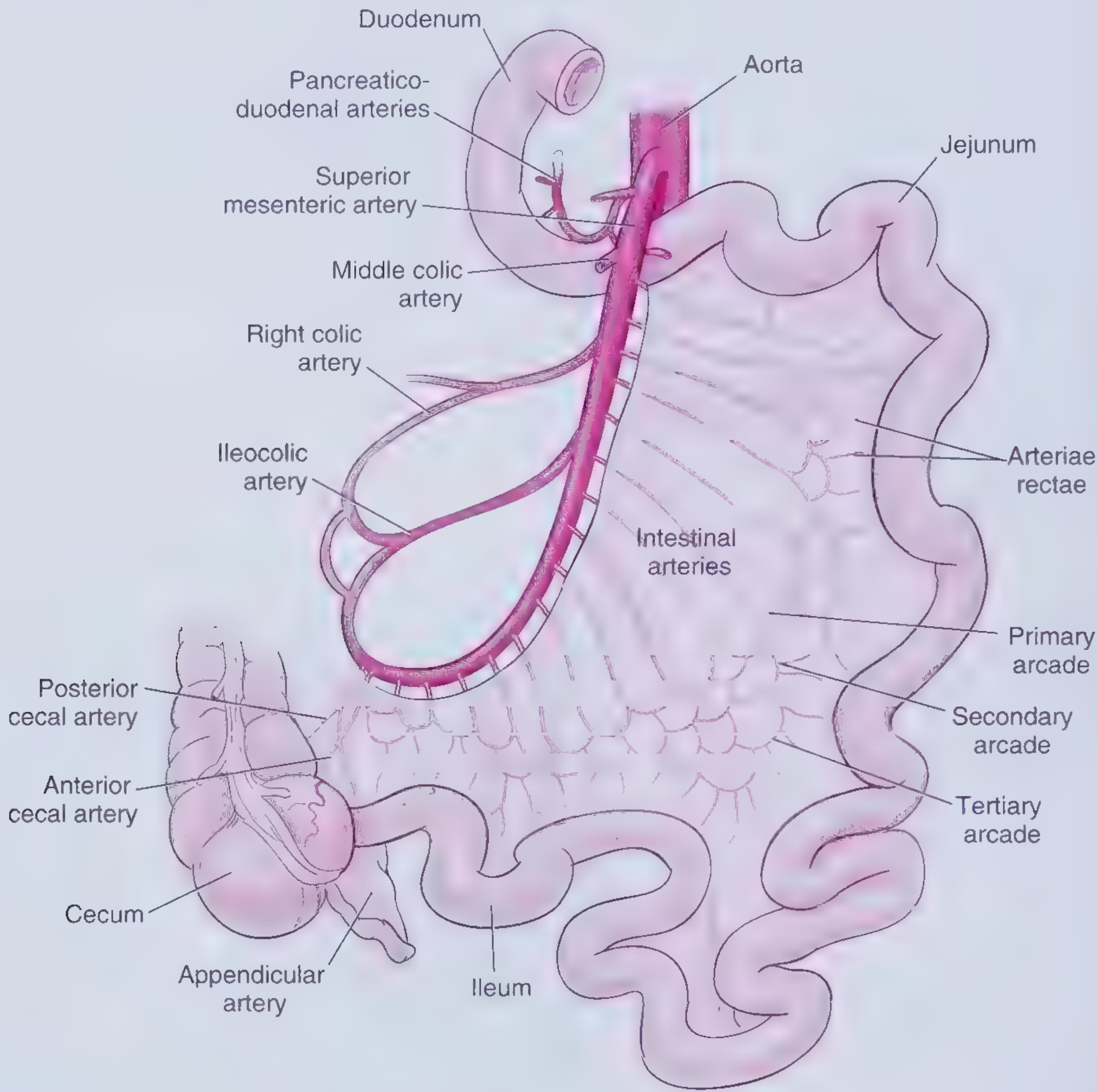
The SMA tapers just beyond the first few jejunal branches, so emboli typically lodge more proximally, near or just distal to the middle colic artery origin.
Classification
| Type | Subtypes |
|---|---|
| Acute Mesenteric Ischemia (AMI) | SMA embolus (SMAE) · SMA thrombosis / Mesenteric arterial occlusive disease (MAOD) · Nonocclusive mesenteric ischemia (NOMI) · Mesenteric venous thrombosis (MVT) · Focal segmental ischemia (FSI) |
| Chronic Mesenteric Ischemia (CMI) | Atherosclerotic CMI · Chronic mesenteric venous thrombosis |
ACUTE MESENTERIC ISCHEMIA (AMI)
Causes and Relative Frequencies
| Cause | Frequency |
|---|---|
| SMA embolus (SMAE) | 40-50% (historically cited as >50%) |
| Mesenteric arterial occlusive disease / SMA thrombosis | 25-35% |
| Nonocclusive mesenteric ischemia (NOMI) | 20% |
| Mesenteric venous thrombosis (MVT) | 5-15% |
| Focal segmental ischemia | ~5% |
1. SMA Embolism (SMAE)
Pathophysiology:
- Most common cause of AMI
- Embolic source is almost always cardiac: atrial fibrillation/flutter, left atrial/ventricular thrombus (after MI or in cardiomyopathy), valvular disease (mitral stenosis, endocarditis), aortic mural thrombus
- Up to 95% of SMAE patients have a documented cardiac disease history
- Up to 68% have simultaneous embolic events in other beds (cerebrovascular, peripheral)
- Emboli preferentially enter the SMA due to its oblique angle off the aorta
- ~50% lodge just distal to the middle colic artery origin - this is the classic site; spares proximal jejunum and the right colon (supplied by branches proximal to the embolus)
- ~15% lodge at the SMA origin itself
- Mortality without intervention: ~54%
Risk factors: Atrial fibrillation, recent MI, heart failure with low ejection fraction, valvular disease, dilated cardiomyopathy, aortic atherosclerosis
2. SMA Thrombosis / Mesenteric Arterial Occlusive Disease (MAOD)
Pathophysiology:
- Second most common arterial cause; accounts for 20-35%
- Occurs as acute-on-chronic event: in situ thrombosis superimposed on pre-existing severe atherosclerotic stenosis, most commonly within 2 cm of the SMA origin
- Because the occlusion is proximal (before the middle colic artery), it causes more extensive ischemia than embolism - involving both jejunum and ileum, and often right colon
- 20-50% of patients have a prodromal history of CMI (postprandial pain, weight loss) for weeks to months before the acute event
- Other thrombotic causes: hypercoagulable syndromes (Factor V Leiden, antiphospholipid syndrome, prothrombin gene mutations, antithrombin III deficiency, protein C/S deficiency), aortic dissection extending to SMA, in-stent or graft thrombosis
- Mortality without intervention: ~77% (higher than embolism due to larger bowel involvement)
3. Nonocclusive Mesenteric Ischemia (NOMI)
Pathophysiology:
- Results from mesenteric vasospasm - no fixed mechanical obstruction
- Triggered by low cardiac output states that activate the sympathoadrenal axis causing intense splanchnic vasoconstriction
- Common trigger situations: septic shock, cardiogenic shock, post-cardiac surgery, haemodialysis, vasopressor use (norepinephrine, vasopressin)
- The vascular bed goes into persistent spasm even after the precipitating cause is corrected
- Angiographic hallmark: diffuse vasoconstriction of SMA and its branches without a discrete filling defect
- Predominantly affects critically ill patients in the ICU
- Incidence has declined in recent decades due to better ICU monitoring, prompt correction of hypotension, and systemic vasodilator use
4. Mesenteric Venous Thrombosis (MVT)
Pathophysiology:
- Accounts for 5-15% of AMI; involves the superior mesenteric vein (SMV) in 95% of cases
- IMA is only rarely involved
- Leads to venous outflow obstruction → bowel wall oedema → arterial inflow impairment → mucosal ischaemia
- Primary MVT: no identifiable aetiology (~20%)
- Secondary MVT: identifiable aetiological factor (~80%):
- Inherited thrombophilias: Factor V Leiden, prothrombin gene mutation, protein C/S deficiency, antithrombin III deficiency
- Acquired: antiphospholipid syndrome, oral contraceptives, nephrotic syndrome, polycythaemia vera, paroxysmal nocturnal haemoglobinuria
- Local intra-abdominal: cirrhosis/portal hypertension, pancreatitis, abdominal sepsis, inflammatory bowel disease, postoperative state
- Malignancy
- More indolent course than arterial AMI; may present subacutely over days to weeks
Clinical Features of AMI
Symptoms
| Feature | Details |
|---|---|
| Abdominal pain | The cardinal symptom - sudden-onset, severe, colicky, out of proportion to physical findings (especially early); mid-abdominal |
| Nausea, vomiting | Common accompaniments |
| Diarrhoea | May be present; bloody stools suggest established infarction |
| Delirium | Particularly in the elderly |
The hallmark of AMI is severe abdominal pain that is disproportionate to the degree of tenderness on examination - this "pain-sign discrepancy" is pathognomonic early on.
Physical Examination
- Early (mucosal/submucosal ischaemia): minimal or absent signs - abdomen soft, non-tender or mildly tender
- Late (transmural infarction): peritonitis - abdominal rigidity, guarding, rebound tenderness, abdominal distension, absent bowel sounds, fever, tachycardia, hypotension, passage of bloody stools
The transition from pain-sign discrepancy to peritonitis signals bowel infarction and constitutes a surgical emergency.
Temporal Pathology
- Mucosal sloughing begins within 3 hours of onset
- Full-thickness intestinal infarction by 6 hours
Investigations
Biochemistry
| Marker | Notes |
|---|---|
| WBC | Usually >15,000/mm³; but normal WBC does not exclude early AMI |
| Serum lactate | Elevated - indicates late-stage disease; poor for early diagnosis |
| Metabolic acidosis | Low bicarbonate, raised anion gap |
| D-dimer | Raised coagulation activity; good diagnostic accuracy |
| Intestinal fatty acid binding protein (I-FABP) | Reflects mucosal damage; promising early marker |
| Ischemia-modified albumin | Promising marker in systematic reviews |
| Procalcitonin | May be helpful |
| Neutrophil-to-lymphocyte ratio (NLR) | Accurate when >9.9; enhanced with RDW |
| LDH, amylase | Elevated in severe disease |
No single serum marker reliably diagnoses early ischaemic bowel injury.
Imaging
Plain abdominal X-ray (sensitivity ~30%):
- Often normal early
- Later: formless loops of small bowel, ileus, thumbprinting of small bowel or ascending colon
- Ominous late signs: pneumatosis linearis (air in bowel wall), portal/mesenteric vein gas - both presume intestinal gangrene
- Pneumoperitoneum = perforation - immediate laparotomy
CT Angiography (CTA) - primary diagnostic modality:
- High sensitivity and specificity
- Identifies: arterial/venous occlusions, bowel wall thickening, pneumatosis, portal venous gas, free fluid/perforation
- SMAE: filling defect in SMA distal to middle colic artery
- MAOD: occlusion within 2 cm of SMA origin; surrounding calcification; prominent collaterals suggest chronic nature
- MVT: enlarged SMV with central lucency (thrombus), rim enhancement, bowel wall thickening, mesenteric oedema, collateral veins
- NOMI: diffuse SMA/branch narrowing without a discrete filling defect
Catheter-based mesenteric angiography (gold standard):
- Now largely replaced by CTA for initial diagnosis
- Still used when: diagnosis uncertain, endovascular treatment planned, NOMI (for intra-arterial vasodilator infusion)
- Findings vary by aetiology (see management section)
Doppler ultrasonography:
- Can evaluate proximal SMA/SMV; limited by bowel gas, obesity
- Moderate sensitivity; cannot image smaller mesenteric vessels reliably
- Useful in chronic disease screening; less practical in acute setting
MRI/MR venography:
- MVT: sensitivity and specificity ~100% and 98% respectively
- Limited by availability, time, and cost
Management of AMI
General Principles (All Causes)
- Immediate resuscitation: IV fluids, correct haemodynamic instability
- Nil by mouth; nasogastric tube if distended
- Broad-spectrum antibiotics - to cover gram-negative aerobes and anaerobes (bacterial translocation from ischaemic gut)
- Anticoagulation with systemic heparin - prevents thrombus propagation; initiated once ischaemia is confirmed (unless contraindicated by bleeding)
- Avoid vasopressors where possible (exacerbate splanchnic vasoconstriction); if unavoidable for haemodynamics, minimise and optimise
- Urgent imaging (CTA) followed by intervention - "time is bowel"
SMA Embolism - Specific Management
Endovascular-first approach (preferred when no peritonitis):
- Catheter-directed thromboaspiration or thrombolysis via angiographic catheter placed in SMA
- Intra-arterial papaverine (vasodilator) infused at SMA catheter site even after embolectomy to prevent/treat associated vasospasm (30-60 mg/h)
- Balloon angioplasty ± stenting of associated stenosis
Open surgical embolectomy:
- Indicated when: peritonitis present, endovascular failure, or bowel infarction
- Transverse arteriotomy of SMA; Fogarty balloon catheter embolectomy
- Bowel assessment: resect clearly non-viable bowel; leave questionable bowel
- Planned second-look laparotomy at 24-48 hours - reassess bowel viability after revascularisation
- Papaverine infusion continued intra- and post-operatively via SMA catheter
MAOD/SMA Thrombosis - Specific Management
- Larger bowel involvement; higher mortality; typically requires revascularisation
- Endovascular: retrograde open mesenteric stenting (ROMS) - SMA catheterised retrogradely via infracolic segment, balloon-expandable covered stent deployed with flare into aortic ostium; or antegrade transfemoral approach with PTA ± stent
- Open surgical revascularisation:
- Antegrade aortomesenteric bypass (from supracoeliac aorta - less often diseased) using Dacron or PTFE graft
- Retrograde aortomesenteric bypass (from infra-renal aorta or iliac artery) - more accessible but higher risk of graft kinking
- Transaortic endarterectomy - for proximal SMA and coeliac lesions
- Bowel resection and second-look laparotomy as needed
- Pre- and post-procedure: antiplatelet therapy + statin; dual antiplatelet therapy (DAPT) for 6-12 weeks post-stenting
NOMI - Specific Management
- Correct the underlying cause (treat shock, optimise cardiac output)
- Intra-arterial papaverine infusion via SMA catheter (30-60 mg/h) - the cornerstone of pharmacological treatment; dramatic reversal of vasospasm seen on repeat angiography after 24-48 hours
- Alternatively: prostaglandin E1 (PGE1) intra-arterially
- Infusion continued for 24 hours; repeat angiography to confirm resolution of vasospasm
- No operation unless peritoneal signs are present; if surgery needed, papaverine infusion is continued during and after exploration
- Leave bowel of questionable viability; second-look operation preferred over massive enterectomy
MVT - Specific Management
- Systemic anticoagulation with heparin (full-dose) is the mainstay - reduces thrombus propagation, allows collateral recanalization, reduces mortality from ~80% to ~20-30%
- Transition to long-term oral anticoagulation (warfarin or DOAC) for 3-6 months minimum; lifelong if underlying thrombophilia or idiopathic
- Thrombolysis/catheter-directed therapy: reserved for progressive disease despite anticoagulation (transhepatic or trans-splenic catheter-directed thrombolysis into SMV)
- Surgery: reserved for bowel infarction (peritonitis or CT evidence of transmural necrosis); resect non-viable bowel, second-look laparotomy
- Test for underlying thrombophilia; treat predisposing conditions
Second-Look Laparotomy
Performed 24-48 hours after initial revascularisation and resection when:
- Bowel viability was uncertain at the first operation
- Marginal bowel was left in situ
- General principle: better to perform a second look than to resect excessively - compromised but viable bowel often improves with revascularisation and supportive care
CHRONIC MESENTERIC ISCHEMIA (CMI)
Pathophysiology
- Results from atherosclerotic stenosis of two or more major splanchnic arteries (coeliac, SMA, IMA) - rarely symptomatic with single-vessel disease due to collateral compensation
- Post-prandial mesenteric blood flow demand (up to 25% of cardiac output) cannot be met by the stenotic vessels - "intestinal angina"
- The insidious development allows extensive collateral formation, so bowel infarction is rare; however, it is a precursor to acute thrombosis
Clinical Features
| Feature | Details |
|---|---|
| Post-prandial abdominal pain | Crampy, epigastric pain beginning 15-30 min after eating, lasting 1-3 hours; classic |
| "Food fear" (sitophobia) | Aversion to eating due to anticipated pain |
| Weight loss | Dramatic and progressive; often misattributed to malignancy |
| Nausea, vomiting, diarrhoea | Variable |
| Abdominal bruit | May be audible in ~50% |
Patients often undergo extensive negative workup for malignancy before the correct diagnosis is made.
Investigations
- Duplex ultrasonography: first-line screening; elevated peak systolic velocities in SMA (>275 cm/s) and coeliac (>200 cm/s) indicate significant stenosis; limited by body habitus and bowel gas
- CT angiography (CTA): most commonly used confirmatory test; demonstrates stenosis, calcification, collaterals (prominent arc of Riolan or marginal artery = clue to significant occlusion)
- Catheter-based angiography: gold standard; required pre-operatively for surgical planning
Management
Revascularisation is the definitive treatment. All symptomatic patients warrant intervention.
Endovascular (preferred first-line):
- Percutaneous transluminal angioplasty (PTA) + stent placement (balloon-expandable stents for ostial lesions; self-expanding for body lesions)
- Lower perioperative morbidity/mortality than open surgery
- Higher long-term restenosis rates (~30-40% at 3 years) vs. open surgery (~10-15%)
- Post-stenting: DAPT for 6-12 weeks, long-term single antiplatelet + statin
- Surveillance: CTA or duplex US at 1 month, then biannually for 2 years, then annually
Open surgical revascularisation:
- Better long-term durability
- Antegrade bypass from supracoeliac aorta (preferred; less atherosclerotic inflow)
- Retrograde bypass from infra-renal aorta/iliac
- Transaortic endarterectomy - suitable for proximal, discrete lesions
- Preferred in younger, fit patients, or after endovascular failure/recurrence
MESENTERIC VENOUS THROMBOSIS (MVT) - Chronic Form
- Chronic MVT involves the portal or splenic veins
- Often asymptomatic (extensive collateral venous drainage)
- May develop portal hypertension → oesophagogastric varices, splenomegaly, hypersplenism
- Some present with bleeding from oesophageal varices
- Management: anticoagulation; endoscopic variceal management; TIPS or surgical portosystemic shunting in refractory portal hypertension
Complications of AMI
| Complication | Notes |
|---|---|
| Bowel infarction / gangrene | Transmural necrosis; major cause of mortality |
| Perforation and peritonitis | Surgical emergency |
| Sepsis | Bacterial translocation |
| Short bowel syndrome | After extensive small bowel resection; most common adult aetiology; requires long-term TPN if <60 cm remaining with intact colon, or <100 cm without colon |
| Multi-organ failure | End-stage consequence; drives high mortality |
| Recurrent ischaemia | In-stent restenosis; recurrent thromboembolism |
Prognosis
| Scenario | Mortality |
|---|---|
| AMI overall (requiring intervention) | 17-21% |
| SMAE (without intervention) | ~54% |
| MAOD/SMA thrombosis (without intervention) | ~77% |
| AMI - open repair (1999-2010 data) | Improved from 43% → 33% |
| AMI - endovascular repair | Improved from 20% → 15% |
The key determinant of outcome is time to diagnosis and revascularisation - a high index of suspicion in at-risk patients (age >50, AF, recent MI, heart failure, known vascular disease, unexplained severe abdominal pain) is the most important clinical tool for reducing mortality.
Sources: Sabiston Textbook of Surgery 21e, Ch. 106 | Sleisenger & Fordtran's Gastrointestinal and Liver Disease, Ch. 118 | Schwartz's Principles of Surgery 11e, Ch. 28 | Frameworks for Internal Medicine, Ch. 18
This is a shared conversation. Sign in to Orris to start your own chat.