
Pectoralis major
Reading File
The pectoralis major is a large, fan-shaped chest muscle with two heads (clavicular and sternocostal), responsible for shoulder flexion, adduction, and medial rotation. Let me pull the full textbook coverage now.
Searching sources...
Finding Sources
Finding Sources
Reading File
Reading File
Reading File
Reading File
Reading File
Loading Image
Loading Image
Here is a complete overview of the Pectoralis Major drawn from Gray's Anatomy for Students, the THIEME Atlas of Anatomy, Imaging Anatomy Vol. 3, and Miller's Review of Orthopaedics.
Pectoralis Major
The pectoralis major is the largest and most superficial muscle of the anterior chest wall. It lies directly deep to the breast, separated from it by deep fascia and the loose connective tissue of the retromammary space. Its inferior margin forms the anterior axillary fold.
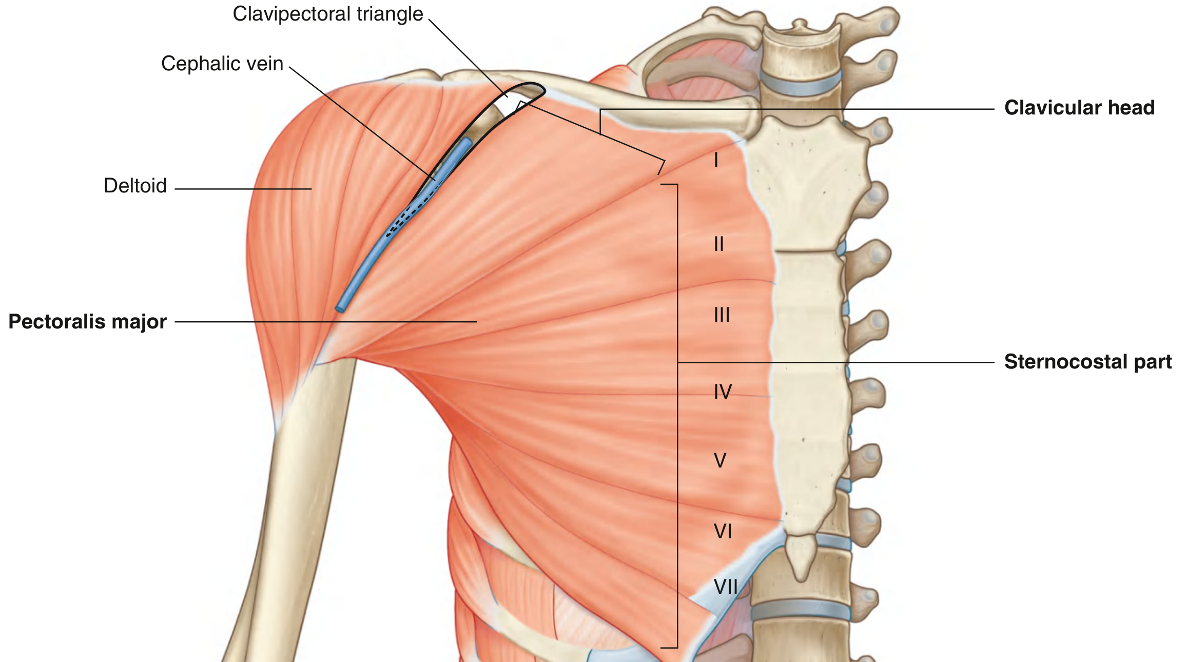
Origin (three parts)
| Part | Origin |
|---|---|
| Clavicular | Medial half of the clavicle |
| Sternocostal | Manubrium, body of sternum, and costal cartilages of ribs 2-6 (some sources: 1-7) |
| Abdominal | Anterior layer of the rectus sheath / aponeurosis of external oblique |
Insertion
The muscle fibers converge laterally into a bilaminar, U-shaped (horseshoe) tendon that inserts into the lateral lip of the intertubercular sulcus (bicipital groove) of the humerus. The tendon footprint spans approximately 4-6 cm on the humeral diaphysis.
A key anatomical detail: the tendon fibers twist on themselves so that:
- The clavicular and manubrial fibers insert into the anterior limb (lower on the humerus)
- The sternocostal fibers insert into the inferior cup
- The abdominal fibers insert into the posterior limb (highest on the humerus)
This arrangement - analogous to an unfolded hand-held fan - means that as the arm is elevated, the fibers become untwisted and stretched, increasing the force the muscle can exert.
Innervation
Medial and lateral pectoral nerves (brachial plexus roots C5-T1)
- The lateral pectoral nerve pierces pectoralis minor and ramifies on the deep surface of pectoralis major
- The medial pectoral nerve supplies both pectoralis minor and pectoralis major
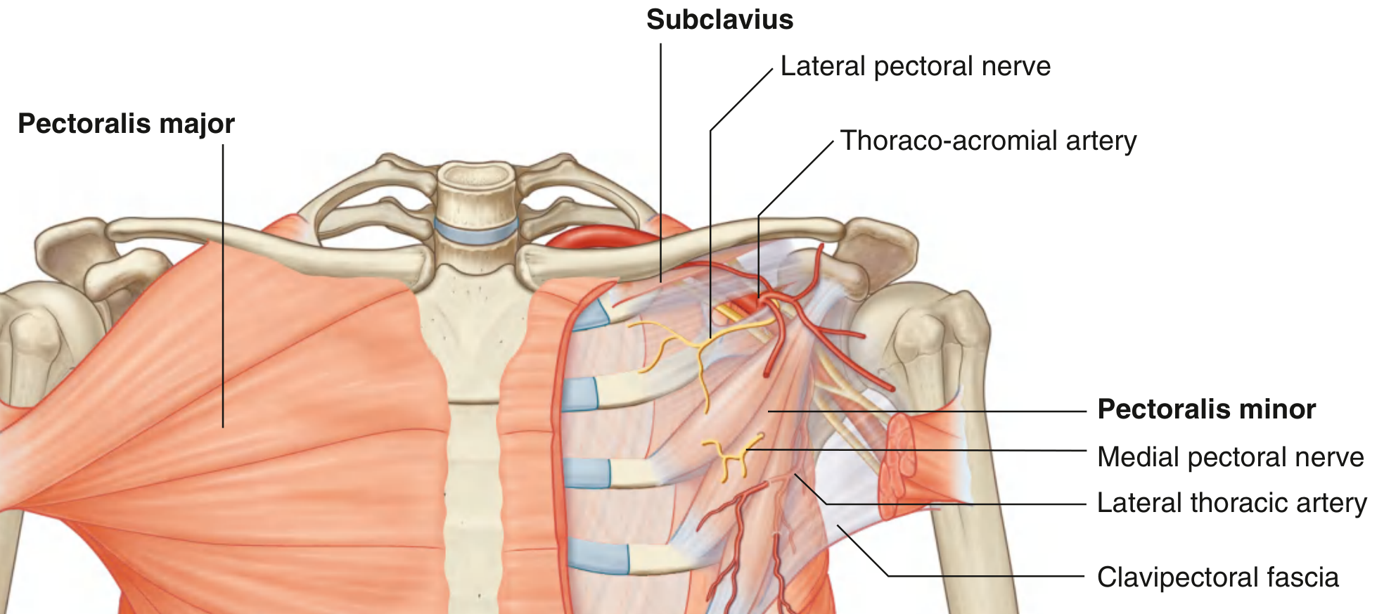
Blood Supply
Supplied by two branches of the axillary artery:
- Thoracoacromial artery (pectoral branch)
- Lateral thoracic artery
Each segment of the muscle has a distinct neurovascular supply running in the epimysium on its posterior surface.
Actions
| Action | Part Responsible |
|---|---|
| Adduction of the arm | Entire muscle |
| Medial (internal) rotation of the arm | Entire muscle |
| Flexion of the arm | Clavicular + sternocostal parts |
| Extension from flexion (against resistance) | Sternocostal head specifically |
| Assists respiration | When the shoulder girdle is fixed (accessory respiratory muscle) |
The clavicular head flexes the arm from an extended position; the sternocostal head extends the arm from a flexed position, particularly against resistance - the two heads therefore have partially antagonistic roles.
Relations
- Directly posterior (deep) to it: pectoralis minor, subclavius, and the clavipectoral fascia
- The cephalic vein runs in the deltopectoral groove (clavipectoral triangle) at the lateral border
- The axillary contents lie posterior to its inferior margin
Variations
- Poland's syndrome: Rare congenital absence of the sternal portion of pectoralis major, sometimes with ipsilateral symbrachydactyly (short, webbed fingers)
- Accessory muscles: Several are described, including the pectoralis quartus and chondroepitroclearis, which originate from the caudal pectoralis major or from ribs 5-6
Clinical Relevance: Rupture
- Typically caused by excessive eccentric load on a maximally contracted muscle; classically occurs in weightlifters (bench press)
- The shoulder must be in an abducted, extended, and externally rotated position with maximal muscle tension for rupture to occur
- Most commonly a tendinous avulsion from the humeral insertion
- Signs: localized swelling, ecchymosis, palpable defect / axillary webbing, weakness of adduction and internal rotation
- Complete ruptures: surgical repair to bone
- Partial ruptures / myotendinous tears: conservative (nonoperative) management
- Ruptures are infrequently reported in women
Sources: Gray's Anatomy for Students (9780323934237) | THIEME Atlas of Anatomy - General Anatomy & Musculoskeletal System (9781626237186) | Imaging Anatomy Vol. 3 (9781626239845) | Miller's Review of Orthopaedics 9th Ed. (9780443112140)
This is a shared conversation. Sign in to Orris to start your own chat.